 Tools
Tools
 Gram-Positive Bacteria
Introduction
Health professionals need to understand the important difference between gram-positive and gram-negative bacteria. Gram-positive bacteria are classified by the color they turn in the Gram stain. Hans Christian Gram developed the staining method in 1884. The staining method uses crystal violet dye, which is retained by the thick peptidoglycan cell wall of gram-positive organisms. This reaction gives gram-positive organisms a blue color when viewed under a microscope. Although gram-negative organisms classically have an outer membrane, they have a thinner peptidoglycan layer that does not retain the blue dye used in the initial dyeing process. Other information used to differentiate bacteria is the shape. Gram-positive bacteria comprise cocci, bacilli, or branching filaments.
Etiology
Register For Free And Read The Full Article
 Search engine and full access to all medical articles
- 10 free questions in your specialty
- Free CME/CE Activities
- Free daily question in your email
- Save favorite articles to your dashboard
- Emails offering discounts
Learn more about a Subscription to StatPearls Point-of-Care
Etiology
Gram-positive cocci include Staphylococcus (catalase-positive), which forms clusters, and Streptococcus (catalase-negative), which forms chains. The staphylococci are further subdivided into coagulase-positive (S. aureus) and coagulase-negative (S epidermidis and S saprophyticus) species. Streptococcus bacteria are subdivided into S pyogenes (group A), S agalactiae (group B), enterococci (group D), S viridans, and S pneumoniae.
Gram-positive bacilli (rods) are subdivided based on their ability to form spores. Bacillus and Clostridia are spore-forming rods, while Listeria and Corynebacterium are not. Spore-forming rods can survive in environments for many years. Also, the branching filament rods encompass Nocardia and Actinomyces. Gram-positive organisms have a thicker peptidoglycan cell wall than gram-negative bacteria. It is a 20 to 80 nm-thick polymer, whereas the peptidoglycan layer of the gram-negative cell wall is 2-3 nm thick and covered by an outer lipid bilayer membrane.
Epidemiology
Bloodstream infection mortality rates have increased by 78% in just two decades[1]. Gram-positive organisms have highly variable growth and resistance patterns. The SCOPE project (Surveillance and Control of Pathogens of Epidemiologic Importance) found that gram-positive organisms in those with an underlying malignancy accounted for 62% of all bloodstream infections in 1995 and 76% in 2000, while gram-negative organisms accounted for 22% and 14% of infections for these years.[2]
Pathophysiology
Gram-positive Cocci
Staphylococcus aureus is a gram-positive, catalase-positive, coagulase-positive coccus that occurs in clusters. S aureus can cause inflammatory diseases, including skin infections, pneumonia, endocarditis, septic arthritis, osteomyelitis, and abscesses. S aureus can also cause toxic shock syndrome (TSST-1), scalded skin syndrome (exfoliative toxin, and food poisoning (enterotoxin).
Staphylococcus epidermidis is a gram-positive, catalase-positive, coagulase-negative coccus that occurs in clusters and is novobiocin-sensitive. S epidermidis commonly infects prosthetic devices and IV catheters, producing biofilms. Staphylococcus saprophyticus is novobiocin resistant and is a normal flora of the genital tract and perineum. S saprophyticus accounts for the second most common cause of uncomplicated urinary tract infections (UTI).
Streptococcus pneumoniae is a gram-positive, encapsulated, lancet-shaped diplococcus that most commonly causes otitis media, pneumonia, sinusitis, and meningitis. Streptococcus viridans consists of S mutans and S mitis, found in the normal flora of the oropharynx, and commonly causes dental caries and subacute bacterial endocarditis (S sanguinis). Streptococcus pyogenes is a gram-positive group A cocci that can cause pyogenic infections (pharyngitis, cellulitis, impetigo, erysipelas), toxigenic infections (scarlet fever, necrotizing fasciitis), and immunologic infections (glomerulonephritis and rheumatic fever). ASO titer detects S pyogenes infections.
Streptococcus agalactiae is a gram-positive group B cocci that colonizes the vagina and is found mainly in babies. Pregnant women need screening for group B strep (GBS) at 35 to 37 weeks of gestation. Enterococci are gram-positive group D cocci found mainly in the colonic flora and can cause biliary tract infections and UTIs. Vancomycin-resistant enterococci (VRE) are an important cause of nosocomial infections.
Gram-Positive Rods
Clostridia are gram-positive, spore-forming rods, including C tetani, C botulinum, C perfringens, and C difficile. C difficile is often secondary to antibiotic use (clindamycin/ampicillin), PPI use, and recent hospitalization. Treatment involves primarily oral vancomycin. Bacillus anthracis is a gram-positive spore-forming rod that produces anthrax toxin, resulting in an ulcer with a black eschar. Bacillus cereus is a gram-positive rod that can be acquired from spores that survive in undercooked or reheated rice. Symptoms include nausea, vomiting, and watery non-bloody diarrhea.
Corynebacterium diphtheria is a gram-positive club-shaped rod that can cause pseudomembranous pharyngitis, myocarditis, and arrhythmias. Toxoid vaccines prevent diphtheria. Listeria monocytogenes is a gram-positive rod acquired through ingestion of cold deli meats and unpasteurized dairy products or through vaginal transmission during birth. Listeria can cause neonatal meningitis, meningitis in immunocompromised patients, gastroenteritis, and septicemia. Treatment includes ampicillin.
History and Physical
It is important to identify patients with sepsis and order necessary blood cultures and labs.
Physical
- Bullous impetigo
- Draining sinus tracts
- Erythema
- Fever
- Murmur if endocarditis is present
- Petechiae if toxic shock syndrome is present
- Superficial abscesses
- Warmth
Evaluation
Once a gram-positive organism infection is suspected, these laboratory studies are useful:
- Complete blood count
- Electrolytes
- Blood cultures
- Procalcitonin level
- Echocardiogram if endocarditis is suspected
- Joint aspiration if a septic joint is suspected
Treatment / Management
Penicillin, discovered by Alexander Fleming in 1928 and introduced into clinical practice during World War II, was the first widely used antibiotic. Natural penicillins have excellent activity against many streptococcal species, but limited activity against most S aureus strains due to penicillinase production, and they do not reliably cover Enterococcus species. Penicillinase-resistant penicillins, including nafcillin, oxacillin, cloxacillin, and dicloxacillin, are active against methicillin-susceptible S aureus (MSSA) and many streptococcal species. Antipseudomonal penicillins, including piperacillin and ticarcillin, have activity against many gram-positive organisms, gram-negative organisms, Pseudomonas aeruginosa, and, when combined with a β-lactamase inhibitor, many anaerobes. Carbapenems provide broad-spectrum activity against many gram-positive and gram-negative organisms, as well as anaerobes.[3][4][5](B3)
Trimethoprim/sulfamethoxazole, clindamycin, and doxycycline are oral antibiotics used for mild to moderate MRSA infections. It is important to note that trimethoprim/sulfamethoxazole increases warfarin levels, leading to increased INR. Vancomycin, linezolid, daptomycin, and tigecycline cover moderate-to-severe community- and hospital-acquired MRSA. Vancomycin requires renal dosing with trough levels between 15 to 20. Linezolid is an option for patients allergic to vancomycin. A complete blood count should be checked weekly to monitor for bone marrow suppression, neutropenia, thrombocytopenia, and anemia. Linezolid, daptomycin, and tigecycline are options to treat vancomycin-resistant enterococci.[6][7][8]
Differential Diagnosis
The differential diagnosis includes the following:
- Bacteremia
- Bronchiectasis imaging
- Chemical burns
- Diarrhea
- Electrical injuries in emergency medicine
- Emergent management of acute otitis
- Emergent management of thermal burns
- Empyema imaging
- Fever in the infant and toddler
- Fever without a focus
- Henoch-schonlein purpura
- Hospital-acquired infections
- Ingrown nails
- Necrotizing enterocolitis imaging
Prognosis
The prognosis following infection with gram-positive organisms is variable. The highest mortality rates are in elderly persons who tend to have suppressed immune systems and less physiologic reserve.
Enhancing Healthcare Team Outcomes
Health professionals, including doctors, nurses, and pharmacists, need to be aware of risk factors to treat patients with selected antibiotics properly. Pharmacists need to accurately monitor vancomycin trough levels to avoid mortality in patients with S aureus. They also need to review medications for dosing and interactions and counsel patients to complete all prescribed antibiotics. Infection control nurses evaluate nosocomial infections and implement appropriate policies. An interprofessional approach will produce the best outcomes. Outcomes: Screening for MRSA risk factors enhances infection control. MRSA risk factors include patients who are older than 65, have a urinary catheter, have had previous antibiotic treatment in the past 3 months, have trauma, and are admitted from a long-term facility.[9]
Media
(Click Image to Enlarge)
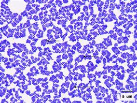
Gram Stain of Staphylococcus aureus.
Contributed by S Jones, MD
References
. National Nosocomial Infections Surveillance (NNIS) system report, data summary from January 1992-April 2000, issued June 2000. American journal of infection control. 2000 Dec:28(6):429-48 [PubMed PMID: 11114613]
Wisplinghoff H, Seifert H, Wenzel RP, Edmond MB. Current trends in the epidemiology of nosocomial bloodstream infections in patients with hematological malignancies and solid neoplasms in hospitals in the United States. Clinical infectious diseases : an official publication of the Infectious Diseases Society of America. 2003 May 1:36(9):1103-10 [PubMed PMID: 12715303]
Righi E, Carnelutti A, Bassetti M. Current role of oxazolidinones and lipoglycopeptides in skin and soft tissue infections. Current opinion in infectious diseases. 2019 Apr:32(2):123-129. doi: 10.1097/QCO.0000000000000529. Epub [PubMed PMID: 30664028]
Level 3 (low-level) evidenceZamoner W, Prado IRS, Balbi AL, Ponce D. Vancomycin dosing, monitoring and toxicity: Critical review of the clinical practice. Clinical and experimental pharmacology & physiology. 2019 Apr:46(4):292-301. doi: 10.1111/1440-1681.13066. Epub 2019 Feb 19 [PubMed PMID: 30623980]
Bento D, Tobin EH. Listeria monocytogenes Infection (Listeriosis). StatPearls. 2026 Jan:(): [PubMed PMID: 30521259]
Rostkowska KA, Szymanek-Pasternak A, Simon KA. Spontaneous bacterial peritonitis - therapeutic challenges in the era of increasing drug resistance of bacteria. Clinical and experimental hepatology. 2018 Dec:4(4):224-231. doi: 10.5114/ceh.2018.80123. Epub 2018 Dec 3 [PubMed PMID: 30603669]
Gashaw M, Berhane M, Bekele S, Kibru G, Teshager L, Yilma Y, Ahmed Y, Fentahun N, Assefa H, Wieser A, Gudina EK, Ali S. Emergence of high drug resistant bacterial isolates from patients with health care associated infections at Jimma University medical center: a cross sectional study. Antimicrobial resistance and infection control. 2018:7():138. doi: 10.1186/s13756-018-0431-0. Epub 2018 Nov 19 [PubMed PMID: 30479751]
Bolia IK, Tsiodras S, Chloros GD, Kaspiris A, Sarlikiotis T, Savvidou OD, Papagelopoulos PJ. A Review of Novel Antibiotic Regimens for the Treatment of Orthopedic Infections. Orthopedics. 2018 Nov 1:41(6):323-328. doi: 10.3928/01477447-20181024-02. Epub [PubMed PMID: 30452066]
Callejo-Torre F, Eiros Bouza JM, Olaechea Astigarraga P, Coma Del Corral MJ, Palomar Martínez M, Alvarez-Lerma F, López-Pueyo MJ. Risk factors for methicillin-resistant Staphylococcus aureus colonisation or infection in intensive care units and their reliability for predicting MRSA on ICU admission. Le infezioni in medicina. 2016 Sep 1:24(3):201-9 [PubMed PMID: 27668900]