 Tools
Tools
 Embryology, Gastrulation
Introduction
Gastrulation is a critical process during week 3 of human development. Gastrulation is an early developmental process in which an embryo transforms from a 1-dimensional layer of epithelial cells (a blastula) into a multilayered, multidimensional structure called the gastrula. In triploblastic organisms such as reptiles, birds, and mammals, gastrulation results in a 3-layered organism composed of endoderm, mesoderm, and ectoderm. Each germ layer corresponds to the development of specific primitive systems during organogenesis. In addition to setting the embryo up for organ formation, gastrulation provides a mechanism for developing a multilevel body plan that demarcates anatomical axis formation. These axes are the dorsal/ventral axis, also termed the anterior/posterior or rostral/posterior axis, and the cranial/caudal or superior/inferior axis. Gastrulation also promotes the retention of global left-right symmetry and the loss of bilateral symmetry in specific organs, such as the heart.
Development
Register For Free And Read The Full Article
 Search engine and full access to all medical articles
- 10 free questions in your specialty
- Free CME/CE Activities
- Free daily question in your email
- Save favorite articles to your dashboard
- Emails offering discounts
Learn more about a Subscription to StatPearls Point-of-Care
Development
After fertilization, the single-celled zygote undergoes multiple mitotic cleavages of the blastomeres to change from a 2-celled to a 16-celled ball or morula. The morula begins as a solid mass of totipotent blastomeres that undergo compaction and cavitation to form the blastula (non-mammalian term) or the blastocyst (human development). Within the blastocyst, 2 tissue layers form: an outer shell, known as the trophoblast, and an inner collection of cells, known as the inner cell mass (ICM). Cells within the outer shell bind together via gap junctions and desmosomes to undergo compaction, ultimately forming a water-tight shell called the trophoblast.[1]
The outer trophoblast develops into structures that provide nutrients, facilitate implantation of the growing embryo in the uterine lining, and contribute to the formation of the placenta. Additionally, the trophoblast cells are essential in the cavitation of the solid morula into a hollow ball of cells with an internal cavity. Trophoblast cells utilize active sodium ion transport and water osmosis to form a fluid-filled cavity known as the blastocoel.[2] The cells remaining after blastocoel formation are pluripotent ICM progenitor cells that give rise to the distinctive structures of the fetus. Rather than being arranged as a solid sphere of cells, the ICM is pushed off to 1 side of the trophoblast sphere. Together, the trophoblastic layer, blastocoel, and inner cell mass are the characteristic features of the human blastocyst.[3]
From zygote to blastocyst formation, the organism is surrounded by the zona pellucida, a layer of the extracellular matrix that helps protect against implantation in the uterine tubes. During blastocyst formation, the zona pellucida begins to disappear, allowing the ball of cells to proliferate, differentiate, change shape, and eventually implant into the uterine wall. During implantation, the trophoblastic layer surrounding the blastocyst further differentiates into 2 functionally distinct layers. The outer trophoblast, known as the syncytiotrophoblast, releases proteolytic enzymes to assist with endometrial implantation. This layer also releases human chorionic gonadotropin (hCG), a hormone necessary for regulating progesterone secretion, the protein used in many pregnancy tests.[4] The inner trophoblast layer, known as the cytotrophoblast, is a single layer of cells that surrounds the extraembryonic mesoderm. Within the cytotrophoblast is the ICM. During the second week of human development, the cells of the ICM spread into a flattened tissue layer and differentiate into a 2-layered tissue comprising the epiblast (columnar epithelial cells) and the hypoblast (cuboidal epithelial cells), collectively known as the bilaminar embryonic disc.[5]
The formation of the bilaminar embryonic disc establishes the dorsal/ventral axis, with the epiblast cell layer positioned dorsal to the hypoblast. The anatomical location of the bilaminar disc is found between the amniotic cavity and the primitive yolk sac. The cells of the epiblast stretch to form a semi-sphere known as the amniotic cavity, while the cells of the hypoblast extend to surround the yolk sac. On the hypoblast is a raised area of columnar cells known as the prechordal plate; this is the earliest delineation of cranial from caudal. Development of the bilaminar embryonic disc directly precedes gastrulation, the stage in week 3 of development that involves the transformation of the human blastocyst into a multilayered gastrula with endoderm, mesoderm, and ectoderm.
Cellular
The beginning of gastrulation is marked by the appearance of the primitive streak, a groove at the caudal end of the epiblast.[6] Thus, the formation of the primitive streak firmly establishes the cranial/caudal axis. The primitive streak initially forms via a thickening of cells near the connecting stalk. As cells proliferate and migrate toward the midline of the embryo, the thickening elongates to become linear in shape, thus the term primitive streak. The cranial end of the embryo seems to play an important role in initiating gastrulation. At the cranial end of the primitive streak, epiblast cells ingress at a greater rate, forming a circular cavity known as the primitive pit. As the primitive streak elongates, migrating epiblast cells join the streak at the cranial end, forming a mass of cells around the primitive pit. This mass is called the primitive node, which becomes the primary tissue organizer in which transcription factors and chemical signaling drive tissue induction. Known signaling factors and pathways in primitive streak formation include transforming growth factor-beta (TGFB), Wnt, Nodal, and bone morphogenetic protein (BMP), which are discussed in more detail in the molecular section.
Epithelial cells at the lateral edge of the epiblast undergo an epithelial-to-mesenchymal transition, delaminate (detach), and migrate down or into the primitive streak.[7] The movement of epiblastic mesenchymal cells down the primitive streak is known as ingression. The first cells to move down the primitive streak integrate into the hypoblast and form the endoderm, the first of the 3 germ layers. The second set of cells to detach and ingress fills the space between the endoderm and epiblast layer to form the second germ layer, mesoderm. Multiple mesodermal structures develop: cells that move into the body stalk help form the extraembryonic mesoderm and, later, the umbilical cord; cells passing through the primitive pit become the notochord or paraxial mesoderm; and other cells passing through the primitive streak become the lateral plate or extraembryonic mesoderm. Finally, the remaining epiblast cells transform into the final germ layer, ectoderm. Cell proliferation and ingression continue in all directions as the embryo grows; however, the primitive streak always expands directionally from the caudal to the cranial end and then regresses in the opposite fashion. Regression occurs after the formation of the intra-embryonic mesoderm, and the primitive streak should completely disappear by the end of the fourth week. A lack of regression of the primitive streak results in clinical abnormalities.
After the 3 germ layers have formed, the newly formed structure (the trilaminar embryonic disc or gastrula) is primed for organ system formation, which relies heavily on direct interactions and induction between the endoderm, mesoderm, and ectoderm. Cells continue to invaginate through what is now called the primitive node. The cells begin to form a hollow tube extending from the cranial end to the prechordal plate, known as the notochordal process. As the embryo grows in all directions, the notochordal process elongates until it fuses with the endoderm to form the notochordal plate. Once the fusion is complete, there is a free passageway between the amniotic cavity and the yolk sac, known as the neurenteric canal, or canal of Kovalevsky.[8] It is theorized that the neuroenteric canal forms to maintain pressure equilibrium between both chambers. Later in development, the 2 edges of the notochordal plate fuse into a solid mesodermal rod known as the notochord. The notochord is a critical embryologic structure that provides structural support and marks the midline of the embryo. Chemical and physical interactions between the notochord and dorsally situated ectoderm give rise to neuroectoderm and, eventually, the nervous system.
Biochemical
RNA helicase A (RHA) can function as a helicase for both RNA and DNA. The sequence and biochemical conservation of RHA and its homologs in humans suggest an evolutionarily conserved function. Normal gastrulation depends on RNA helicase A activity, as a lack of proper RHA signaling results in ectodermal cell death with clear alterations in differentiation.[9] The differentiation of pluripotent stem cells into lineage-specified cells within the endoderm, mesoderm, and ectoderm is marked by the downregulation of pluripotency markers (ie, Oct4, Nanog, or Sox2) and the activation of lineage-specific gene expression, including microRNAs. MicroRNAs (miRNAs) have been demonstrated to be enriched in germ layers, specifically targeting TGFB to promote mesoderm and restrict or block neuroectoderm.[10]
Molecular Level
Primitive Streak
The initiation of the primitive streak is based on a system of signaling pathways that both positively and negatively regulate downstream expression. The combination of TGFB, Wnt, Nodal, and BMPs is important in primitive streak development.[11][12][13][14][15] The interplay between Wnt and TGFB signaling appears to induce the formation of the primitive streak. Specifically, Vg1 (a member of the TGFB family) has been shown to induce streak formation and to prevent formation with Vg1 misexpression at the posterior marginal zone.[16] Vg1 acts on Nodal to continue the chemical cascade to streak formation. To ensure the proper location of the streak on the epiblast, the hypoblast releases antagonists of Nodal signaling.[17] Additionally, the induction of streak formation can be regulated by Wnt factors; not only has upregulation of Wnt induced streak formation, but the use of Wnt antagonists such as Dkk-1 and Crescent prevents streak formation.[11] Finally, BMP signaling has been shown to regulate streak formation. Closer to the streak, BMP concentration is low, with the surrounding embryo exhibiting higher levels of active BMP. In addition to this, BMP inhibitors cause the formation of a streak in chick embryos. As seen in BMP and other signaling, concentration gradients are typical throughout most of gastrulation. Different concentrations of signaling factors allow cells to differentiate into unique tissues.
Endoderm
Endoderm is the embryonic precursor to the thyroid, lungs, pancreas, liver, and intestines, which evolve from 4 consecutive developmental steps: proliferation and induction of pluripotent stem cells, the separation of stem cell-derived endoderm versus mesoderm germ layers, anterior-posterior patterning, and bifurcation of the liver and pancreas. Cells near the anterior portion of the primitive streak express Forkhead box A2 (Foxa2) and give rise to definitive endoderm. The definitive endoderm pattern itself into the foregut, midgut, and hindgut via mesodermal induction during embryonic folding, with foregut cells expressing Hhex, Sox2, and Foxa2, and hindgut cells expressing the homeobox genes Cdx1, Cdx2, and Cdx4. The upregulation of TGFB signaling promotes pancreas formation, with BMP and FGF/MAPK signaling specifying the liver.[18] The specification of the respiratory bud begins with the expression of the Nbx1-2 gene. Complex signaling between the respiratory bud epithelium and mesoderm involves FGF and FGFR interactions to promote respiratory bud growth.[19]
Mesoderm
Epiblast cells invaginating through the primitive streak that express high levels of fibroblast growth factor 2 (FGF2) are fated to become mesodermal cells. More specifically, they end up as paraxial, intermediate, or lateral plate mesoderm, which correlates with different tissues as the embryo develops.[20]
Notochord
Progenitor cells from the primitive node and primitive pit migrate to initiate notochord formation. Epiblast cells from the floor plate of the amniotic cavity fill in the notochord, forming a thick, rod-like structure along the midline of the embryo. Providing support and serving as an induction center for surrounding cells, the notochord in vertebrates extends the entire length of the vertebral column and reaches as far as the midbrain. The notochord develops first, and then mesodermal cells grow medially to surround it. The notochord is present only in developing organisms and serves the primary goal of patterning the surrounding tissues. The notochord secretes Sonic Hedgehog, Chordin, and Noggin in a morphogenetic gradient, with the highest concentration near the notochord and diffusion outward. These bind to receptors on target cells to induce specification and differentiation events in the neural plate, somites, and ectoderm.[21]
Mesoderm divides into 3 main categories: paraxial (or axial), intermediate, and lateral (or lateral plate) mesoderm, which are the embryonic precursors to a large variety of cells and tissues, including smooth, cardiac, and skeletal muscle, kidney, reproductive organs, the muscles of the tongue and the pharyngeal arches, connective tissue, bone, cartilage, the dermis and subcutaneous layers of the skin, dura mater, vascular endothelium, blood cells, microglia, and adrenal cortex. Cells of the paraxial mesoderm first organize to form somitomeres. As the somitomeres develop into somites in a cranial-to-caudal fashion, the outer cells undergo a mesenchymal-to-epithelial transition, which serves as a distinct boundary between individual somites. Individual somites then separate into cranial and caudal portions, followed by the cranial portion of each fusing with the caudal portion of the somite directly anterior to it. Distinct regions of each somite (sclerotome, dermatome, myotome) give rise to specific tissue and cell types as the body matures. The skull, vertebral column, and brain meninges develop from the mesoderm surrounding the neural tube and notochord. The intermediate mesoderm connects the paraxial mesoderm with the lateral plate and differentiates into urogenital structures. The lateral plate mesoderm splits into a parietal (or somatic) layer to aid lateral body fold wall formation and a visceral (or splanchnic) layer involved in gut tube formation.
Ectoderm
The interplay between BMPs and Hox genes is integral to the differentiation of the remaining epiblast into ectoderm. This is especially important for the surface ectoderm and for what becomes the neuroectoderm, which gives rise to the brain and spinal cord.[13] The notochord is the main inductive tissue delineating neuroectoderm from the remaining ectoderm that becomes skin.[22] The entire presumptive ectoderm plate expresses BMP and TGFB. Noggin and Chordin secretion from the notochord diffuses into the ectoderm directly anterior to the notochord and binds to receptors in the overlying ectoderm to block BMP. The BMP blockade specifies the tissue as neural ectoderm, while the remaining ectoderm, which still expresses BMP, becomes skin.
Function
Gastrulation occurs during week 3 of human development. The process of gastrulation generates the 3 primary germ layers: ectoderm, endoderm, and mesoderm. Gastrulation primes the system for organogenesis and is one of the most critical steps of development. The endoderm is the innermost layer, which gives rise to the gastrointestinal tract, the lining of the gut, the liver, the pancreas, and portions of the lungs and glandular tissues. The mesoderm gives rise to the musculoskeletal system, including its connective tissue, the non-epidermal portions of the integumentary system, the circulatory system, the kidney, and the internal sex organs. The ectoderm is the outer layer of the embryo, which gives rise to the external ectoderm (epidermis, hair, nails) and the neuroectoderm (neural crest and neural tube), along with the lens of the eyes and the inner ear. Another important function of gastrulation is to establish directionality within the developing embryo. Cranial/caudal directionality is established by the placement of the prechordal plate and the path of the primitive groove. The layering of the epiblast and hypoblast establishes the dorsal/ventral axis.
Mechanism
Gastrulation involves a complex series of cellular morphogenesis, cellular movements, and cell signaling via transcription factors, chemical morphogenetic gradients, and differential gene expression to induce germ cell layer formation, which orchestrates the initiation of eventual organ system development.
Clinical Significance
Miscarriage is the most frequent type of pregnancy loss, according to the American College of Obstetricians and Gynecologists. Spontaneous abortion is defined as embryonic or fetal death or passage of the products of conception before 20 weeks of gestation, with early miscarriage occurring in the first thirteen weeks. Estimates are that 10% to 25% of all clinically recognized pregnancies end in miscarriage.[23][24] Investigators report that most defects related to gastrulation are incompatible with life, with select instances where mothers can carry fetuses with associated teratologies to term. Morphogenetic processes between the blastocyst stage and gastrulation can be altered, resulting in structural abnormalities, including patterns of multiple congenital anomalies (MCAs) arising from developmental field defects. Severe damage may cause the death of the product of conception, or embryonic stem cells may repair the damage and allow development to continue.
Teratoma is defined as a solid mass or germ cell tumor comprised of a combination of tissues from all 3 germ layers. Teratoma is often a result of the abnormal persistence (or lack of full regression) of the primitive streak. The most commonly diagnosed fetal teratomas are sacrococcygeal teratoma (Altman types I, II, and III) and cervical (neck) teratoma.[25] Because these teratomas protrude from the fetal body into the surrounding amniotic fluid, they can be visible on a routine prenatal ultrasound. Teratomas within the fetal body are less apparent on ultrasound; an MRI of the pregnant uterus is more informative.
Sirenomelia (or mermaid syndrome) is a congenital abnormality consisting of a total or partial fusion of the lower limbs, often accompanied by urogenital and gastrointestinal malformation.[26] Sirenomelia incidence rates in the literature range from 1 per 60,000 to 1 per 100,000, with a male-to-female bias.[27] There are 2 hypotheses regarding the development of this pathology. The first, the vascular steal theory, posits that insufficient blood flow to the caudal mesoderm impairs the formation of midline structures, thereby preventing the precise development of the lower limb.[27] The second, the blastogenesis hypothesis, explains dysfunction at the caudal mesoderm. This hypothesis posits that a dysfunctional event during gastrulation damages the caudal mesoderm.[27]
Tethered cord syndrome is a rare neurological disorder (related to spina bifida) resulting from congenital abnormalities surrounding gastrulation or injury later in life. Symptoms arise from malformations in tissue attachments that stretch the spinal cord, eventually restricting movement, inducing loss of feeling, or causing pain and autonomic symptoms.[28] Errors in gastrulation, neurulation, or primitive streak regression can result in tethered cord syndrome.[29] Specifically, temporary cell-to-cell communication between the yolk sac and the external amniotic membrane at the neurenteric canal provides critical signaling during normal gastrulation, which ends upon successful completion of gastrulation. Continued communication between the yolk sac and amniotic sac results in persistent progenitor cell proliferation, leading to duplication of neuronal tissue (split cord malformation of the vertebral column) or cyst formation (neurenteric cyst).[30]
Media
(Click Image to Enlarge)
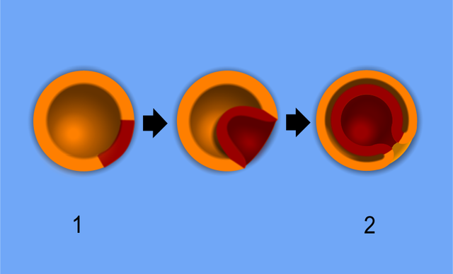
Diagram of gastrulation of a blastula. Contributed From Wikimedia by Wiki Commons user Pidalka44.
(Click Image to Enlarge)
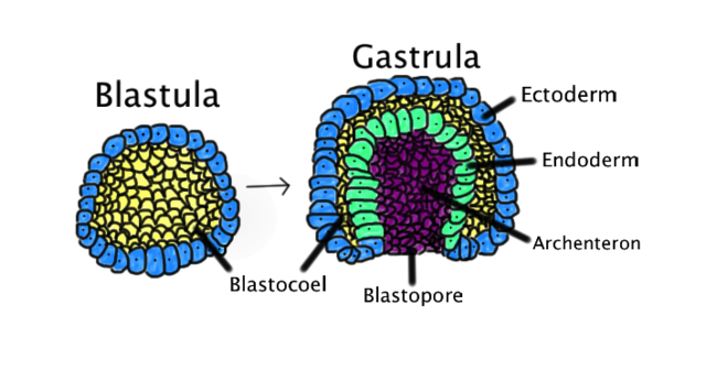
Comparison of blastula to gastrula. Contributed From Wikimedia by Abigail Pyne.
References
Zhang HT, Hiiragi T. Symmetry Breaking in the Mammalian Embryo. Annual review of cell and developmental biology. 2018 Oct 6:34():405-426. doi: 10.1146/annurev-cellbio-100617-062616. Epub 2018 Aug 10 [PubMed PMID: 30095292]
Câmara DR, Kastelic JP, Thundathil JC. Role of the Na(+)/K(+)-ATPase ion pump in male reproduction and embryo development. Reproduction, fertility, and development. 2017 Aug:29(8):1457-1467. doi: 10.1071/RD16091. Epub [PubMed PMID: 27456939]
Marikawa Y, Alarcón VB. Establishment of trophectoderm and inner cell mass lineages in the mouse embryo. Molecular reproduction and development. 2009 Nov:76(11):1019-32. doi: 10.1002/mrd.21057. Epub [PubMed PMID: 19479991]
Level 3 (low-level) evidenceBastida-Ruiz D, Yart L, Wuillemin C, Ribaux P, Morris N, Epiney M, Martinez de Tejada B, Cohen M. The fine-tuning of endoplasmic reticulum stress response and autophagy activation during trophoblast syncytialization. Cell death & disease. 2019 Sep 9:10(9):651. doi: 10.1038/s41419-019-1905-6. Epub 2019 Sep 9 [PubMed PMID: 31501418]
Niu Y, Sun N, Li C, Lei Y, Huang Z, Wu J, Si C, Dai X, Liu C, Wei J, Liu L, Feng S, Kang Y, Si W, Wang H, Zhang E, Zhao L, Li Z, Luo X, Cui G, Peng G, Izpisúa Belmonte JC, Ji W, Tan T. Dissecting primate early post-implantation development using long-term in vitro embryo culture. Science (New York, N.Y.). 2019 Nov 15:366(6467):. pii: eaaw5754. doi: 10.1126/science.aaw5754. Epub 2019 Oct 31 [PubMed PMID: 31672917]
Mikawa T, Poh AM, Kelly KA, Ishii Y, Reese DE. Induction and patterning of the primitive streak, an organizing center of gastrulation in the amniote. Developmental dynamics : an official publication of the American Association of Anatomists. 2004 Mar:229(3):422-32 [PubMed PMID: 14991697]
Level 3 (low-level) evidenceSimunovic M, Metzger JJ, Etoc F, Yoney A, Ruzo A, Martyn I, Croft G, You DS, Brivanlou AH, Siggia ED. A 3D model of a human epiblast reveals BMP4-driven symmetry breaking. Nature cell biology. 2019 Jul:21(7):900-910. doi: 10.1038/s41556-019-0349-7. Epub 2019 Jul 1 [PubMed PMID: 31263269]
Rulle A, Tsikolia N, de Bakker B, Drummer C, Behr R, Viebahn C. On the Enigma of the Human Neurenteric Canal. Cells, tissues, organs. 2018:205(5-6):256-278. doi: 10.1159/000493276. Epub 2018 Nov 27 [PubMed PMID: 30481762]
Skariah G, Perry KJ, Drnevich J, Henry JJ, Ceman S. RNA helicase Mov10 is essential for gastrulation and central nervous system development. Developmental dynamics : an official publication of the American Association of Anatomists. 2018 Apr:247(4):660-671. doi: 10.1002/dvdy.24615. Epub 2018 Jan 18 [PubMed PMID: 29266590]
Level 3 (low-level) evidenceIvey KN, Srivastava D. microRNAs as Developmental Regulators. Cold Spring Harbor perspectives in biology. 2015 Jul 1:7(7):a008144. doi: 10.1101/cshperspect.a008144. Epub 2015 Jul 1 [PubMed PMID: 26134312]
Level 3 (low-level) evidenceSkromne I, Stern CD. Interactions between Wnt and Vg1 signalling pathways initiate primitive streak formation in the chick embryo. Development (Cambridge, England). 2001 Aug:128(15):2915-27 [PubMed PMID: 11532915]
Level 3 (low-level) evidenceFaure S, de Santa Barbara P, Roberts DJ, Whitman M. Endogenous patterns of BMP signaling during early chick development. Developmental biology. 2002 Apr 1:244(1):44-65 [PubMed PMID: 11900458]
Level 3 (low-level) evidenceDurston AJ. What are the roles of retinoids, other morphogens, and Hox genes in setting up the vertebrate body axis? Genesis (New York, N.Y. : 2000). 2019 Jul:57(7-8):e23296. doi: 10.1002/dvg.23296. Epub 2019 Apr 25 [PubMed PMID: 31021058]
Tam PP, Loebel DA. Gene function in mouse embryogenesis: get set for gastrulation. Nature reviews. Genetics. 2007 May:8(5):368-81 [PubMed PMID: 17387317]
Level 3 (low-level) evidenceWei S, Wang Q. Molecular regulation of Nodal signaling during mesendoderm formation. Acta biochimica et biophysica Sinica. 2018 Jan 1:50(1):74-81. doi: 10.1093/abbs/gmx128. Epub [PubMed PMID: 29206913]
Shah SB, Skromne I, Hume CR, Kessler DS, Lee KJ, Stern CD, Dodd J. Misexpression of chick Vg1 in the marginal zone induces primitive streak formation. Development (Cambridge, England). 1997 Dec:124(24):5127-38 [PubMed PMID: 9362470]
Level 3 (low-level) evidenceBertocchini F, Stern CD. The hypoblast of the chick embryo positions the primitive streak by antagonizing nodal signaling. Developmental cell. 2002 Nov:3(5):735-44 [PubMed PMID: 12431379]
Level 3 (low-level) evidenceLoh KM, Ang LT, Zhang J, Kumar V, Ang J, Auyeong JQ, Lee KL, Choo SH, Lim CY, Nichane M, Tan J, Noghabi MS, Azzola L, Ng ES, Durruthy-Durruthy J, Sebastiano V, Poellinger L, Elefanty AG, Stanley EG, Chen Q, Prabhakar S, Weissman IL, Lim B. Efficient endoderm induction from human pluripotent stem cells by logically directing signals controlling lineage bifurcations. Cell stem cell. 2014 Feb 6:14(2):237-52. doi: 10.1016/j.stem.2013.12.007. Epub 2014 Jan 9 [PubMed PMID: 24412311]
Level 3 (low-level) evidenceThotakura S, Basova L, Makarenkova HP. FGF Gradient Controls Boundary Position Between Proliferating and Differentiating Cells and Regulates Lacrimal Gland Growth Dynamics. Frontiers in genetics. 2019:10():362. doi: 10.3389/fgene.2019.00362. Epub 2019 May 28 [PubMed PMID: 31191595]
Naticchia MR, Laubach LK, Tota EM, Lucas TM, Huang ML, Godula K. Embryonic Stem Cell Engineering with a Glycomimetic FGF2/BMP4 Co-Receptor Drives Mesodermal Differentiation in a Three-Dimensional Culture. ACS chemical biology. 2018 Oct 19:13(10):2880-2887. doi: 10.1021/acschembio.8b00436. Epub 2018 Sep 14 [PubMed PMID: 30157624]
Yang C, Li S, Li X, Li H, Li Y, Zhang C, Lin J. Effect of sonic hedgehog on motor neuron positioning in the spinal cord during chicken embryonic development. Journal of cellular and molecular medicine. 2019 May:23(5):3549-3562. doi: 10.1111/jcmm.14254. Epub 2019 Mar 4 [PubMed PMID: 30834718]
Anderson C, Stern CD. Organizers in Development. Current topics in developmental biology. 2016:117():435-54. doi: 10.1016/bs.ctdb.2015.11.023. Epub 2016 Feb 12 [PubMed PMID: 26969994]
Wang X, Chen C, Wang L, Chen D, Guang W, French J. Conception, early pregnancy loss, and time to clinical pregnancy: a population-based prospective study. Fertility and sterility. 2003 Mar:79(3):577-84 [PubMed PMID: 12620443]
Level 2 (mid-level) evidenceZinaman MJ, Clegg ED, Brown CC, O'Connor J, Selevan SG. Estimates of human fertility and pregnancy loss. Fertility and sterility. 1996 Mar:65(3):503-9 [PubMed PMID: 8774277]
Kremer MEB, Althof JF, Derikx JPM, van Baren R, Heij HA, Wijnen MHWA, Wijnen RMH, van der Zee DC, van Heurn LWE. The incidence of associated abnormalities in patients with sacrococcygeal teratoma. Journal of pediatric surgery. 2018 Oct:53(10):1918-1922. doi: 10.1016/j.jpedsurg.2018.01.013. Epub 2018 Jan 31 [PubMed PMID: 29453131]
Valenzano M, Paoletti R, Rossi A, Farinini D, Garlaschi G, Fulcheri E. Sirenomelia. Pathological features, antenatal ultrasonographic clues, and a review of current embryogenic theories. Human reproduction update. 1999 Jan-Feb:5(1):82-6 [PubMed PMID: 10333372]
Level 3 (low-level) evidenceSamal SK, Rathod S. Sirenomelia: The mermaid syndrome: Report of two cases. Journal of natural science, biology, and medicine. 2015 Jan-Jun:6(1):264-6. doi: 10.4103/0976-9668.149227. Epub [PubMed PMID: 25810681]
Level 3 (low-level) evidenceKanda S, Akiyama T, Chikuda H, Yamaguchi T, Saita K. An Unusual Presentation of Adult Tethered Cord Syndrome Associated with Severe Chest and Upper Back Pain. Case reports in orthopedics. 2015:2015():926185. doi: 10.1155/2015/926185. Epub 2015 Sep 9 [PubMed PMID: 26442162]
Level 3 (low-level) evidenceAcharya UV, Pendharkar H, Varma DR, Pruthi N, Varadarajan S. Spinal dysraphism illustrated; Embroyology revisited. The Indian journal of radiology & imaging. 2017 Oct-Dec:27(4):417-426. doi: 10.4103/ijri.IJRI_451_16. Epub [PubMed PMID: 29379236]
Dias M, Partington M, SECTION ON NEUROLOGIC SURGERY. Congenital Brain and Spinal Cord Malformations and Their Associated Cutaneous Markers. Pediatrics. 2015 Oct:136(4):e1105-19. doi: 10.1542/peds.2015-2854. Epub [PubMed PMID: 26416933]