 Tools
Tools
 Clavicle Fracture
Introduction
Fractures of the clavicle are quite common, accounting for up to 10% of all fractures.[1] A clavicle fracture is the most common fracture of childhood and is most frequently caused by a fall onto the lateral shoulder (see Image. Clavicle Fracture After a Fall from a Ladder). Radiographs confirm the diagnosis and aid in further evaluation and treatment. While most clavicle fractures are treated conservatively, recent literature suggests that operative treatment may more effectively prevent nonunion in displaced fractures.[2]
Etiology
Register For Free And Read The Full Article
 Search engine and full access to all medical articles
- 10 free questions in your specialty
- Free CME/CE Activities
- Free daily question in your email
- Save favorite articles to your dashboard
- Emails offering discounts
Learn more about a Subscription to StatPearls Point-of-Care
Etiology
In 87% of reported cases, a clavicle fracture results from a fall directly onto the lateral shoulder. Less commonly, fractures may result from direct trauma to the clavicle or from a fall onto an outstretched hand.
Epidemiology
Clavicle fractures represent 2% to 10% of all fractures and affect approximately 1 in 1000 people per year. They are the most common fractures in childhood, with approximately two-thirds occurring in men. There is a bimodal age distribution among men with clavicle fractures, with peaks at ages younger than 25 years (most commonly due to sports-related injuries) and older than 55 years (most commonly due to falls).[3] Approximately 20% of women and more than one-third of men with clavicular fractures are between 13 and 20 years old.[4]
The middle third of the clavicle is fractured in 69% of cases, the distal third is fractured in 28% of cases, and the proximal third is fractured in 3% of cases.[4] The middle third of the clavicle accounts for 95% of fractures in children. In children younger than 10, these fractures are frequently nondisplaced, whereas in children older than 10, the majority are displaced. Clavicle fractures represent 95% of fractures seen during childbirth.[5][6][7]
Pathophysiology
The clavicle is an S-shaped bone and is the only osseous link between the upper extremity and the trunk. The clavicle articulates distally with the acromion at the acromioclavicular joint and articulates proximally with the sternum at the sternoclavicular joint. Due to its superficial subcutaneous location and the numerous ligamentous and muscular forces applied to it, the clavicle is easily fractured. Because the midshaft of the clavicle is the thinnest segment and does not contain ligamentous attachments, it is the most easily fractured aspect of the clavicle.
Fractures of the clavicle are typically classified according to the Allman classification system, which divides the clavicle into 3 groups based on location. Fractures of the middle third, or midshaft, are classified as Group I (the most common); distal or lateral third as Group II; and proximal or medial third as Group III.[8] Neer has further revised the Allman classification and includes the following:
- Type 1 fracture is where there is minimal displacement. These fractures occur just lateral to the intact coracoclavicular ligament and are managed nonsurgically.
- Type 2 fracture occurs when the medial fragment is separated from the coracoclavicular complex. The fragment is displaced inferiorly due to the pull of the sternocleidomastoid muscle. The distal fragment is displaced cranially. This fracture results in an obvious deformity and is associated with a high rate of nonunion.
- Type 3 fracture is characterized by nondisplacement of the fracture but by its extension into the acromioclavicular joint. Again, these fractures are typically treated nonsurgically. However, late acromioclavicular degenerative changes can occur and may require excision of the distal clavicular segment.[9]
Emerging classification systems such as Xue and Cho, which stratify fractures by location, displacement, and comminution to guide surgical decision-making, alongside outcome-focused thresholds described by von Rüden et al, support a more personalized approach to clavicle fracture treatment planning.[10][11] In a multicenter retrospective study of 1075 patients with distal clavicle fractures, Xue et al proposed a novel classification system that stratifies fractures based on their anatomical relationship to coracoclavicular ligament footprints and the presence of ligament disruption. This system, which divides injuries into 2 primary types with multiple subtypes, demonstrated high inter and intraobserver reliability and excellent agreement in treatment choice. Importantly, the classification directly informs management. Type I fractures, which involve minimal displacement and preserved ligament integrity, are often treated nonoperatively, whereas Types II and certain Type I subtypes with significant displacement or ligament disruption more frequently require surgical fixation. These findings support the clinical relevance of this system in standardizing and personalizing treatment planning. Many important structures are adjacent to the clavicle and thus are at risk of injury in a fracture. Notably, the subclavian artery passes anterior to the first rib and is in close proximity to the middle segment of the clavicle, while the brachial plexus also courses behind the clavicle and is at risk when there is a fracture of the middle clavicle.
More than 85% of clavicular fractures occur from a fall onto the shoulder.[12] Most of these fractures among the young occur in traffic accidents or are sports-related.[4] Approximately 40% of injuries caused by traffic accidents occur in cyclists, more than 25% in car drivers or passengers, 17% in motorcyclists, and 17% in pedestrians.[4] There seems to be no correlation between the clavicular fracture site and the mechanism of injury.[12] Clavicle fractures do occur in isolation, but when there is a high-energy injury, one should always look for associated injuries, including pneumothorax, hemothorax, and head trauma.
History and Physical
Patients with clavicle fractures typically present with well-localized pain over the fracture site. The affected extremity is typically held close to the body. Patients may report a snapping or cracking sound at the time of injury. The most common reported mechanism is a fall onto the lateral shoulder (see Image. Clavicle Fracture). A direct blow to the clavicle or a fall on an outstretched hand are less common mechanisms of injury.
On physical examination, the patient may present with a visible or palpable deformity over the fracture site. The shoulder is typically pulled downward in patients with fractures of the middle third of the clavicle, due to the effect of the pectoralis major and latissimus dorsi muscles on the distal fragment. The sternocleidomastoid displaces the proximal fragment upward. There may be localized tenderness, crepitus, ecchymoses, or edema over the clavicle. Severe angulation or displacement of the fracture may result in skin tenting, indicating a high risk of progression to an open fracture.
Because of the proximity of the brachial plexus and subclavian vessels to the clavicle, it is important to perform a complete neurovascular examination. Decreased distal pulses, discoloration, or edema may be present in a subclavian vessel injury. Brachial plexus injury may result in distal neurologic findings. A complete lung examination should also be performed, as an injury to the lung apex may rarely result in pneumothorax or hemothorax. Shortness of breath or diminished breath sounds may be a clinical clue. Palpation of the surrounding ribs and scapula should be performed to evaluate for possible associated rib or scapular fractures. Repetitive stress on the proximal clavicle from different activities may lead to a stress fracture in patients with no history of acute trauma.[13][14][15]
Evaluation
A standard anteroposterior clavicle radiograph should be obtained in all patients who present with suspected injury to the clavicle. A second 45° cephalic tilt view radiograph improves the assessment of the degree of clavicle displacement. This additional view also minimizes overlap between the first rib and the scapula. While most clavicle fractures are visible using these views, a computed tomography scan may be necessary to guide treatment in the less frequently fractured proximal or distal thirds of the clavicle to evaluate intraarticular involvement.[16] An expiratory posteroanterior chest radiograph should be obtained if there is a clinical concern for possible pneumothorax or rib injury. If there is a concern for neurovascular injury, arteriography, ultrasonography, and computed tomography may be used to guide further management.
Evaluation of proximal clavicular stress fractures begins with plain radiographs and proceeds to computed tomography when indicated. Magnetic resonance imaging (MRI) is particularly useful for assessing ligamentous injuries and associated acromioclavicular joint disruption in high-energy mechanisms. In cases of significant trauma, computed tomography angiography is recommended to rule out subclavian vessel injury.[17] Ipsilateral midshaft clavicle and acromioclavicular joint injuries are frequently underdiagnosed in high-energy trauma.[18] Routine advanced imaging, especially MRI or computed tomography, can identify these complex patterns and improve management outcomes.
Treatment / Management
Immediate orthopedic consultation should be obtained for patients with neurovascular compromise, open fractures, tenting of the skin, severe angulation or displacement, or any break in the skin near the fracture, which are absolute indications for surgery. Relative surgery indications include Neer Type II displaced distal-third fractures, fracture-shortening above 1.5 cm, or 15% of the contralateral side, floating shoulder, polytrauma, significant seizure or neuromuscular disorder, and cosmetic issues due to displacement.[19][20][21] After a complete evaluation for associated injuries and ruling out surgical indications, the mainstay of treatment for clavicle fractures is analgesia, immobilization, and proper orthopedic follow-up.
In group I midshaft clavicle fractures, conservative nonoperative management is the most common approach. Treatment of these fractures consists of supportive or reductive measures. Supportive treatment involves the placement of a sling or sling and swathe, while reductive treatment includes the use of a figure-of-eight brace. Similar union rates have been achieved using either method. In uncomplicated nondisplaced midshaft fractures, patients treated nonoperatively with these conservative measures have fewer complications and a faster recovery than those treated operatively. However, in patients at higher risk of nonunion (due to fracture displacement, clavicle shortening, or fracture comminution), surgical fixation yields improved patient outcomes compared with nonoperative management. Emerging data from the most recent systematic review suggest that early fixation (<2 weeks postinjury) of displaced fractures reduces the rates of nonunion and reoperation.[22] Conservative treatment remains viable for fractures with minimal shortening (<1 cm), but operative management has demonstrated improved alignment and lower rates of malunion. In completely displaced and comminuted midshaft fractures, operative management has demonstrated improved alignment, higher union rates, lower risk of malunion, and faster return to function than nonoperative care.[2][23][Clinical outcome of conservatively managed midshaft clavicle fractures in adults, 2024] Surgical fixation is achieved with open reduction with plate fixation or intramedullary fixation.[24](A1)
In group II distal clavicle fractures, patients should be immobilized with a simple sling or sling and swathe. Figure-of-eight braces should be avoided as they may increase fracture displacement. Given that nonunion occurs in approximately 30% of cases, orthopedic referral is necessary. Treatment remains controversial, with some studies supporting surgical fixation and others reporting comparable outcomes with nonoperative care. A 2021 multicenter randomized trial reported that operative management significantly increased union rates (95% vs 64%) for Neer Type II fractures, although 44% required hardware removal.[18][Clinical outcome of conservatively managed midshaft clavicle fractures in adults, 2024] Locking plates may yield better outcomes than hook plates in these patients.[2](A1)
Nondisplaced, proximal, group III clavicle fractures are treated conservatively, with a sling used for support and comfort. Pain control with analgesics and early range-of-motion exercises is encouraged. Significantly displaced proximal clavicle fractures are rare due to strong ligamentous support. Serious associated injuries are found in approximately 90% of displaced proximal clavicle fractures. If signs of neurovascular compromise are present, displaced proximal fractures should be reduced immediately. These patients should be evaluated carefully for severe intrathoracic injury.[25] Treatment for children is similar to that for adults. Because of the great potential for periosteal regeneration in children, healing occurs more quickly than in adults. Callus formation can be prominent in children, and parents should be educated on this normal finding.
Differential Diagnosis
The differential diagnosis of a clavicle fracture includes acromioclavicular joint injury, rib fracture, scapular fracture, shoulder dislocation, rotator cuff injury, and sternoclavicular joint injury. Possible complications of clavicle fractures must also be fully evaluated, including pneumothorax, brachial plexus injury, and subclavian vessel injury. An inflammatory or neoplastic process could mimic a clavicular stress fracture.
Prognosis
The prognosis for most clavicle fractures is good. Most clavicle fractures are treated conservatively and nonoperatively. Patients are immobilized in a sling or figure-of-eight brace until the clinical union is achieved. This typically occurs between 6 and 12 weeks in adults and between 3 and 6 weeks in children. Patients should perform range-of-motion and strengthening exercises under the supervision of physical therapy once immobilization is no longer necessary. Patients typically may resume full daily activity approximately 6 weeks after injury. Return to full contact sports requires 2 to 4 months of rehabilitation and should be preceded by radiographic evidence of bony healing, no tenderness to palpation, a full range of motion, and normal shoulder strength.
Complications
Serious complications from clavicle fractures are uncommon. Injury to the brachial plexus or subclavian vessels may occur at the time of trauma or during the healing process as callus forms. Excessive callus formation can compress the brachial plexus, potentially resulting in peripheral neuropathy.
The most common complication of clavicle fractures is malunion, or when the clavicle fracture heals with angulation, shortening, or a poor cosmetic appearance. Patients with clavicle malunion typically retain full function, and most cases are clinically insignificant. Some malunions may cause neurologic or functional deficits, particularly when shortening exceeds 2 cm.[26] In patients with persistent pain, decreased range of motion, or reduced strength secondary to malunion, delayed surgical correction may be considered.
Nonunion refers to a failure of fracture healing within 4 to 6 months. Among nonoperatively treated middle-third clavicle fractures, the nonunion rate is approximately 6%, rising to 15% in displaced fractures. For distal third clavicle fractures, nonunion rates range from 28% to 44%. Risk factors for nonunion include advanced age, female sex, smoking, significant fracture displacement or shortening, comminution, and inadequate immobilization. Many patients with clavicle fracture nonunion are asymptomatic and do not require further treatment. Symptomatic patients may experience persistent pain, reduced range of motion, or functional limitation and should be referred to an orthopedic surgeon for consideration of surgical management.[27]
Proximal-third clavicle fracture complications include nonunion and posttraumatic arthritis. Acutely, proximal clavicle fractures displaced inwardly may result in severe intrathoracic injuries, including brachial plexus injury, subclavian vessel injury, and pneumothorax.[28] Fractures of the distal third of the clavicle have the highest incidence of nonunion; however, many of these nonunions are asymptomatic.[29] Degenerative arthritis within the acromioclavicular joint can be a late complication.
Enhancing Healthcare Team Outcomes
Management of clavicular fracture cases involves an interprofessional team that includes an orthopedic surgeon, an emergency department clinician, a primary care clinician, and a physical therapist to optimize evaluation, treatment, and rehabilitation. Most patients with clavicular fractures first present to the emergency department, so it is important to consult with the radiologist for the appropriate imaging studies. A thorough neurovascular and lung exam is necessary, as bone damage can compromise these structures (see Image. Right Clavicle Fracture).
Most clavicular fractures are managed conservatively.[30][31] However, immediate orthopedic consultation should be obtained for patients with neurovascular compromise, open fractures, tenting of the skin, or any break in the skin near the fracture.[32][33] The orthopedic nurse should inform the patient that a visible prominence may persist for months and is a normal part of the healing process. If the patient continues to have pain and difficulty with motion, then nonunion should be suspected. The patient should be informed that a return to sports should occur only after complete healing. The fracture may take 8 to 12 weeks to heal, and most patients have a good outcome. However, a few patients may have chronic shoulder pain and limited shoulder range of motion.
Media
(Click Image to Enlarge)
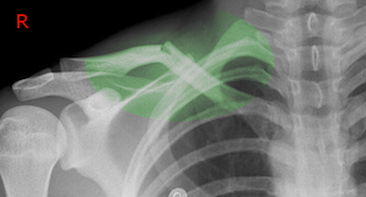
Clavicle Fracture. Radiograph of clavicle fracture after a fall from a ladder.
Contributed by S Bhimji, MD
(Click Image to Enlarge)
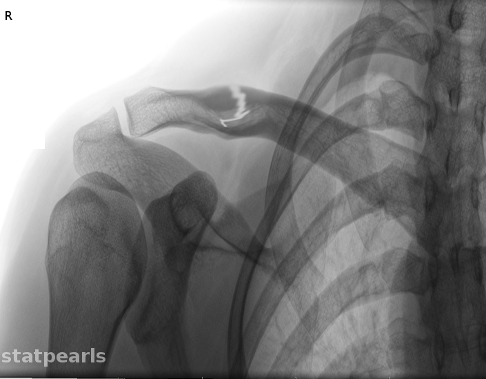
Clavicle Fracture.
Contributed by O Chaigasame, MD
(Click Image to Enlarge)
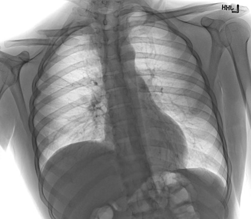
Right Clavicle Fracture. Chest radiograph demonstrating a right clavicle fracture with elevation of the right hemidiaphragm, consistent with associated phrenic nerve injury.
Contributed by Zhongyu Li, MD, PhD
References
Ropars M, Thomazeau H, Huten D. Clavicle fractures. Orthopaedics & traumatology, surgery & research : OTSR. 2017 Feb:103(1S):S53-S59. doi: 10.1016/j.otsr.2016.11.007. Epub 2016 Dec 30 [PubMed PMID: 28043849]
von Rüden C, Rehme-Röhrl J, Augat P, Friederichs J, Hackl S, Stuby F, Trapp O. Evidence on treatment of clavicle fractures. Injury. 2023 Oct:54 Suppl 5():110818. doi: 10.1016/j.injury.2023.05.049. Epub 2023 May 15 [PubMed PMID: 37217399]
Wiesel B, Nagda S, Mehta S, Churchill R. Management of Midshaft Clavicle Fractures in Adults. The Journal of the American Academy of Orthopaedic Surgeons. 2018 Nov 15:26(22):e468-e476. doi: 10.5435/JAAOS-D-17-00442. Epub [PubMed PMID: 30180095]
Robinson CM. Fractures of the clavicle in the adult. Epidemiology and classification. The Journal of bone and joint surgery. British volume. 1998 May:80(3):476-84 [PubMed PMID: 9619941]
Level 2 (mid-level) evidenceHughes K, Kimpton J, Wei R, Williamson M, Yeo A, Arnander M, Gelfer Y. Clavicle fracture nonunion in the paediatric population: a systematic review of the literature. Journal of children's orthopaedics. 2018 Feb 1:12(1):2-8. doi: 10.1302/1863-2548.12.170155. Epub [PubMed PMID: 29456747]
Level 1 (high-level) evidenceBan I, Troelsen A. Risk profile of patients developing nonunion of the clavicle and outcome of treatment--analysis of fifty five nonunions in seven hundred and twenty nine consecutive fractures. International orthopaedics. 2016 Mar:40(3):587-93. doi: 10.1007/s00264-016-3120-8. Epub 2016 Feb 4 [PubMed PMID: 26847264]
Robinson CM, Court-Brown CM, McQueen MM, Wakefield AE. Estimating the risk of nonunion following nonoperative treatment of a clavicular fracture. The Journal of bone and joint surgery. American volume. 2004 Jul:86(7):1359-65 [PubMed PMID: 15252081]
Allman FL Jr. Fractures and ligamentous injuries of the clavicle and its articulation. The Journal of bone and joint surgery. American volume. 1967 Jun:49(4):774-84 [PubMed PMID: 6026010]
Neer CS 2nd. Fractures of the distal third of the clavicle. Clinical orthopaedics and related research. 1968 May-Jun:58():43-50 [PubMed PMID: 5666866]
Xue C, Yang W, Rui Y, Shi H, Zheng X, Song L, Li X, Fang J. A new classification system for distal clavicle fractures: based on fracture location and ligament integrity. Archives of orthopaedic and trauma surgery. 2024 Dec 12:145(1):8. doi: 10.1007/s00402-024-05633-7. Epub 2024 Dec 12 [PubMed PMID: 39666058]
Cho CH, Kim BS, Kim DH, Choi CH, Dan J, Lee H. Distal clavicle fractures: A new classification system. Orthopaedics & traumatology, surgery & research : OTSR. 2018 Dec:104(8):1231-1235. doi: 10.1016/j.otsr.2018.05.015. Epub 2018 Nov 2 [PubMed PMID: 30393071]
Stanley D, Trowbridge EA, Norris SH. The mechanism of clavicular fracture. A clinical and biomechanical analysis. The Journal of bone and joint surgery. British volume. 1988 May:70(3):461-4 [PubMed PMID: 3372571]
Abbot AE, Hannafin JA. Stress fracture of the clavicle in a female lightweight rower. A case report and review of the literature. The American journal of sports medicine. 2001 May-Jun:29(3):370-2 [PubMed PMID: 11394611]
Level 3 (low-level) evidenceFallon KE, Fricker PA. Stress fracture of the clavicle in a young female gymnast. British journal of sports medicine. 2001 Dec:35(6):448-9 [PubMed PMID: 11726488]
Level 3 (low-level) evidencePeebles CR, Sulkin T, Sampson MA. 'Cable-maker's clavicle': stress fracture of the medial clavicle. Skeletal radiology. 2000 Jul:29(7):421-3 [PubMed PMID: 10963430]
Level 3 (low-level) evidenceSambandam B, Gupta R, Kumar S, Maini L. Fracture of distal end clavicle: A review. Journal of clinical orthopaedics and trauma. 2014 Jun:5(2):65-73. doi: 10.1016/j.jcot.2014.05.007. Epub 2014 Jun 19 [PubMed PMID: 25983473]
Chalidis B, Davitis V, Papadopoulos P, Pitsilos C. Subclavian vessels injury: An underestimated complication of clavicular fractures. World journal of critical care medicine. 2024 Dec 9:13(4):98579. doi: 10.5492/wjccm.v13.i4.98579. Epub 2024 Dec 9 [PubMed PMID: 39655299]
Hall JA, Schemitsch CE, Vicente MR, Dehghan N, Nauth A, Nowak LL, Schemitsch EH, McKee MD, Canadian Orthopaedic Trauma Society (COTS). Operative Versus Nonoperative Treatment of Acute Displaced Distal Clavicle Fractures: A Multicenter Randomized Controlled Trial. Journal of orthopaedic trauma. 2021 Dec 1:35(12):660-666. doi: 10.1097/BOT.0000000000002211. Epub [PubMed PMID: 34128498]
Level 1 (high-level) evidencePandya NK, Namdari S, Hosalkar HS. Displaced clavicle fractures in adolescents: facts, controversies, and current trends. The Journal of the American Academy of Orthopaedic Surgeons. 2012 Aug:20(8):498-505. doi: 10.5435/JAAOS-20-08-498. Epub [PubMed PMID: 22855852]
Zenni EJ Jr, Krieg JK, Rosen MJ. Open reduction and internal fixation of clavicular fractures. The Journal of bone and joint surgery. American volume. 1981 Jan:63(1):147-51 [PubMed PMID: 7451517]
Fanter NJ, Kenny RM, Baker CL 3rd, Baker CL Jr. Surgical treatment of clavicle fractures in the adolescent athlete. Sports health. 2015 Mar:7(2):137-41. doi: 10.1177/1941738114566381. Epub [PubMed PMID: 25984259]
Sanchez PH, Fleury IG, Parker EA, Davison J, Westermann R, Kopp B, Willey MC, Buckwalter JA. Early Versus Delayed Surgery for Midshaft Clavicle Fractures: A Systematic Review. The Iowa orthopaedic journal. 2023:43(1):151-160 [PubMed PMID: 37383857]
Level 1 (high-level) evidenceHan X, Zhang Y, Zhang X, Tan J. Comprehensive comparison between conservative therapy and surgical management for completely displaced and comminuted mid-shaft clavicle fractures. International orthopaedics. 2024 Jul:48(7):1871-1877. doi: 10.1007/s00264-024-06198-1. Epub 2024 May 7 [PubMed PMID: 38713287]
Coppa V, Dei Giudici L, Cecconi S, Marinelli M, Gigante A. Midshaft clavicle fractures treatment: threaded Kirschner wire versus conservative approach. Strategies in trauma and limb reconstruction. 2017 Nov:12(3):141-150. doi: 10.1007/s11751-017-0293-7. Epub 2017 Aug 20 [PubMed PMID: 28825169]
Anderson K. Evaluation and treatment of distal clavicle fractures. Clinics in sports medicine. 2003 Apr:22(2):319-26, vii [PubMed PMID: 12825533]
McKee MD, Wild LM, Schemitsch EH. Midshaft malunions of the clavicle. The Journal of bone and joint surgery. American volume. 2003 May:85(5):790-7 [PubMed PMID: 12728026]
Luo TD, Ashraf A, Larson AN, Stans AA, Shaughnessy WJ, McIntosh AL. Complications in the treatment of adolescent clavicle fractures. Orthopedics. 2015 Apr:38(4):e287-91. doi: 10.3928/01477447-20150402-56. Epub [PubMed PMID: 25901621]
Bishop JY, Flatow EL. Pediatric shoulder trauma. Clinical orthopaedics and related research. 2005 Mar:(432):41-8 [PubMed PMID: 15738802]
Banerjee R, Waterman B, Padalecki J, Robertson W. Management of distal clavicle fractures. The Journal of the American Academy of Orthopaedic Surgeons. 2011 Jul:19(7):392-401 [PubMed PMID: 21724918]
Vautrin M, Kaminski G, Barimani B, Elmers J, Philippe V, Cherix S, Thein E, Borens O, Vauclair F. Does candidate for plate fixation selection improve the functional outcome after midshaft clavicle fracture? A systematic review of 1348 patients. Shoulder & elbow. 2019 Feb:11(1):9-16. doi: 10.1177/1758573218777996. Epub 2018 Jun 4 [PubMed PMID: 30719093]
Level 1 (high-level) evidenceLenza M, Buchbinder R, Johnston RV, Ferrari BA, Faloppa F. Surgical versus conservative interventions for treating fractures of the middle third of the clavicle. The Cochrane database of systematic reviews. 2019 Jan 22:1(1):CD009363. doi: 10.1002/14651858.CD009363.pub3. Epub 2019 Jan 22 [PubMed PMID: 30666620]
Level 1 (high-level) evidenceCalbiyik M, Taskoparan M, Ipek D. Surgical treatment of displaced clavicle fractures with a novel intramedullary device; comparison of less-invasive versus standard technique. Acta orthopaedica Belgica. 2018 Sep:84(3):331-337 [PubMed PMID: 30840576]
Dong WW, Zhao X, Mao HJ, Yao LW. [Minimally-invasive internal fixation for mid-lateral 1/3 clavicle fracture with distal clavicular anatomic locking plate]. Zhongguo gu shang = China journal of orthopaedics and traumatology. 2019 Jan 25:32(1):28-32. doi: 10.3969/j.issn.1003-0034.2019.01.006. Epub [PubMed PMID: 30813664]