 Tools
Tools
 Fascial Dehiscence
Introduction
Fascial dehiscence is the separation of the edges of abdominal fascia that has been sutured together following a surgery. This technical issue is a concerning complication of open surgical intervention, which often results in the need for additional surgical intervention; dehiscence also represents a significant influence on postoperative morbidity and mortality. High clinical suspicion is essential for early identification and treatment to prevent short- and long-term complications, eg, chronic wounds, hernias, and evisceration. Early fascial dehiscence may be detectable only on radiologic imaging. Optimizing patient factors and surgical techniques can limit the rates of development of fascial dehiscence and thus spare the patient the associated morbidities and need for additional surgical procedures.
Etiology
Register For Free And Read The Full Article
 Search engine and full access to all medical articles
- 10 free questions in your specialty
- Free CME/CE Activities
- Free daily question in your email
- Save favorite articles to your dashboard
- Emails offering discounts
Learn more about a Subscription to StatPearls Point-of-Care
Etiology
The primary technical pathways for fascial dehiscence are rupture of the suture, knot slippage, inappropriate tension of sutures placed, and the development of hematomas and seromas that place tension on the fascial repair. Traumatic mechanisms, surgical site infections, poor nutrition (albumin <3), comorbidities (ie, diabetes, chronic steroid use), and prolonged critical care course all increase the risk of fascial dehiscence.[1] Fascial dehiscence places patients at risk of hernias and their associated short- and long-term complications, including obstruction and strangulation. Additionally, progression to evisceration represents a true surgical emergency.[2][3][4][5]
Epidemiology
Fascial dehiscence is a complication of both elective and emergent surgeries. Rates of fascial dehiscence following open elective surgery are approximately 1% to 3%. Emergent operations have a higher rate of fascial dehiscence at 5% to 50%; when trauma laparotomy is performed, rates range from 5% to 10% for definitive laparotomy. Damage-control procedures, whether trauma or emergency general surgery, are associated with fascial dehiscence rates of 13% to 50%.
The associated mortality rate with this complication is reported to be greater than 21%, and even 5-year survival has been reportedly reduced.[1][2][3][4][5][6] Given the number of open operations performed yearly (approximately 2 million per year in the United States from 2009 to 2013), a notable number of patients have their postoperative course complicated by fascial dehiscence with resultant increases in costs, length of stay, need for additional interventions, and complications.[7]
Pathophysiology
Research has identified several mechanisms for the development of fascial dehiscence, secondary to both modifiable and nonmodifiable factors. Traumatic injury requiring exploratory laparotomy is itself a risk factor; presumably, the damage-control nature of the surgery, the open abdomen, and reexploration (with associated potential for loss of domain), and the often critical nature and course of these patients present likely mechanisms for poor fascial healing. Emergency nontrauma surgical intervention confers a similar risk. The high rates of associated sepsis and intraabdominal contamination, prolonged critical care requirements, including the need for vasopressors for shock states, elevated rates of ileus and pulmonary complications, need for planned or unplanned reexploration, and development or continuation of malnutrition states present a myriad of factors influencing the breakdown of the fascial closure.
Surgical site infection is a well-documented cause of dehiscence; bacteria and inflammatory factors present in infected sites are detrimental to wound healing. Nutritional factors, including preoperative malnutrition or prolonged delay in resuming postoperative enteral or parenteral nutrition, can deprive the body of necessary substrates for adequate healing. Hypoalbuminemia (<3.5 g/dL) has been associated with fascial dehiscence as well.[8][9]
Chronic steroid use is a well-known inhibitor of tissue repair; chronic diseases such as end-stage renal disease and diabetes, and their associated microvascular complications, are similarly well-published independent risk factors for failure of wound healing and surgical site infections. Importantly, clinicians cannot ignore technical errors as causes; loose knots, tissue strangulation, or inadequate tissue incorporation in suture bites can lead to failure of fascial closure and tissue dehiscence. Smoking is also a well-known inhibitor of wound healing.[10] Obesity has correlations with poor wound healing and skin dehiscence; evidence has not directly linked obesity with fascial dehiscence; however, extrapolation of data suggests a trend in increased fascial complications in obese patients.[11] Seroma/hematoma may increase the risk of superficial wound dehiscence.[1][12]
The physiology of wound healing involves a well-choreographed pathway. Local tissue factors promote platelet aggregation, acute hemostasis, and chemotaxis. The inflammatory response recruits neutrophils, macrophages, and fibroblasts, which clear cellular debris and begin the formation of a regenerative matrix. Angiogenesis occurs, extracellular matrix components get deposited, and collagen forms and remodels. This process is complex and requires multiple cofactors, enzymatic signals, adequate vascularity, and adequate tissue oxygenation. The process commences immediately upon injury, or, in this case, upon surgical incision and closure, and continues for months, with remodeling and maturation. Collagen production begins within 12 to 72 hours of injury and peaks within 7 to 21 days; 80% of tissue strength returns at 3 months. Due to the complexity of wound healing, multiple opportunities for factors that are detrimental to the process exist.
Ultimately, dehiscence results from a failure or inadequacy in the wound-healing pathway, typically occurring between postoperative days 3 and 7.[1] Deprivation of tissue repair factors and substrates, overabundance of inflammatory cells and products, presence of clinically significant bacteria load, or malperfusion (from tissue strangulation, microvascular disease, or critical illness) all act at the cellular level to impair the healing pathway; reducing the risk of fascial dehiscence relies upon the optimization of these factors as able given the patient’s clinical status and course.
History and Physical
As with any condition, a thorough history and physical is necessary. The history may be limited in critically ill postoperative individuals, particularly if intubated; bedside nurse reports can be essential in this situation. New or markedly increased wound drainage or a new abdominal wall bulge are indicators of dehiscence of the fascia. The clinician should ask the patient about obstructive symptoms, including nausea, vomiting, or obstipation. Patients may report a pulling or popping sensation as the inciting symptom; this may occur during coughing, straining, or retching.
The clinician should also elicit a surgical history, with particular attention to recent surgical procedures, as dehiscence is most likely within the first 1 to 2 weeks postoperatively. The medical history should also have a specific focus on diabetes, conditions requiring chronic steroid use (eg, chronic obstructive pulmonary disease, adrenal insufficiency), connective tissue disorders, and malnutrition. A smoking history and an updated medical history are also necessary.
A focused abdominal examination should follow. The prior incision site should undergo inspection for signs of infection, including erythema or drainage. If drainage is present, the character is important; purulent fluid raises suspicion of an underlying surgical site infection. Clear or serous-appearing drainage may represent decompressing seroma or may be normal in small quantities as healing progresses. The classic “salmon-colored” drainage is concerning for fascial dehiscence. The abdomen should be palpated to evaluate for any masses or bulging, which could signify seroma, abscess, or herniation of abdominal contents. Peritoneal signs (guarding, rebound tenderness) may be present if an organ space infection or bowel ischemia is present.
Surgical closures, including staples, sutures, or negative-pressure wound therapy devices, may be present. If the skin closure remains intact, removal of sutures/staples before imaging, or the presence of a surgeon, is not recommended, as opening the incision may convert fascial dehiscence to evisceration requiring emergent surgical intervention. Probing of the wound with sterile swabs (ie, between staples at areas of persistent skin separation) may be performed to evaluate the integrity of the fascial closure and attempt to detect fascial separation. However, this should be left to the surgeon's discretion.
In some instances, patients present with an apparent or suspected dehiscence, as noted during dressing changes (packing or negative-pressure wound therapy), when superficial tissues have been left open to heal by secondary intention. Fascial defects, with or without visualized underlying bowel loops, may be visible. The diagnosis of fascial dehiscence should be clear in this subset of cases. Bowel evisceration, if noted, should prompt emergent surgical evaluation.
Evaluation
Early involvement of a surgical clinician, preferably the surgeon who performed the fascial closure, is prudent for the evaluation and management of suspected dehiscence. In stable individuals with no evidence of evisceration, imaging should be obtained to evaluate the presence and size of the fascial defect; abdominopelvic computed tomography (CT) with contrast provides excellent anatomic detail and can also identify additional pathology, including superficial and deep abscesses and organ-space abscesses, as well as herniation of abdominal contents through the fascial defect. As noted previously, opening the incision is not recommended, as it can lead to evisceration and necessitate an emergent operative procedure. Instead, radiologic evaluation can guide decision-making as to whether elective/urgent operative intervention is necessary.
Treatment / Management
Several clinical factors guide treatment decisions for fascial dehiscence. If a patient experiences evisceration, the bowel should be wrapped in gauze moistened with warm saline to prevent fluid losses, and the patient should be prepared for emergent abdominal exploration to return the bowel to the abdominal cavity and provide overlying closure. In a stable individual with dehiscence and no evidence of underlying infectious complications, nonoperative management can be considered if the skin overlying the fascial defect remains closed to prevent evisceration.
For patients with a prolonged open abdomen course, abdominal contents may become frozen; these patients are at relatively low risk of bowel herniation and may not require future repair of the defect. For patients who do not have a frozen abdomen, the defect can be monitored and allowed to develop into a controlled hernia, which may be repaired at a future date in an elective fashion once the patient has recovered from the index procedure, and acute inflammation has resolved; importantly, no signs of peritonitis, strangulation, or obstruction should be present if nonoperative management is to merit consideration. Additionally, the clinician must weigh the risk of bowel strangulation posed by minor defects.
Indications for operative intervention include evisceration, evidence of bowel strangulation/ischemia, bowel obstruction, or undrained infectious collection. Return of herniated bowel contents to the abdominal cavity should occur. Superficial, deep, or organ-space infections should be drained and irrigated. Nonviable tissues require debridement; bowel resection may be necessary if bowel segments are irreversibly ischemic.
Multiple techniques are available for closure of the fascial defect. If the dehiscence is minor and the fascia is healthy, the edges can be debrided and primary closure performed. Augmentation of the repair with mesh has been shown to improve outcomes and reduce recurrence.[13] Recent evidence supports the safety of prophylactic mesh augmentation in appropriately selected individuals and demonstrates an association with improved hernia-free survival.[14][15] Surgeons should avoid mesh in infected cases or in cases requiring bowel resection, although biologic mesh repair is reasonable. Large defects that do not readily approximate may require management with progressive wound closure, including an open abdomen with vacuum therapy, sequential tightening of progressive closure devices (such as a Wittmann patch), or creation of a planned hernia by developing cutaneous flaps and closing them over the defect. In severe cases, the application of a biologic graft for eventual skin grafting may be necessary.(A1)
Abdominal component separation may be useful for some large defects; however, care must be taken not to compromise future reconstructive options, as patients who experience fascial dehiscence requiring reoperation are at high risk of incisional hernia formation. Consequently, component separation in the acute setting is not routinely recommended. Patients with large defects are likely better treated in the acute setting in a nonoperative manner if able, or with a nonfascial disrupting technique as mentioned above if surgical intervention is needed; staged repair with various component separation techniques can then be pursued in both the expectantly managed groups and those who develop subsequent incisional hernias with greater options for fascial release and closure.[16][17]
If closure does not take place immediately and incisional hernia is allowed to develop, then reclosure can be pursued at a later time to prevent complications related to hernia incarceration, obstruction, or strangulation. Adequate time, usually at least 2 to 3 months, should elapse to allow for healing from the initial procedure. The patient’s clinical status should be optimized, including attention to nutritional factors and adequate management of comorbidities. Ideally, patients will have returned to their baseline functional status before elective fascial repair.
The above-listed techniques are all applicable to reclosure, with the additional benefit of a clean field and the opportunity to place nonabsorbable mesh. No difference has been identified in subsequent wound complications with continuous vs interrupted suture repair of defects or with the type of suture material used.[18][19] Studies have also investigated whether the use of triclosan-coated sutures during fascial closure mitigates the risk of fascial dehiscence. Still, these studies have shown only a decrease in the rate of surgical site infections, not in fascial dehiscence itself.[20] Mesh repair has been associated with multiple complications, including abscess formation, fistula, adhesive bowel obstruction, and bowel erosion; however, the markedly lower recurrence rate of incisional hernia after mesh repair has been shown to outweigh the potential risks of mesh use.[21] Both open and laparoscopic repairs are viable options with low recurrence rates when mesh is used, compared with primary fascial repair alone.[21][22](A1)
Differential Diagnosis
Differential diagnosis includes postoperative subcutaneous seroma or hematoma, which may decompress through the incision with exertion or if it develops to a substantial size. Surgical site infection with abscess formation is also in the differential diagnosis and may coexist with fascial dehiscence, given the association between infection and fascial closure breakdown. Preexisting hernias may also be present and may not have been addressed in the index operation, so abdominal bulging away from the incision may represent a hernia, which could be preexisting or enlarging.
Pertinent Studies and Ongoing Trials
Several landmark studies have shaped the modern approach to fascial dehiscence. Results from extensive cohort analyses and risk factor studies have established chronic illness, infection, and technical errors as key predictors of dehiscence. Randomized controlled trials' results have demonstrated that small-bite suture techniques and appropriate mesh augmentation—particularly in clean abdominal closures—significantly reduce recurrence rates and long-term hernia recurrence.
The adoption of machine learning models based on clinical and perioperative variables has further enhanced surgical decision-making and individualized risk prediction. Current ongoing trials are evaluating optimal suture materials (including triclosan-coated sutures), the role of prophylactic mesh in complex and contaminated cases, and the impact of early, protocol-driven diagnostic strategies. Prospective multicenter studies and international registries are expected to provide high-level evidence to further guide prevention and management in the coming years.[1][9][23][9][1][24][25][26]
Prognosis
Fascial dehiscence after abdominal surgery is a major contributor to patient morbidity and mortality. Subsequent complications following dehiscence are estimated to be as high as 75%; published reports on mortality range from 15% to 50%.[1] Prolonged hospital course (15 to 26 days), increased intensive care requirements, and higher healthcare costs are all associated with abdominal wall complications following laparotomy. While prompt recognition and treatment are important, the development of fascial dehiscence presents a substantial morbidity and mortality burden for the patient, irrespective of the timing of subsequent intervention.[27][28][29]
Complications
As noted above, the development of fascial dehiscence places a considerable morbidity and mortality burden on the affected patient. The development of subsequent infections is a concern, with an approximate 10% risk. Abdominal washout should be considered, and skin/subcutaneous tissues may be left open to heal by secondary intention when urgent/emergent reoperation is necessary. The surgeon encounters frankly contaminated or necrotic tissue.[30]
Complications include death, evisceration, and associated infections in the acute setting; bowel herniation can occur, and an incisional hernia can form with the development of subsequent bowel obstruction and/or strangulation in both the short- and long-term settings. The need for additional procedures to repair the defect or hernia, reduce or resect involved bowel, and drain accumulated fluid collections potentiate the risks of anesthesia-, surgical- and hospital-associated complications including pneumonia, hospital-acquired infection, prolonged intensive care unit courses, deconditioning, adhesive small bowel obstruction, venous thromboembolic events, and need for short- or long-term rehab/nursing care. Notably, fascial dehiscence can significantly affect a patient’s quality of life and survival. Consequently, modifiable risk factors (both patient- and technical-related) should be managed to improve patient outcomes.[27][28][29]
Deterrence and Patient Education
Multiple modifiable factors exist that can alter the outcome of abdominal surgeries and can influence the prevention (or formation) of wound complications, including fascial dehiscence. For the elective patient setting, smoking cessation before surgical intervention is optimal.[31][32] Blood glucose should be controlled in the pre-, peri-, and postoperative periods.[33] Steroid use should be avoided or minimized as much as possible; however, this may not be feasible in patients on chronic steroids. Nutrition requires supplementation, and nutritional parameters may be measured if preoperative malnutrition is suspected, as outcomes are worse with an albumin <3.5 g/dL. Intraabdominal distention secondary to ileus, bowel obstruction, or urinary retention should be managed with decompression (ie, nasogastric tube placement, catheterization). Pulmonary interventions to reduce atelectasis and promote oxygenation should be implemented.
For patients with damage-control or open abdomen, closure of the abdomen as soon as clinically safe is recommended; more than 4 reoperations before closure have been associated with a significant increase in the rate of failure of primary fascial closure.[34] Delayed closure in the setting of resuscitative efforts can result in bowel edema, which increases intraabdominal pressure upon closure of the fascia. Loss of the abdominal wall domain can occur, and fascial edges may not be approximated with minimal tension, thereby increasing the risk of closure breakdown. In the setting of loss of domain, multiple strategies exist to facilitate closure, including Clostridium botulinum toxin, sequential closure with hook-and-loop devices, component separation, progressive pneumoperitoneum, and mesh utilization; in severe cases, skin flaps may be raised and closed without fascial reapproximation, thereby creating a controlled hernia.[35][36]
Incision type has been considered a risk factor for dehiscence. Data are limited; however, trends indicate a lower rate of fascial complications with transverse or oblique incisions compared with midline or paramedian incisions.[37] However, the type of incision appropriate for the planned procedure often dictates incision placement and orientation.
For all fascial closures, attention to the surgical technique is essential. Care should be taken to snugly approximate fascial edges without strangulating tissue. Knots should be securely tied. Monofilament suture is associated with lower rates of subsequent dehiscence as compared to multifilament suture.[38] No difference in dehiscence rates has been identified between absorbable and nonabsorbable sutures. Additionally, no difference in dehiscence rates has been observed between continuous and interrupted sutures or between mass and layered fascial closure.[38][39][40][19]
Limited evidence suggests that a “small-bites” technique, with 5 to 8 mm fascial bites placed every 5 mm, may reduce the incidence of complications.[41] The closure (or nonclosure) of the peritoneal layer is not associated with the development of fascial dehiscence.[42][40] Evidence suggests that a suture-to-wound length ratio of 4:1 in continuous suture closure reduces fascial closure breakdown.[43] Drains have not been shown to prevent fascial dehiscence; however, drains or ostomies should not be placed within the midline incision, as these can increase the incidence of fascial complications.[42]
The use of retention sutures (internal or external) is a long-standing topic of discussion in surgery, particularly regarding fascial dehiscence and evisceration. Classic teaching states that retention sutures prevent evisceration; however, they do not prevent fascial dehiscence. A recent small randomized series contradicts this finding, reporting a decrease in dehiscence rates in the retention-suture group.[44] Ultimately, retention suture use is left to the discretion of the operating surgeon, though it likely confers benefit only in patients at high risk of wound complications.[44]
Patients should be educated on wound care and concerning findings, such as wound appearance and drainage. Many surgeons will implement activity restrictions following abdominal surgery to prevent failure of the fascial closure. These tend to vary in length by surgeon and procedure. No substantial evidence exists to support these recommendations, as unavoidable activities of daily living (ie, getting up from a chair, coughing, straining with Valsalva) may equal or exceed the intra-abdominal pressures generated with lifting; nevertheless, counseling patients to avoid straining, heavy lifting, and activities that cause incisional pain for several weeks following surgery is likely prudent.[45] Patients should also receive instruction regarding concerning features that may develop, including increased or large-volume wound drainage, focal bulging, or evidence of wound infection. Early notification of their primary surgeon and clinical evaluation (in the office or emergency department) may prevent subsequent development of more serious wound complications.
Pearls and Other Issues
Fascial dehiscence is a life-threatening postoperative complication that most commonly occurs between postoperative days 3 and 10 and is frequently heralded by sudden salmon-colored (serosanguinous) wound drainage or a reported “popping” sensation. Early recognition is essential to prevent progression to evisceration. Risk is heightened by increased intraabdominal pressure, wound infection, malnutrition, poor glycemic control, and chronic pulmonary disease. Meticulous midline fascial closure using continuous, slowly absorbable monofilament suture with an adequate suture-to-wound length ratio remains the cornerstone of prevention. At the same time, mesh augmentation in clean fields significantly reduces the risk of recurrence.
Fascial dehiscence constitutes a surgical emergency requiring prompt surgical consultation and operative management. Patients should undergo hemodynamic stabilization and receive broad-spectrum antibiotics when infection is suspected. When the diagnosis is uncertain, and the skin remains intact, computed tomography is preferred over opening the wound at the bedside to avoid precipitating evisceration. Definitive management involves urgent return to the operating room for re-exploration and secure fascial closure, with temporary abdominal closure or mesh reinforcement considered in unstable, contaminated, or high-risk cases.
Common pitfalls include dismissing early warning signs as superficial wound complications, delaying operative intervention, or attempting bedside wound exploration. Failure to recognize occult fascial disruption, especially in obese individuals, can lead to catastrophic evisceration. Inadequate suture technique, excessive tension, and inability to address contributors such as infection, ileus, cough, ascites, or poor nutrition substantially increase the risk of recurrence.
Prevention relies on both perioperative optimization and sound surgical technique. Key measures include optimizing nutrition, glycemic control, and pulmonary function; minimizing postoperative ileus and coughing; and early identification and treatment of wound infection. Prophylactic mesh reinforcement should be considered in high-risk patients with clean operative fields. Close postoperative wound surveillance and appropriate patient education regarding activity restrictions further reduce morbidity associated with fascial dehiscence.
Enhancing Healthcare Team Outcomes
Fascial dehiscence involves the separation of previously sutured abdominal fascia and remains a serious postoperative complication associated with increased morbidity, hernia formation, and potential evisceration. Early recognition depends on careful assessment of wound drainage, abdominal wall changes, focal symptoms, and targeted radiologic evaluation. Risk factors such as infection, malnutrition, chronic steroid use, diabetes, and prolonged critical illness further heighten the need for vigilant monitoring. Evidence-based management ranges from urgent operative repair for evisceration or obstruction to selective nonoperative strategies for stable individuals without infection or signs of strangulation.
An interprofessional approach strengthens prevention, detection, and recovery. Physicians, advanced practitioners, and emergency or outpatient clinicians must maintain a high level of suspicion and rapidly coordinate surgical consultation. Nurses and wound-care specialists provide frontline evaluation of changes in drainage, abdominal distention, respiratory complications, and overall healing. Pharmacists contribute by optimizing antimicrobial regimens, reconciling medications, and communicating safety concerns. Respiratory therapists support pulmonary hygiene to reduce postoperative strain. Close communication, shared decision-making, and coordinated monitoring across the care continuum enhance patient-centered care, reduce complications, and improve surgical outcomes.
Media
(Click Image to Enlarge)
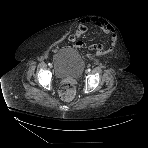
Ventral Hernia, Abdomen CT.
Contributed by S Dulebohn, MD
References
Parsa H, Haji Maghsoudi L, Mohammadzadeh A, Hosseini M. The evaluation of risk factors in fascia dehiscence after abdominal surgeries. Annals of medicine and surgery (2012). 2024 Sep:86(9):4984-4989. doi: 10.1097/MS9.0000000000002335. Epub 2024 Jul 19 [PubMed PMID: 39239048]
Harvin JA, Wray CJ, Steward J, Lawless RA, McNutt MK, Love JD, Moore LJ, Wade CE, Cotton BA, Holcomb JB. Control the damage: morbidity and mortality after emergent trauma laparotomy. American journal of surgery. 2016 Jul:212(1):34-9. doi: 10.1016/j.amjsurg.2015.10.014. Epub 2015 Dec 15 [PubMed PMID: 26754456]
Harvin JA, Sharpe JP, Croce MA, Goodman MD, Pritts TA, Dauer ED, Moran BJ, Rodriguez RD, Zarzaur BL, Kreiner LA, Claridge JA, Holcomb JB. Effect of damage control laparotomy on major abdominal complications and lengths of stay: A propensity score matching and Bayesian analysis. The journal of trauma and acute care surgery. 2019 Aug:87(2):282-288. doi: 10.1097/TA.0000000000002285. Epub [PubMed PMID: 30939584]
O'Meara L, Ahmad SB, Glaser J, Diaz JJ, Bruns BR. Outcomes of primary fascial closure after open abdomen for nontrauma emergency general surgery patients. American journal of surgery. 2015 Dec:210(6):1126-30; discussion 1130-1. doi: 10.1016/j.amjsurg.2015.06.030. Epub 2015 Sep 26 [PubMed PMID: 26520871]
George MJ, Adams SD, McNutt MK, Love JD, Albarado R, Moore LJ, Wade CE, Cotton BA, Holcomb JB, Harvin JA. The effect of damage control laparotomy on major abdominal complications: A matched analysis. American journal of surgery. 2018 Jul:216(1):56-59. doi: 10.1016/j.amjsurg.2017.10.044. Epub 2017 Nov 11 [PubMed PMID: 29157889]
Stropnicky PV, Kandemir F, Schäffer M, Pochhammer J. Abdominal fascia dehiscence: is there a connection to a special microbial spectrum? Hernia : the journal of hernias and abdominal wall surgery. 2023 Jun:27(3):549-556. doi: 10.1007/s10029-022-02679-7. Epub 2022 Sep 22 [PubMed PMID: 36138267]
Carney MJ, Weissler JM, Fox JP, Tecce MG, Hsu JY, Fischer JP. Trends in open abdominal surgery in the United States-Observations from 9,950,759 discharges using the 2009-2013 National Inpatient Sample (NIS) datasets. American journal of surgery. 2017 Aug:214(2):287-292. doi: 10.1016/j.amjsurg.2017.01.001. Epub 2017 Jan 12 [PubMed PMID: 28202162]
Sharma R, Lonare SB, Arora P, Al-Dwlai H, Vadher A, Hersi M. Risk Factors and Predictive Accuracy of the Rotterdam Risk Index for Wound Dehiscence Following Abdominal Surgery. Cureus. 2025 Jan:17(1):e76769. doi: 10.7759/cureus.76769. Epub 2025 Jan 1 [PubMed PMID: 39748882]
Murugavel J, Vajiravelu Thirunavukkarasu A, Gnana Chellaiyan V, Sridharan V. A Prospective Study on the Outcome After Mass Closure of Post-laparotomy Wound Dehiscence in a Tertiary Care Hospital, Tamil Nadu, India. Cureus. 2024 May:16(5):e59642. doi: 10.7759/cureus.59642. Epub 2024 May 4 [PubMed PMID: 38832180]
Tillou A, Weng J, Alkousakis T, Velmahos G. Fascial dehiscence after trauma laparotomy: a sign of intra-abdominal sepsis. The American surgeon. 2003 Nov:69(11):927-9 [PubMed PMID: 14627249]
Sandy-Hodgetts K, Carville K, Leslie GD. Determining risk factors for surgical wound dehiscence: a literature review. International wound journal. 2015 Jun:12(3):265-75. doi: 10.1111/iwj.12088. Epub 2013 May 21 [PubMed PMID: 23692188]
Sandy-Hodgetts K, Watts R. Effectiveness of negative pressure wound therapy/closed incision management in the prevention of post-surgical wound complications: a systematic review and meta-analysis. JBI database of systematic reviews and implementation reports. 2015 Jan:13(1):253-303. doi: 10.11124/jbisrir-2015-1687. Epub [PubMed PMID: 26447018]
Level 1 (high-level) evidenceJakob MO, Spari D, Zindel J, Pinworasarn T, Candinas D, Beldi G. Prophylactic, Synthetic Intraperitoneal Mesh Versus No Mesh Implantation in Patients with Fascial Dehiscence. Journal of gastrointestinal surgery : official journal of the Society for Surgery of the Alimentary Tract. 2018 Dec:22(12):2158-2166. doi: 10.1007/s11605-018-3873-z. Epub 2018 Jul 23 [PubMed PMID: 30039450]
Jensen TK, Kvist M, Damkjær MB, Burcharth J. Short-term outcomes in mesh versus suture-only treatment of burst abdomen: a case-series from a university hospital. Hernia : the journal of hernias and abdominal wall surgery. 2025 Feb 18:29(1):100. doi: 10.1007/s10029-025-03279-x. Epub 2025 Feb 18 [PubMed PMID: 39966188]
Level 3 (low-level) evidenceLima HVG, Rasslan R, Novo FCF, Lima TMA, Damous SHB, Bernini CO, Montero EFS, Utiyama EM. Prevention of Fascial Dehiscence with Onlay Prophylactic Mesh in Emergency Laparotomy: A Randomized Clinical Trial. Journal of the American College of Surgeons. 2020 Jan:230(1):76-87. doi: 10.1016/j.jamcollsurg.2019.09.010. Epub 2019 Oct 28 [PubMed PMID: 31672681]
Level 1 (high-level) evidenceLópez-Cano M, García-Alamino JM, Antoniou SA, Bennet D, Dietz UA, Ferreira F, Fortelny RH, Hernandez-Granados P, Miserez M, Montgomery A, Morales-Conde S, Muysoms F, Pereira JA, Schwab R, Slater N, Vanlander A, Van Ramshorst GH, Berrevoet F. EHS clinical guidelines on the management of the abdominal wall in the context of the open or burst abdomen. Hernia : the journal of hernias and abdominal wall surgery. 2018 Dec:22(6):921-939. doi: 10.1007/s10029-018-1818-9. Epub 2018 Sep 3 [PubMed PMID: 30178226]
Piccoli M, Agresta F, Attinà GM, Amabile D, Marchi D, “Complex abdominal wall study” Italian Collaborative Group. "Complex abdominal wall" management: evidence-based guidelines of the Italian Consensus Conference. Updates in surgery. 2019 Jun:71(2):255-272. doi: 10.1007/s13304-018-0577-6. Epub 2018 Sep 25 [PubMed PMID: 30255435]
van't RM, De Vos Van Steenwijk PJ, Bonjer HJ, Steyerberg EW, Jeekel J. Incisional hernia after repair of wound dehiscence: incidence and risk factors. The American surgeon. 2004 Apr:70(4):281-6 [PubMed PMID: 15098775]
Level 2 (mid-level) evidencePolychronidis G, Rahbari NN, Bruckner T, Sander A, Sommer F, Usta S, Hermann J, Albers MB, Sargut M, Knebel P, Klotz R. Continuous versus interrupted abdominal wall closure after emergency midline laparotomy: CONTINT: a randomized controlled trial [NCT00544583]. World journal of emergency surgery : WJES. 2023 Oct 17:18(1):51. doi: 10.1186/s13017-023-00517-4. Epub 2023 Oct 17 [PubMed PMID: 37848901]
Level 1 (high-level) evidenceTimmer AS, Wolfhagen N, Pianka F, Knebel P, Justinger C, Stravodimos C, Ichida K, Rikiyama T, Baracs J, Vereczkei A, Gianotti L, Sandini M, Ruiz-Tovar J, Marc-Hernández A, Nakamura T, Dijkgraaf MGW, Boermeester MA, de Jonge SW. The Effect of Fascial Closure With Triclosan-Coated Sutures on the Incidence of Abdominal Wall Dehiscence: An Individual Participant Data Meta-Analysis. Journal of abdominal wall surgery : JAWS. 2024:3():13337. doi: 10.3389/jaws.2024.13337. Epub 2024 Sep 18 [PubMed PMID: 39360221]
Level 1 (high-level) evidenceKokotovic D, Bisgaard T, Helgstrand F. Long-term Recurrence and Complications Associated With Elective Incisional Hernia Repair. JAMA. 2016 Oct 18:316(15):1575-1582. doi: 10.1001/jama.2016.15217. Epub [PubMed PMID: 27750295]
Petersson P, Montgomery A, Petersson U. Wound dehiscence: outcome comparison for sutured and mesh reconstructed patients. Hernia : the journal of hernias and abdominal wall surgery. 2014 Oct:18(5):681-9. doi: 10.1007/s10029-014-1268-y. Epub 2014 Jun 12 [PubMed PMID: 24916421]
Level 2 (mid-level) evidenceFukai S, Mizusawa Y, Noda H, Tsujinaka S, Maeda Y, Hasebe R, Eguchi Y, Kanemitsu R, Matsuzawa N, Abe I, Endo Y, Fukui T, Takayama Y, Ichida K, Inoue K, Muto Y, Watanabe F, Futsuhara K, Miyakura Y, Rikiyama T. Superiority trial for the development of an ideal method for the closure of midline abdominal wall incisions to reduce the incidence of wound complications after elective gastroenterological surgery: study protocol for a randomized controlled trial. Trials. 2024 May 17:25(1):327. doi: 10.1186/s13063-024-08167-w. Epub 2024 May 17 [PubMed PMID: 38760769]
Level 1 (high-level) evidenceMehdorn M, Groos L, Kassahun W, Jansen-Winkeln B, Gockel I, Moulla Y. Interrupted sutures prevent recurrent abdominal fascial dehiscence: a comparative retrospective single center cohort analysis of risk factors of burst abdomen and its recurrence as well as surgical repair techniques. BMC surgery. 2021 Apr 26:21(1):208. doi: 10.1186/s12893-021-01219-x. Epub 2021 Apr 26 [PubMed PMID: 33902549]
Level 2 (mid-level) evidenceFrassini S, Calabretto F, Granieri S, Fugazzola P, Massaro M, Sargenti B, Schiavone L, Zanghì S, Dal Mas F, Ansaloni L, Cobianchi L. Prophylactic mesh augmentation after laparotomy for elective and emergency surgery: meta-analysis. BJS open. 2023 Jul 10:7(4):. doi: 10.1093/bjsopen/zrad060. Epub [PubMed PMID: 37504969]
Level 1 (high-level) evidenceCole J, Hughey S, Metzger A, Geiger P, Fluke L, Booth GJ. Machine Learning to Predict Fascial Dehiscence after Exploratory Laparotomy Surgery. The Journal of surgical research. 2021 Dec:268():514-520. doi: 10.1016/j.jss.2021.06.068. Epub 2021 Aug 26 [PubMed PMID: 34455314]
Rodríguez-Hermosa JI, Codina-Cazador A, Ruiz B, Roig J, Gironès J, Pujadas M, Pont J, Aldeguer X, Acero D. [Risk factors for acute abdominal wall dehiscence after laparotomy in adults]. Cirugia espanola. 2005 May:77(5):280-6 [PubMed PMID: 16420934]
Level 2 (mid-level) evidenceGili-Ortiz E, González-Guerrero R, Béjar-Prado L, Ramírez-Ramírez G, López-Méndez J. [Postoperative dehiscence of the abdominal wound and its impact on excess mortality, hospital stay and costs]. Cirugia espanola. 2015 Aug-Sep:93(7):444-9. doi: 10.1016/j.ciresp.2015.02.005. Epub 2015 May 6 [PubMed PMID: 25956459]
Hahler B. Surgical wound dehiscence. Medsurg nursing : official journal of the Academy of Medical-Surgical Nurses. 2006 Oct:15(5):296-300; quiz 301 [PubMed PMID: 17128900]
Norman G, Atkinson RA, Smith TA, Rowlands C, Rithalia AD, Crosbie EJ, Dumville JC. Intracavity lavage and wound irrigation for prevention of surgical site infection. The Cochrane database of systematic reviews. 2017 Oct 30:10(10):CD012234. doi: 10.1002/14651858.CD012234.pub2. Epub 2017 Oct 30 [PubMed PMID: 29083473]
Level 1 (high-level) evidenceDeLancey JO, Blay E Jr, Hewitt DB, Engelhardt K, Bilimoria KY, Holl JL, Odell DD, Yang AD, Stulberg JJ. The effect of smoking on 30-day outcomes in elective hernia repair. American journal of surgery. 2018 Sep:216(3):471-474. doi: 10.1016/j.amjsurg.2018.03.004. Epub 2018 Mar 6 [PubMed PMID: 29559083]
Borad NP, Merchant AM. The effect of smoking on surgical outcomes in ventral hernia repair: a propensity score matched analysis of the National Surgical Quality Improvement Program data. Hernia : the journal of hernias and abdominal wall surgery. 2017 Dec:21(6):855-867. doi: 10.1007/s10029-017-1664-1. Epub 2017 Sep 1 [PubMed PMID: 28864961]
Level 2 (mid-level) evidenceBerríos-Torres SI, Umscheid CA, Bratzler DW, Leas B, Stone EC, Kelz RR, Reinke CE, Morgan S, Solomkin JS, Mazuski JE, Dellinger EP, Itani KMF, Berbari EF, Segreti J, Parvizi J, Blanchard J, Allen G, Kluytmans JAJW, Donlan R, Schecter WP, Healthcare Infection Control Practices Advisory Committee. Centers for Disease Control and Prevention Guideline for the Prevention of Surgical Site Infection, 2017. JAMA surgery. 2017 Aug 1:152(8):784-791. doi: 10.1001/jamasurg.2017.0904. Epub [PubMed PMID: 28467526]
Frazee RC, Abernathy SW, Jupiter DC, Smith RW. The number of operations negatively influences fascia closure in open abdomen management. American journal of surgery. 2012 Dec:204(6):996-8; discussion 998-9. doi: 10.1016/j.amjsurg.2012.07.029. Epub 2012 Sep 28 [PubMed PMID: 23022246]
Level 2 (mid-level) evidenceKoss W, Ho HC, Yu M, Edwards K, Ghows M, Tan A, Takanishi DM Jr. Preventing loss of domain: a management strategy for closure of the "open abdomen" during the initial hospitalization. Journal of surgical education. 2009 Mar-Apr:66(2):89-95. doi: 10.1016/j.jsurg.2008.12.003. Epub [PubMed PMID: 19486872]
Demetriades D, Salim A. Management of the open abdomen. The Surgical clinics of North America. 2014 Feb:94(1):131-53. doi: 10.1016/j.suc.2013.10.010. Epub [PubMed PMID: 24267502]
Brown SR, Goodfellow PB. Transverse verses midline incisions for abdominal surgery. The Cochrane database of systematic reviews. 2005 Oct 19:2005(4):CD005199 [PubMed PMID: 16235395]
Level 1 (high-level) evidencePatel SV, Paskar DD, Nelson RL, Vedula SS, Steele SR. Closure methods for laparotomy incisions for preventing incisional hernias and other wound complications. The Cochrane database of systematic reviews. 2017 Nov 3:11(11):CD005661. doi: 10.1002/14651858.CD005661.pub2. Epub 2017 Nov 3 [PubMed PMID: 29099149]
Level 1 (high-level) evidencePeponis T, Bohnen JD, Muse S, Fuentes E, van der Wilden GM, Mejaddam A, Alam H, Kaafarani HMA, Fagenholz PJ, King DR, Yeh DD, Velmahos GC, de Moya MA. Interrupted versus continuous fascial closure in patients undergoing emergent laparotomy: A randomized controlled trial. The journal of trauma and acute care surgery. 2018 Sep:85(3):459-465. doi: 10.1097/TA.0000000000001970. Epub [PubMed PMID: 29787547]
Level 1 (high-level) evidenceLe Huu Nho R, Mege D, Ouaïssi M, Sielezneff I, Sastre B. Incidence and prevention of ventral incisional hernia. Journal of visceral surgery. 2012 Oct:149(5 Suppl):e3-14. doi: 10.1016/j.jviscsurg.2012.05.004. Epub 2012 Nov 9 [PubMed PMID: 23142402]
Millbourn D, Cengiz Y, Israelsson LA. Effect of stitch length on wound complications after closure of midline incisions: a randomized controlled trial. Archives of surgery (Chicago, Ill. : 1960). 2009 Nov:144(11):1056-9. doi: 10.1001/archsurg.2009.189. Epub [PubMed PMID: 19917943]
Level 1 (high-level) evidenceGurusamy KS, Cassar Delia E, Davidson BR. Peritoneal closure versus no peritoneal closure for patients undergoing non-obstetric abdominal operations. The Cochrane database of systematic reviews. 2013 Jul 4:2013(7):CD010424. doi: 10.1002/14651858.CD010424.pub2. Epub 2013 Jul 4 [PubMed PMID: 23828487]
Level 1 (high-level) evidenceMuysoms FE, Antoniou SA, Bury K, Campanelli G, Conze J, Cuccurullo D, de Beaux AC, Deerenberg EB, East B, Fortelny RH, Gillion JF, Henriksen NA, Israelsson L, Jairam A, Jänes A, Jeekel J, López-Cano M, Miserez M, Morales-Conde S, Sanders DL, Simons MP, Śmietański M, Venclauskas L, Berrevoet F, European Hernia Society. European Hernia Society guidelines on the closure of abdominal wall incisions. Hernia : the journal of hernias and abdominal wall surgery. 2015 Feb:19(1):1-24. doi: 10.1007/s10029-014-1342-5. Epub 2015 Jan 25 [PubMed PMID: 25618025]
Khorgami Z, Shoar S, Laghaie B, Aminian A, Hosseini Araghi N, Soroush A. Prophylactic retention sutures in midline laparotomy in high-risk patients for wound dehiscence: a randomized controlled trial. The Journal of surgical research. 2013 Apr:180(2):238-43. doi: 10.1016/j.jss.2012.05.012. Epub 2012 May 24 [PubMed PMID: 22677612]
Level 1 (high-level) evidenceGuttormson R, Tschirhart J, Boysen D, Martinson K. Are postoperative activity restrictions evidence-based? American journal of surgery. 2008 Mar:195(3):401-3; discussion 403-4. doi: 10.1016/j.amjsurg.2007.12.014. Epub [PubMed PMID: 18207126]