 Tools
Tools
 Esophageal Varices
Introduction
Esophageal varices are dilated submucosal distal esophageal veins connecting the portal and systemic circulations. Varices develop as a sequela of elevated portal vein pressure, increased portal venous inflow, and resistance to portal blood flow. The most common cause of portal hypertension is cirrhosis, followed by portal vein thrombosis. The most common fatal complication of cirrhosis is variceal hemorrhage. The severity of liver disease correlates with the presence of varices and risk of bleeding. The portal vein circulates over 1500 mL/min of blood, and obstruction results in elevated portal venous pressure. The physiologic response to increased venous pressure is the development of collaterals. These portosystemic collaterals divert blood from the portal venous system to the inferior and superior vena cava. Gastroesophageal collaterals drain into the azygos vein and contribute to the development of esophageal varices. When these varices get enlarged, rupture can occur, producing severe hemorrhage. Bleeding from esophageal varices is the third most common cause of upper gastrointestinal bleeding, after duodenal and gastric ulcers. (See Image. Esophageal Varices.)
Etiology
Register For Free And Read The Full Article
 Search engine and full access to all medical articles
- 10 free questions in your specialty
- Free CME/CE Activities
- Free daily question in your email
- Save favorite articles to your dashboard
- Emails offering discounts
Learn more about a Subscription to StatPearls Point-of-Care
Etiology
Causes of Portal Hypertension
- Prehepatic: Portal vein obstruction or massive splenomegaly with increased splenic vein blood flow
- Posthepatic: Severe right-sided heart failure, constrictive pericarditis, and hepatic vein obstruction (Budd-Chiari syndrome)
- Intrahepatic: Cirrhosis accounts for most cases of portal hypertension
Less frequent causes include schistosomiasis, massive fatty change, and diseases affecting portal microcirculation, including nodular regenerative hyperplasia and diffuse fibrosing granulomatous disease, including sarcoidosis.[1] Other rare causes of portal hypertension include:
- Wilson disease
- A-1 antitrypsin deficiency
- Primary biliary cholangitis
- Tuberculosis
Epidemiology
Incidence
- At diagnosis, 30% of patients with cirrhosis have varices, and the prevalence increases to 90% over 10 years.
- The 1-year rate of first variceal bleeding is 5% for small varices and 15% for large varices.
- Portal hypertension is common in pediatric chronic liver disease.
Prevalence
- Esophageal varices are more common in men than in women. Fifty percent of patients with esophageal varices experience bleeding at some point.
- Variceal bleeding has a 10% to 20% mortality rate in the 6 weeks following the episode.
- In the Western Hemisphere, the 2 most common causes of portal hypertension are alcohol and viral hepatitis. In Asia and Africa, the most common causes of portal hypertension include schistosomiasis as well as hepatitis B and C.[1]
Pathophysiology
Portal hypertension causes portocaval anastomoses to develop and decompress the portal circulation. Normal portal pressure is between 5 and 10 mm Hg, but portal obstruction can increase portal pressure to 15 to 20 mm Hg. Since the portal venous system has no valves, resistance at any level between the splanchnic vessels and the right side of the heart results in retrograde flow and elevated pressure. The collaterals slowly enlarge and connect the systemic circulation to the portal venous system. Over time, the process leads to a congested submucosal venous plexus with tortuous dilated veins in the distal esophagus. Variceal rupture results in hemorrhage.
Pathophysiology of Portal Hypertension
Increased resistance to portal flow at the level of hepatic sinusoids is caused by:
- Intrahepatic vasoconstriction due to decreased nitric oxide production and increased release of endothelin-1, angiotensinogen, and eicosanoids
- Sinusoidal remodeling disrupts blood flow
- Increased portal flow is caused by hyperdynamic circulation due to splanchnic arterial vasodilation through mediators such as nitric oxide, prostacyclin, and tumor necrosis factor [2]
Risk factors for variceal bleeding:
- Varix size; larger varices have a greater potential for rupture
- Advanced Child-Pugh score, which increases the risk of hemorrhage
- Red colored markings on the varices during endoscopy, which are associated with an increased risk for rupture
- Active alcohol consumption [3]
History and Physical
The first indication of varices is often a gastrointestinal (GI) bleeding episode, including hematemesis, melena, or hematochezia. Occult bleeding (anemia) is uncommon.
History
Variceal bleeding can be the initial presentation of previously undiagnosed cirrhosis. Risk factors include alcoholism and exposure to blood-borne viruses. Symptoms can include hematemesis, melena, or hematochezia. Rapid upper GI bleeding can present as rectal bleeding. Additional findings in patients with chronic liver disease include weight loss, anorexia, abdominal discomfort, jaundice, pruritus, as well as encephalopathic symptoms such as altered mental status and muscle cramps.
Physical Examination
- Assess hemodynamic stability, including hypotension and tachycardia, in the presence of active bleeding.
- Perform an abdominal examination, including liver palpation and percussion (often small and firm with cirrhosis).
- Assess for splenomegaly and ascites (including shifting dullness and fluid wave).
- Inspect for visible abdominal periumbilical collateral circulation (caput medusae).
- Evaluate for peripheral stigmata of alcohol use disorder, including spider angiomata on the chest and back, palmar erythema, testicular atrophy, and gynecomastia.
- Auscultate for venous hums.
- Assess for anal varices and blood on rectal examination.
- Evaluate for hepatic encephalopathy and asterixis.
Evaluation
Evaluation for esophageal varices is warranted when there is evidence of portal hypertension or cirrhosis, as indicated by clinical signs or laboratory findings, to prevent complications such as variceal bleeding.
Initial Laboratory Testing and Imaging
- Anemia: Hemoglobin may be normal during active bleeding and may take 6 to 24 hours to equilibrate. Other causes of anemia are common in patients with cirrhosis.
- Thrombocytopenia is the most sensitive and specific laboratory parameter associated with portal hypertension and large esophageal varices.
- Elevated aspartate aminotransferase, alanine aminotransferase, alkaline phosphatase, and bilirubin levels, along with prolonged prothrombin time and low albumin, suggest cirrhosis.
- Blood urea nitrogen levels are often elevated in gastrointestinal bleeding.
- Sodium levels may drop in patients treated with terlipressin.
- Additional laboratory evaluation can include a coagulation profile, renal function testing, arterial blood gas analysis, and hepatitis serology.
Esophagogastroduodenoscopy
Esophagogastroduodenoscopy (EGD) can identify actively bleeding varices, large varices, and stigmata of recent bleeding. EGD can also treat bleeding with esophageal band ligation (preferred to sclerotherapy), prevent rebleeding, detect gastric varices and portal hypertensive gastropathy, and diagnose alternative bleeding sites. EGD can also identify and treat nonbleeding varices (protruding submucosal veins in the distal third of the esophagus).
Diagnostic Procedures
Transient elastography can identify patients with chronic liver disease at risk of developing clinically significant portal hypertension. A hepatic venous pressure gradient greater than 10 mm Hg is the gold standard for diagnosing clinically significant portal hypertension (normal result is 1 to 5 mm Hg). A hepatic venous pressure gradient response of 10% or greater, or to 12 mm Hg or less, with intravenous propranolol may identify responders to nonselective β-blocker therapy and is associated with a significant decrease in the risk of variceal bleeding. Video capsule endoscopy screening may be an alternative to traditional endoscopy. Doppler sonography (second line) demonstrates patency, diameter, and flow in the portal and splenic veins, and collaterals; Doppler sonography is sensitive for gastric varices and can document patency after ligation or transjugular intrahepatic portosystemic shunt (TIPS). Computed tomography or magnetic resonance angiography (second-line, not routine) demonstrates large vascular channels in the abdomen and mediastinum and can assess patency of the intrahepatic portal and splenic veins. Venous-phase celiac arteriography demonstrates the portal vein and collaterals and can diagnose hepatic vein occlusion. (See Image. Portal Venogram via Transjugular Intrahepatic Portosystemic Shunt.) Portal pressure measurement can be performed using a retrograde catheter in the hepatic vein. Ultrasonography of the abdomen may reveal biliary obstruction (eg, cancer).
Treatment / Management
Acute management focuses on stabilizing the patient, controlling active bleeding, and preventing complications, while chronic management aims to prevent recurrent bleeding through pharmacologic and endoscopic interventions. The interprofessional team must manage underlying cirrhotic comorbidities. Hepatic encephalopathy and infection often complicate variceal bleeding.
Active Bleeding
- Establish intravenous (IV) access, hemodynamic resuscitation
- Avoid overtransfusion because it increases portal pressure and increases rebleeding risk
- Treat coagulopathy as necessary. Fresh frozen plasma may increase blood volume and increase rebleeding risk
- Monitor mental status. Avoid sedation, nephrotoxic drugs, and β-blockers acutely
- Administer intravenous octreotide as an adjunct to endoscopic management to lower portal venous pressure (50 μg bolus followed by 50 μg/h infusion)
- Terlipressin (alternative): 2 mg every 4 hours IV for 24 to 48 hours, then 1 mg every 4 hours
- Erythromycin 250 mg IV 30 to 120 minutes before endoscopy
- Urgent upper GI endoscopy for diagnosis and treatment
- If no contraindication, initiate β-blocker (nitrates are an alternative)
Variceal band ligation is preferred to sclerotherapy for bleeding varices and for nonbleeding medium-to-large varices to decrease bleeding risk. Ligation has lower rates of rebleeding, fewer complications, more rapid cessation of bleeding, and a higher rate of variceal eradication.
Repeat Ligation and Sclerosant for Rebleeding
If endoscopic treatment fails, consider self-expanding esophageal metal stents or peroral placement of a Sengstaken-Blakemore-type tube for up to 24 hours to stabilize the patient for TIPS. As many as two-thirds of patients with variceal bleeding develop an infection, most commonly spontaneous bacterial peritonitis, urinary tract infection, or pneumonia. Antibiotic prophylaxis with oral norfloxacin 400 mg or intravenous ceftriaxone, 1 g every 24 hours for up to 1 week, is indicated. With active bleeding, avoid β-blockers, which decrease blood pressure and blunt the physiologic increase in heart rate during acute hemorrhage.
Prevent Recurrence of Acute Bleeding
- Vasoconstrictors: terlipressin, octreotide (reduce portal pressure)
- Endoscopic band ligation: if bleeding recurs or portal pressure measurement shows portal pressure remains greater than 12 mm Hg
- TIPS: Second-line therapy if the above methods fail; TIPS decreases portal pressure by creating communication between the hepatic vein and an intrahepatic portal vein branch.
Medications
First-line
Nonselective β-blockers reduce portal pressure and decrease the risk of the first bleed from 25% to 15% in primary prophylaxis. Carvedilol 6.25 mg daily is more effective than nonselective β-blockers (propranolol and nadolol) in reducing the hepatic venous pressure gradient. In chronic prevention of rebleeding (secondary prevention), nonselective β-blockers and endoscopic band ligation reduce the rate of rebleeding to a similar extent; β-blockers reduce mortality, whereas ligation does not.[1][2][4](B3)
Second-line
- Obliterate varices with esophageal banding for patients who do not tolerate medication prophylaxis.
- During ligation, proton pump inhibitors, such as lansoprazole 30 mg daily, are used until varices are obliterated.
- Management of Budd-Chiari syndrome: anticoagulation, angioplasty/thrombolysis, TIPS, and orthotopic liver transplant.
- Management of extrahepatic portal vein obstruction: anticoagulation; mesenteric-left portal vein bypass (Meso-Rex procedure).
- Refer for endoscopy, liver transplant, and interventional radiology for TIPS.[5][6][7]
- Pneumococcal and hepatitis A and B vaccines need to be considered.
Surgery and Other Procedures
- Esophageal transection: in rare cases of uncontrollable, exsanguinating bleeding
- Liver transplant
- Portosystemic shunt
- Inpatient admission to the intensive care unit to stabilize acute bleeding and hemodynamic status, and therapeutic endoscopy
- Discharge criteria: bleeding cessation, hemodynamic stability, and an appropriate plan for treating comorbidities
Radiology
Percutaneous transhepatic embolization has been used to stop variceal bleeding. However, its effectiveness remains questionable. Percutaneous transhepatic embolization is generally reserved for patients who are not candidates for a surgical procedure. TIPS is a salvage procedure to stop acute variceal bleeding. However, the procedure is also associated with serious complications, including encephalopathy and occlusion of the shunt within 12 months. TIPS may be a bridge to a liver transplant.
Differential Diagnosis
The differential diagnoses include the following:
- Acute gastric erosions
- Duodenal ulcers
- Gastric ulcers
- Gastric cancer
- Mallory-Weiss tear
- Nasogastric tube trauma
- Portal hypertensive gastropathy
Prognosis
Once a patient has a single episode of variceal bleeding, the risk of rebleeding is 70%. At least 30% of rebleeding episodes are fatal. Most deaths occur within the first few days after the bleed. Mortality rates are highest with surgical intervention and during acute variceal bleeding.
Complications
Complications may include the following:
- Aspiration
- Multiorgan failure
- Hepatic encephalopathy
- Perforation of the esophagus
- Death
Deterrence and Patient Education
Lifestyle modifications that can positively impact liver health and potentially reduce portal hypertension include:
- Alcohol abstinence
- Weight management, which can reduce the risk of nonalcoholic fatty liver disease and its progression to cirrhosis
- Dietary changes (eg, a low-sodium diet) can help manage ascites and reduce portal pressure.
- Regular exercise
These lifestyle changes can help stabilize liver function, reduce portal hypertension, and decrease the risk of developing or worsening esophageal varices.
Pearls and Other Issues
Endoscopic variceal ligation should be repeated every 1 to 4 weeks until varices are eradicated. If a TIPS is done, repeat endoscopy to assess rebleeding. Endoscopic screening should be done in patients with known cirrhosis every 2 to 3 years and yearly in patients with decompensated cirrhosis. Patients with liver stiffness less than 20 kPa and platelet counts greater than 150,000 can avoid endoscopic screening and may follow up with annual transient elastography and platelet counts.
Prognosis
- In cirrhosis, 1-year survival is 50% for patients who survive 2 weeks following a variceal bleed.
- In-hospital mortality remains high because of the severity of underlying cirrhosis, ranging from 0% in Child-Pugh A disease to 32% in Child-Pugh C disease
- Prognosis in noncirrhotic portal fibrosis is better than for patients with cirrhosis
Complications
- Formation of gastric varices after eradication of esophageal varices
- Esophageal varices can recur
- Hepatic encephalopathy, renal dysfunction, and hepatorenal syndrome
- Infections after banding or ligation of varices
Enhancing Healthcare Team Outcomes
Esophageal varices are dilated submucosal veins that develop as a consequence of portal hypertension, most commonly from cirrhosis. Progressive elevation in portal pressure leads to the formation of portosystemic collaterals within the distal esophagus, which may rupture and cause life-threatening hemorrhage. Variceal bleeding represents a major cause of upper gastrointestinal bleeding and carries a guarded prognosis, particularly in patients with advanced liver disease. Acute management requires prompt hemodynamic stabilization, vasoactive therapy, infection prophylaxis, and urgent endoscopic intervention, while long-term strategies focus on preventing recurrence and addressing the underlying cause of portal hypertension.
Optimal management requires coordinated interprofessional care. Gastroenterologists perform diagnostic and therapeutic endoscopy, and internists and general practitioners manage comorbidities and chronic liver disease, and intensivists oversee hemodynamic stabilization in monitored settings. Surgeons and interventional radiologists evaluate candidates for TIPS or surgical intervention when endoscopic therapy fails. Clinicians should continuously monitor vital signs and oxygenation. Clinicians should monitor blood work to ensure patients do not develop anemia, renal dysfunction, or hepatic dysfunction. Nurses play a critical role in the continuous monitoring of vital signs, oxygenation, and laboratory trends, and in the early detection of complications such as perforation, infection, renal dysfunction, or encephalopathy, while ensuring deep vein thrombosis and pressure ulcer prophylaxis. Pharmacists guide appropriate vasoactive therapy, antibiotic prophylaxis, and discontinuation of hepatotoxic medications. Regular team communication and shared decision-making enhance patient safety, reduce recurrence, and improve outcomes.
Media
(Click Image to Enlarge)
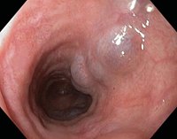
Esophageal Varices. This endoscopic image of the distal esophagus shows large, tortuous, bluish submucosal varices protruding into the lumen, with a smooth mucosal surface.
Contributed by S Bhimji, MD
(Click Image to Enlarge)
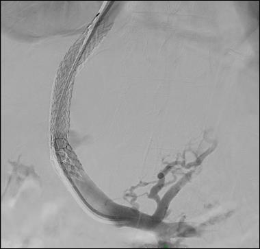
Portal Venogram via Transjugular Intrahepatic Portosystemic Shunt. This fluoroscopic portal venogram demonstrates contrast opacification of the portal venous system and the concomitant retrograde filling of large, tortuous collateral vessels characteristic of esophageal varices.
Contributed by M Young, MD
References
Kaplan DE, Ripoll C, Thiele M, Fortune BE, Simonetto DA, Garcia-Tsao G, Bosch J. AASLD Practice Guidance on risk stratification and management of portal hypertension and varices in cirrhosis. Hepatology (Baltimore, Md.). 2024 May 1:79(5):1180-1211. doi: 10.1097/HEP.0000000000000647. Epub 2023 Oct 23 [PubMed PMID: 37870298]
de Franchis R, Bosch J, Garcia-Tsao G, Reiberger T, Ripoll C, Baveno VII Faculty. Baveno VII - Renewing consensus in portal hypertension. Journal of hepatology. 2022 Apr:76(4):959-974. doi: 10.1016/j.jhep.2021.12.022. Epub 2021 Dec 30 [PubMed PMID: 35120736]
Level 3 (low-level) evidenceSingh S, Chandan S, Vinayek R, Aswath G, Facciorusso A, Maida M. Comprehensive approach to esophageal variceal bleeding: From prevention to treatment. World journal of gastroenterology. 2024 Nov 21:30(43):4602-4608. doi: 10.3748/wjg.v30.i43.4602. Epub [PubMed PMID: 39575399]
Gupta T. Non-invasive assessment of esophageal varices: Status of today. World journal of hepatology. 2024 Feb 27:16(2):123-125. doi: 10.4254/wjh.v16.i2.123. Epub [PubMed PMID: 38495268]
Gralnek IM, Camus Duboc M, Garcia-Pagan JC, Fuccio L, Karstensen JG, Hucl T, Jovanovic I, Awadie H, Hernandez-Gea V, Tantau M, Ebigbo A, Ibrahim M, Vlachogiannakos J, Burgmans MC, Rosasco R, Triantafyllou K. Endoscopic diagnosis and management of esophagogastric variceal hemorrhage: European Society of Gastrointestinal Endoscopy (ESGE) Guideline. Endoscopy. 2022 Nov:54(11):1094-1120. doi: 10.1055/a-1939-4887. Epub 2022 Sep 29 [PubMed PMID: 36174643]
Lee EW, Eghtesad B, Garcia-Tsao G, Haskal ZJ, Hernandez-Gea V, Jalaeian H, Kalva SP, Mohanty A, Thabut D, Abraldes JG. AASLD Practice Guidance on the use of TIPS, variceal embolization, and retrograde transvenous obliteration in the management of variceal hemorrhage. Hepatology (Baltimore, Md.). 2024 Jan 1:79(1):224-250. doi: 10.1097/HEP.0000000000000530. Epub 2023 Jun 30 [PubMed PMID: 37390489]
Wang X, Liu G, Wu J, Xiao X, Yan Y, Guo Y, Yang J, Li X, He Y, Yang L, Luo X. Small-Diameter Transjugular Intrahepatic Portosystemic Shunt versus Endoscopic Variceal Ligation Plus Propranolol for Variceal Rebleeding in Advanced Cirrhosis. Radiology. 2023 Aug:308(2):e223201. doi: 10.1148/radiol.223201. Epub [PubMed PMID: 37606572]