 Tools
Tools
 Erythrasma
Introduction
Erythrasma is a common bacterial skin disorder. The condition was first described in 1859 by Burchardt, who hypothesized a fungal etiology. In 1862, Burchardt’s teacher, Von Barensprung, coined the term "erythrasma" and identified the causative organism as Microsporum minutissimum. Subsequent research established Corynebacterium minutissimum, a gram-positive, catalase-positive, non-spore-forming bacterium, as the true pathogen.[1] Erythrasma primarily affects intertriginous regions and typically presents with itching, scaling, and erythema; however, lesions are often asymptomatic.[2][3]
Etiology
Register For Free And Read The Full Article
 Search engine and full access to all medical articles
- 10 free questions in your specialty
- Free CME/CE Activities
- Free daily question in your email
- Save favorite articles to your dashboard
- Emails offering discounts
Learn more about a Subscription to StatPearls Point-of-Care
Etiology
In 1961, Sarkay et al examined scrapings from erythrasma lesions using microscopy and identified gram-positive rods corresponding to C minutissimum.[4] The bacterium can cause severe disease in immunocompromised patients, including cutaneous fistulas, granulomatous lesions, and subcutaneous abscesses. Corynebacterium species normally exist as commensal skin bacteria.[5][6] Owing in part to the potential for serious complications in immunocompromised hosts, the genome of C minutissimum has been sequenced by organizations within the Centers for Disease Control and Prevention in Atlanta, Georgia.[7] Additional environmental and host factors that increase erythrasma risk include residence in humid climates, excessive sweating, diabetes mellitus, inadequate hygiene, advanced age, coexisting skin disorders, and obesity.[8][9]
Epidemiology
Most erythrasma cases occur in healthy adults. The infection is less common in children. Prevalence increases in hot, humid climates.[10] Institutional living is an additional risk factor, particularly in settings such as college dormitories, military barracks, and nursing facilities.[11] Incidence may rise with age. The condition occurs worldwide and appears more frequent in men. Epidemiologic data suggest erythrasma is most common among African Americans.
Pathophysiology
C minutissimum colonizes the upper stratum corneum under moist, occluded conditions. The organism produces coproporphyrin III, resulting in coral-red fluorescence on Wood lamp examination.[12]
Histopathology
A biopsy is rarely required for erythrasma diagnosis. Microscopic examination of affected skin reveals rod-like organisms in the stratum corneum, although these bacteria may be missed. Coccoid forms may also be present. Histopathology demonstrates hyperkeratosis with a mild lymphohistiocytic perivascular infiltrate in the upper dermis.
Gram staining shows gram-positive rods. Prolonged culture of C minutissimum may result in gram-negative staining and variable morphologies. The organism is not acid-fast. C minutissimum produces acid from dextrose, sucrose, and maltose, but not lactose. In vitro studies demonstrate sensitivity to erythromycin and resistance to penicillin. Visualization is challenging on hematoxylin and eosin–stained slides, but periodic acid-Schiff and Giemsa stains may enhance detection.
History and Physical
Erythrasma is most commonly observed in the toe webs, followed by the groin crease. The condition is less frequently found in the axilla. Affected individuals may report mildly pruritic skin discoloration in these areas. Scaling may also be present. Lesions are typically dark red and moist. Pruritus intensity varies. Lesions present as well-demarcated macular patches. The affected skin often exhibits a wrinkled appearance.
Interdigital Presentation
C minutissimum is the most common cause of bacterial infection in the interdigital spaces of the feet. Infection produces maceration and scaling. The fourth and fifth toes are most often affected. Lesions are usually asymptomatic but may be pruritic. Coinfection with dermatophytes or Candida species occasionally occurs. Potassium hydroxide preparation may be required to assess for coexisting infection.[13]
Intertriginous Variant
Lesions may be mildly pruritic or asymptomatic. The condition presents as patches or plaques in intertriginous areas, producing a "cigarette paper" appearance. Fine scaling may be present (see Image. Axillary Erythrasma).[14]
Disciform Pattern
Disciform erythrasma is a rare variant that may involve any body region. This form occurs more frequently in Black women in tropical climates. Lesions are well-defined, scaly plaques on the trunk and proximal limbs and exhibit coral-red fluorescence under Wood lamp examination.
Evaluation
Wood lamp examination aids in the diagnosis of erythrasma. However, empiric treatment based on clinical evaluation is reasonable in the absence of a lamp. Coral-red fluorescence results from coproporphyrin III produced by C minutissimum (see Image. Erythrasma Under Wood Lamp). Biopsy of suspected erythrasma may reveal rod-like organisms.[15] Visualization is often difficult, and Giemsa or periodic acid-Schiff staining may be required. These limitations render biopsy impractical for routine diagnosis.
Treatment / Management
Erythrasma responds to topical and oral therapies, although recurrence is common. Topical treatments include fusidic acid, clindamycin, and erythromycin. Fusidic acid, an antibiotic unavailable in the United States, inhibits aminoacyl–tRNA transfer during protein synthesis in susceptible bacteria. Researchers at University Hospital in Izmir, Turkey, conducted a randomized, double-blind, placebo-controlled trial involving 151 patients aged 18 years or older. Participants received 1 of 5 regimens: a single 1-g clarithromycin tablet; erythromycin 1 g orally daily for 14 days; a matching oral placebo for 14 days; fusidic acid 2% cream applied twice daily for 14 days; or a matching placebo cream twice daily for 14 days. Fusidic acid cream demonstrated superior reduction in Wood lamp scores compared with oral clarithromycin and erythromycin. Scores were based on coral-red fluorescence intensity, reflecting removal of coproporphyrin III from the stratum corneum. Complete clinical response was achieved in 96% of patients treated with fusidic acid, compared with 66% with clarithromycin and 53% with erythromycin.[16][17][18][19] Extensive erythrasma requires oral therapy. Oral options include a single 1-g dose of clarithromycin or 1 g of erythromycin daily, administered in divided doses for 14 days.(B2)
Clarithromycin offers advantages over erythromycin because it can be administered as a single dose. Clarithromycin is a macrolide that inhibits bacterial protein synthesis by binding to the 50S ribosomal subunit. Hydroxy-to-O-methyl substitution at position 6 of the lactone ring enhances bioavailability relative to erythromycin. Clarithromycin may be more effective for groin erythrasma. Few comparative studies between erythromycin and clarithromycin have been conducted. Adverse effects of both oral therapies include nausea, abdominal pain, metallic taste, hearing loss, ventricular arrhythmias, cholestatic jaundice, and toxic epidermal necrolysis.
Differential Diagnosis
The differential diagnosis for erythrasma includes seborrheic dermatitis, inverse psoriasis, candidiasis, dermatophytosis, tinea versicolor, parapsoriasis, and pityriasis rotunda. Seborrheic dermatitis typically presents with greasy, erythematous lesions along the scalp and nasolabial folds, while inverse psoriasis exhibits shiny plaques. Coral-red fluorescence under Wood lamp examination facilitates distinction of erythrasma from both seborrheic dermatitis and inverse psoriasis. Potassium hydroxide preparation may be required to differentiate erythrasma from dermatophytosis affecting the toe webs. Erythrasma involving the groin is readily distinguished from tinea cruris by the presence of a scaling border in tinea cruris.
Prognosis
Outcomes for most patients with erythrasma are excellent. Recurrence is possible if predisposing factors are not addressed; the infection does not produce residual sequelae in immunocompetent individuals. In contrast, C minutissimum can spread rapidly in immunocompromised patients.
Complications
Complications of erythrasma occur primarily in immunocompromised individuals and include endocarditis, abscess formation, intravascular catheter–associated infection, cellulitis, and pyelonephritis. Prompt recognition and appropriate antimicrobial therapy reduce the risk of systemic spread.
Deterrence and Patient Education
Deterrence and patient education are cornerstones of long-term erythrasma management, as recurrence is common unless predisposing factors are adequately addressed. Education should emphasize that erythrasma is a superficial bacterial infection caused by C minutissimum that proliferates in warm, moist, and occluded skin environments. Therefore, preventive instruction should focus on minimizing moisture and friction in intertriginous regions through regular gentle cleansing, meticulous drying of skin folds, and routine use of nonocclusive, breathable clothing. Intermittent use of absorbent powders or antiseptic washes may benefit individuals with excessive sweating.
Counseling should address systemic risk factors, particularly obesity and diabetes mellitus, because poor glycemic control and increased skin fold maceration significantly contribute to persistence and relapse. Sweat-reduction strategies, including topical antiperspirants, may be offered to individuals with hyperhidrosis. Clear guidance should be provided on the appropriate use and duration of prescribed topical or oral antibacterial therapy, and inappropriate or prolonged use of topical corticosteroids should be discouraged, as it can alter clinical appearance and delay diagnosis. Education should also include recognition of early recurrence and the importance of prompt treatment to prevent disease progression. Reassurance regarding the benign nature of erythrasma, combined with structured preventive guidance and lifestyle modification, supports sustained disease control and reduces the likelihood of repeated infection.
Enhancing Healthcare Team Outcomes
Management of most skin disorders involves an interprofessional team. Cutaneous lesions with unclear diagnosis are commonly encountered in clinical practice, and dermatology consultation is recommended in such cases. Multiple topical and oral treatment options are available for erythrasma. Extensive disease typically requires oral antibiotic therapy. Clinical outcomes in immunocompetent individuals are excellent, though recurrence is common.
Ongoing management may be overseen by primary care clinicians, who should be familiar with the adverse effects of macrolides, which are frequently used in the treatment of erythrasma. Nurses and pharmacists play key roles in patient education on lifestyle modifications that reduce the risk of erythrasma. Counseling should emphasize weight reduction and physical activity, as obesity is a major risk factor for morbidity and mortality. Education should also address personal hygiene and maintenance of dryness in intertriginous areas. Application of talcum powder may be recommended for persistently damp regions. In hot and humid locations, environmental cooling and the use of cotton garments, which absorb moisture more effectively than synthetic fabrics, should be encouraged.
Dietary counseling should include guidance on healthy eating patterns and limitation of refined sugars, given the association between diabetes mellitus and erythrasma. Immunocompromised individuals should be referred to dermatology for closer monitoring and intensified management. Effective communication among interprofessional team members is essential to optimize outcomes.
Media
(Click Image to Enlarge)
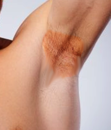
Axillary Erythrasma. A well-demarcated, brownish-red patch with fine scaling is present in the axilla, consistent with erythrasma involving an intertriginous area.
Contributed by S Bhimji, MD
(Click Image to Enlarge)
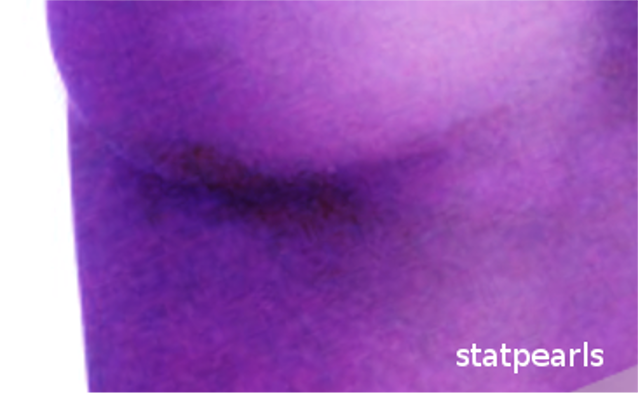
Erythrasma Under Wood Lamp. Coral-red fluorescence is visible in the affected skin, reflecting coproporphyrin III production by C minutissimum.
Contributed by S Bhimji, MD
References
Radhakrishnan S, Logamoorthy R, Karthikeyan K, Mohan R, Mary J JF. Erythrasma: a systematic review of interventions. Clinical and experimental dermatology. 2025 Oct 27:50(11):2139-2146. doi: 10.1093/ced/llaf307. Epub [PubMed PMID: 40635638]
Level 1 (high-level) evidenceDunn C, Applebaum DS, Dao H Jr. Widespread hyperpigmented rash present for 1 year. JAAD case reports. 2018 Sep:4(8):743-745. doi: 10.1016/j.jdcr.2018.04.012. Epub 2018 Sep 12 [PubMed PMID: 30225330]
Level 3 (low-level) evidenceTorres-Navarro I. Erythrasma. Emergencias : revista de la Sociedad Espanola de Medicina de Emergencias. 2018 Ago:30(4):283 [PubMed PMID: 30033708]
Pan S, Ryan MP, Rapini RP. Erythrasma. JAMA dermatology. 2024 Mar 1:160(3):352-353. doi: 10.1001/jamadermatol.2023.5129. Epub [PubMed PMID: 38231505]
Masood M, Usatine RP, Heath CR. Erythrasma. Cutis. 2022 Dec:110(6):338-339. doi: 10.12788/cutis.0662. Epub [PubMed PMID: 36735984]
H V, Madke B, Singh A. Extensive Erythrasma. The Journal of dermatology. 2025 Nov:52(11):e968-e969. doi: 10.1111/1346-8138.17946. Epub 2025 Sep 1 [PubMed PMID: 40888387]
Mitchell BI, Markantonis JE. An underestimated pathogen: Corynebacterium species. Journal of clinical microbiology. 2025 Oct 8:63(10):e0155224. doi: 10.1128/jcm.01552-24. Epub 2025 Aug 20 [PubMed PMID: 40833082]
Greywal T, Cohen PR. Erythrasma: A report of nine men successfully managed with mupirocin 2% ointment monotherapy. Dermatology online journal. 2017 May 15:23(5):. pii: 13030/qt9zh116s1. Epub 2017 May 15 [PubMed PMID: 28537862]
Sand FL, Thomsen SF. Skin diseases of the vulva: Infectious diseases. Journal of obstetrics and gynaecology : the journal of the Institute of Obstetrics and Gynaecology. 2017 Oct:37(7):840-848. doi: 10.1080/01443615.2017.1306696. Epub 2017 Apr 11 [PubMed PMID: 28397528]
Masood M, Heath CR, Usatine RP. Erythrasma. The Journal of family practice. 2022 Dec:71(10):E13-E14. doi: 10.12788/jfp.0528. Epub [PubMed PMID: 36735943]
Zisova L, Valchev V, Kasabov G. Erythrasma in athletes and football players. Wiener medizinische Wochenschrift (1946). 2021 Feb:171(1-2):24-28. doi: 10.1007/s10354-020-00753-2. Epub 2020 May 26 [PubMed PMID: 32458367]
Martínez-Ortega JI, Franco González S. Erythrasma: Pathogenesis and Diagnostic Challenges. Cureus. 2024 Aug:16(8):e68308. doi: 10.7759/cureus.68308. Epub 2024 Aug 31 [PubMed PMID: 39350848]
Trifoi M, Waseh S, Hsu S. Interdigital Toe Web Space Erythrasma in a Young Child: A Case Report. Cureus. 2023 Nov:15(11):e48435. doi: 10.7759/cureus.48435. Epub 2023 Nov 7 [PubMed PMID: 38074027]
Level 3 (low-level) evidenceRiquelme IL, Moyano EG. Axillary and inguinal erythrasma. CMAJ : Canadian Medical Association journal = journal de l'Association medicale canadienne. 2021 Oct 4:193(39):E1535. doi: 10.1503/cmaj.210310. Epub [PubMed PMID: 34607848]
Robles-Silva C, Hidalgo L, Vera-Kellet C, Navarrete-Dechent C. Erythrasma and the role of Wood's light: A simple diagnosis pearl. Medicina clinica. 2023 Oct 13:161(7):320. doi: 10.1016/j.medcli.2023.02.023. Epub 2023 Jul 28 [PubMed PMID: 37517932]
Blasco-Morente G, Arias-Santiago S, Pérez-López I, Martínez-López A. Coral-Red Fluorescence of Erythrasma Plaque. Sultan Qaboos University medical journal. 2016 Aug:16(3):e381-2. doi: 10.18295/squmj.2016.16.03.023. Epub 2016 Aug 19 [PubMed PMID: 27606125]
Ramírez-Hobak L, Moreno-Coutiño G, Arenas-Guzmán R, Gorzelewski A, Fernández-Martínez R. [Treatment of interdigital foot Erythrasma with ozonated olive oil]. Revista medica del Instituto Mexicano del Seguro Social. 2016 Jul-Aug:54(4):458-61 [PubMed PMID: 27197103]
Badri T, Sliti N, Benmously R, Hammami H, Ben Jennet S, Mokhtar I, Fenniche S. [Erythrasma: study of 16 cases]. La Tunisie medicale. 2014 Apr:92(4):245-8 [PubMed PMID: 25224418]
Level 2 (mid-level) evidenceSariguzel FM, Koc AN, Yagmur G, Berk E. Interdigital foot infections: Corynebacterium minutissimum and agents of superficial mycoses. Brazilian journal of microbiology : [publication of the Brazilian Society for Microbiology]. 2014:45(3):781-4 [PubMed PMID: 25477907]