 Tools
Tools
 Histology, Epithelial Cell
Introduction
Epithelial cells make up primary tissues throughout the body. Epithelial cells arise from ectoderm, mesoderm, and endoderm, which explains why they line body cavities and cover most body and organ surfaces.[1] There are many epithelial cell arrangements, such as squamous, cuboidal, and columnar, that are classified as simple, stratified, pseudostratified, and transitional. Since epithelial cells are prevalent throughout the body, their functions vary by location (see Table: Epithelial Cells, Location and Function). For example, epithelial cells in the skin provide protection, whereas they have secretory and absorptive properties in the gut. This topic seeks to explain the anatomical characteristics of epithelial cells and their functions, and describe features evident upon histological staining.[2][3]
Issues of Concern
Register For Free And Read The Full Article
 Search engine and full access to all medical articles
- 10 free questions in your specialty
- Free CME/CE Activities
- Free daily question in your email
- Save favorite articles to your dashboard
- Emails offering discounts
Learn more about a Subscription to StatPearls Point-of-Care
Issues of Concern
Epithelial cells are among the most abundant cells covering the skin, body cavities, and blood vessels. They contribute significantly to several aspects of the human life cycle, from embryogenesis to adulthood. Their highly specialized histologic features are critical to their physiological functions across different organs. Disorders of epithelial cell morphology and function have been confirmed in multiple clinical conditions, including cancer, organ fibrosis, celiac disease, and bullous pemphigoid.[4][5]
Structure
Epithelial cells exhibit structural polarity, with 3 distinct regions (apical, basal, and lateral). The apical domain faces the lumen of an organ or the external environment. This region often contains structures that affect cellular function, such as microvilli, cilia, and stereocilia. Microvilli are finger-like projections with a core of cross-linked actin filaments that are attached to the terminal web, parallel to the apical surface. Cilia are motile projections of the cell surface comprised of 2 central microtubules encompassed by 9 microtubule doublets. Lastly, stereocilia are finger-like projections supported by actin filaments.[6][7]
The basal domain is connected to the basal lamina by hemidesmosomes, which combine with intermediate filaments. The basal lamina separates connective tissue from the epithelium. The lateral domain connects neighboring cells and facilitates intercellular communication. Various junctional complexes connect adjacent cells. Desmosomes anchor/adhere junctions that tightly join cells by integrating with the cytoskeletal structures. Tight junctions are occluding junctions that regulate the movement of fluid and solutes. Gap junctions are intercellular channels found throughout the lateral domain, allowing small molecules and ions to pass between adjacent cells.[8]
Epithelial cells are organized according to their shape and the number of layers. Simple epithelial cells are a single layer, whereas stratified epithelial cells have 2 or more layers. Pseudostratified epithelial cells have a single layer of cells, but the cells vary in size, so they appear stratified or layered. Epithelial cells can be classified into 3 main shapes: squamous, cuboidal, and columnar. Squamous cells are flat, sheet-like; cuboidal cells are cube-like, with equal width, height, and depth; and columnar cells are taller than they are wide, making them rectangular.[9][10]
Function
Epithelial cells are found throughout the body and perform diverse functions, depending on their morphology and location. Structures of the apical domain significantly affect function. Microvilli are involved in fluid transport and absorption. The number of microvilli correlates with the cells' absorptive properties. Also, cilia transport substances across the surface of epithelial cells. Stereocilia are essential for hearing and balance.[7]
Simple squamous epithelium lines blood vessels (endothelium) and body cavities (mesothelium) and facilitates the diffusion of molecules, including gases, in gas exchange. Simple cuboidal cells are secretory and typically line ducts. Simple columnar cells are found throughout the intestines and can have either an absorptive or secretory function.[11][3]
Some stratified forms of these cells have similar functions. For example, stratified cuboidal cells are found in exocrine ducts and retain secretory function. Stratified columnar cells are present in large exocrine glands. Stratified squamous epithelium permits less nutrient diffusion than simple squamous epithelium because nutrients must traverse multiple layers, which provide protection. There are also 2 other categories of the epithelium, pseudostratified columnar and transitional (uroepithelium) epithelial cells. The pseudostratified epithelium is often ciliated to facilitate the transport of luminal contents. Transitional epithelial cells are present within distensible organs.[12][13]
Tissue Preparation
Epithelial cell examination can be performed via biopsy. Generally, after a sample is collected, it is fixed in formalin, embedded in paraffin, sectioned, and stained.[14]
Histochemistry and Cytochemistry
Epithelial cells have specialized cytoskeletons comprised of microtubules, actin filaments, and intermediate filaments. Intermediate filaments provide structural resilience to the cytoskeleton and show tissue-specific expression, unlike microtubules and actin filaments. These intermediate filaments are valuable targets for histochemical staining. There are 5 main groups of intermediate filaments. Glial filaments are found in astrocytes, neurofilaments are found in nerves, desmin filaments are found in muscles, vimentin filaments are seen in the mesenchyme, and keratin, which occurs in epithelial cells. Keratin not only defines the tissue as epithelial but also differentiates among epithelial cell types. For example, keratin 3 is found in the corneal epithelium, whereas keratin 20 is present in Merkel cells and umbrella cells of the urothelium. These keratins are crucial to epithelial cells, and mutations in keratin genes or loss of keratin can cause or predispose individuals to many diseases.[15][16]
Microscopy, Light
Understanding the appearance of normal, healthy epithelial cells is essential for identifying pathology in a tissue specimen. Light microscopy is typically used to visualize stained epithelial cells. Light microscopy can be used to determine the morphology of the epithelium present in the tissue specimen. Columnar epithelial cells tend to be rectangular, cuboidal cells appear square, and squamous cells are long and flat.[17][18]
Microscopy, Electron
Electron microscopy is a form of microscopy that allows a higher magnification than light microscopy. Electron microscopy allows visualization of many ultrastructural features, such as tight junctions, adherens junctions, desmosomes, hemidesmosomes, gap junctions, and the basal lamina, which separates the epithelial cells from connective tissue.[19][20]
Clinical Significance
One of the major concerns regarding epithelial cells is the risk of malignant transformation, including adenocarcinoma and papillary carcinoma. Some of the common adenocarcinomas that have high morbidity and mortality rates are lung, prostate, colon, and breast cancer.[21]
Another clinical concern that relates to epithelial cells is metaplasia. Metaplasia is the conversion of 1 cell type into another in response to environmental stressors or changes. This process can occur in physiological or pathological conditions. For instance, a study of the cervical epithelium in postpartum women has shown that columnar epithelium is replaced by squamous epithelium during the physiologic healing of the postpartum period.[22] Pathologic metaplasia is more likely to be dysplastic and may progress to malignancy. One relatively common example of pathologic metaplasia is Barrett esophagus. The esophagus is typically lined by squamous epithelium. In patients with uncontrolled gastroesophageal reflux disease (GERD), the constant stress induced by gastric acid causes the squamous cells of the esophagus to differentiate into mucin-producing columnar cells. The mucin-producing columnar cells are better equipped to withstand the stress of gastric acid, thereby preventing erosion of the esophagus. If GERD is properly treated, the columnar cells may revert to squamous cells; however, if the stressor remains untreated, metaplasia may progress to dysplasia, which can become malignant. Several studies have demonstrated that Barrett esophagus increases the risk of esophageal adenocarcinoma.[23][24]
In addition to cancer and metaplasia, another important aspect of epithelial cell biology is the epithelial-mesenchymal transition, a functional transition in which fully differentiated, polarized epithelial cells become motile mesenchymal cells. Like metaplasia, epithelial-mesenchymal transition occurs in physiological and pathological circumstances, including embryonic gastrulation, tissue regeneration, inflammation, fibrosis, and cancer metastasis.[5][25]
Furthermore, other forms of epithelial cell disorders are associated with various human diseases, particularly immune-related conditions. Celiac disease and certain bacterial infections can damage the microvilli of intestinal epithelial cells. In the lungs of premature infants, cuboidal alveolar cells (type II pneumocytes) have not fully developed, and surfactant production is impaired, leading to respiratory distress syndrome. In the skin, bullous pemphigoid, an autoimmune subepidermal skin blistering disease, essentially renders hemidesmosomes ineffective. Human papillomavirus strains 1-4 can cause warts on the squamous cells of the epidermis. This virus causes an overgrowth of epithelial tissue on a connective tissue papilla. This vast array of diseases underscores the clinical significance of understanding epithelial cells.[4]
Media
(Click Image to Enlarge)
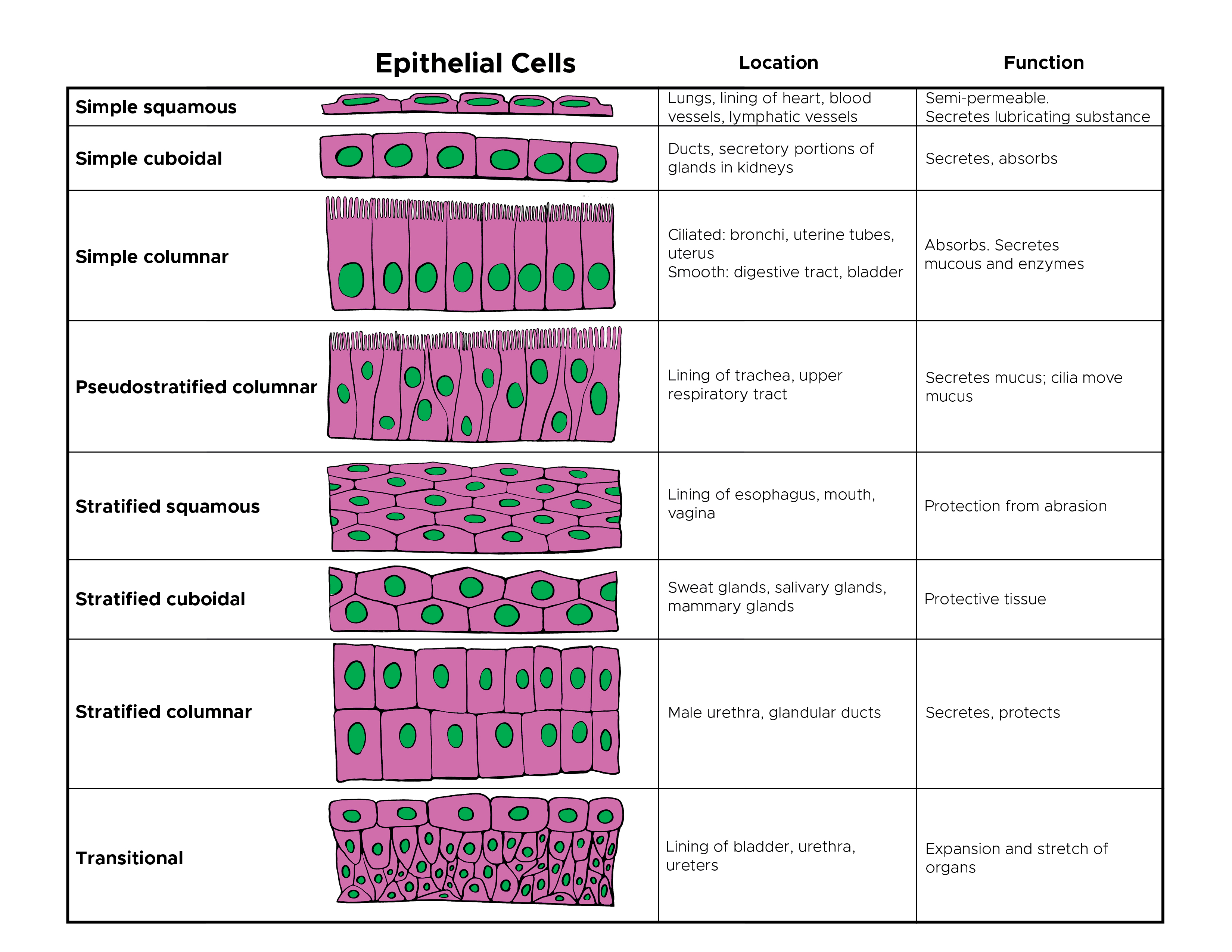
Epithelial Cells, Location and Function. This illustration depicts the various types of epithelial cells and locations in the body.
Illustrated by C Rowe
References
Acloque H, Adams MS, Fishwick K, Bronner-Fraser M, Nieto MA. Epithelial-mesenchymal transitions: the importance of changing cell state in development and disease. The Journal of clinical investigation. 2009 Jun:119(6):1438-49. doi: 10.1172/JCI38019. Epub 2009 Jun 1 [PubMed PMID: 19487820]
Level 3 (low-level) evidenceTadeu AM, Horsley V. Epithelial stem cells in adult skin. Current topics in developmental biology. 2014:107():109-31. doi: 10.1016/B978-0-12-416022-4.00004-4. Epub [PubMed PMID: 24439804]
Kong S, Zhang YH, Zhang W. Regulation of Intestinal Epithelial Cells Properties and Functions by Amino Acids. BioMed research international. 2018:2018():2819154. doi: 10.1155/2018/2819154. Epub 2018 May 9 [PubMed PMID: 29854738]
Rosenbach M, Wanat KA, Lynm C. Bullous pemphigoid. JAMA dermatology. 2013 Mar:149(3):382. doi: 10.1001/jamadermatol.2013.112. Epub [PubMed PMID: 23553008]
Kalluri R, Weinberg RA. The basics of epithelial-mesenchymal transition. The Journal of clinical investigation. 2009 Jun:119(6):1420-8. doi: 10.1172/JCI39104. Epub [PubMed PMID: 19487818]
Level 3 (low-level) evidencePelaseyed T, Bretscher A. Regulation of actin-based apical structures on epithelial cells. Journal of cell science. 2018 Oct 17:131(20):. doi: 10.1242/jcs.221853. Epub 2018 Oct 17 [PubMed PMID: 30333133]
Lange K. Fundamental role of microvilli in the main functions of differentiated cells: Outline of an universal regulating and signaling system at the cell periphery. Journal of cellular physiology. 2011 Apr:226(4):896-927. doi: 10.1002/jcp.22302. Epub [PubMed PMID: 20607764]
Level 3 (low-level) evidenceGarcia MA, Nelson WJ, Chavez N. Cell-Cell Junctions Organize Structural and Signaling Networks. Cold Spring Harbor perspectives in biology. 2018 Apr 2:10(4):. pii: a029181. doi: 10.1101/cshperspect.a029181. Epub 2018 Apr 2 [PubMed PMID: 28600395]
Level 3 (low-level) evidenceBartle EI, Rao TC, Urner TM, Mattheyses AL. Bridging the gap: Super-resolution microscopy of epithelial cell junctions. Tissue barriers. 2018 Jan 2:6(1):e1404189. doi: 10.1080/21688370.2017.1404189. Epub 2018 Feb 8 [PubMed PMID: 29420122]
Ikenouchi J. Roles of membrane lipids in the organization of epithelial cells: Old and new problems. Tissue barriers. 2018:6(2):1-8. doi: 10.1080/21688370.2018.1502531. Epub 2018 Aug 29 [PubMed PMID: 30156967]
Karseladze AI. Impairment of vascularization of the surface covering epithelium induces ischemia and promotes malignization: a new hypothesis of a possible mechanism of cancer pathogenesis. Clinical & translational oncology : official publication of the Federation of Spanish Oncology Societies and of the National Cancer Institute of Mexico. 2015 Jun:17(6):446-53. doi: 10.1007/s12094-014-1255-x. Epub 2014 Nov 19 [PubMed PMID: 25408194]
Shashikanth N, Yeruva S, Ong MLDM, Odenwald MA, Pavlyuk R, Turner JR. Epithelial Organization: The Gut and Beyond. Comprehensive Physiology. 2017 Sep 12:7(4):1497-1518. doi: 10.1002/cphy.c170003. Epub 2017 Sep 12 [PubMed PMID: 28915334]
Yee M, Gelein R, Mariani TJ, Lawrence BP, O'Reilly MA. The Oxygen Environment at Birth Specifies the Population of Alveolar Epithelial Stem Cells in the Adult Lung. Stem cells (Dayton, Ohio). 2016 May:34(5):1396-406. doi: 10.1002/stem.2330. Epub 2016 Mar 7 [PubMed PMID: 26891117]
Wick MR. The hematoxylin and eosin stain in anatomic pathology-An often-neglected focus of quality assurance in the laboratory. Seminars in diagnostic pathology. 2019 Sep:36(5):303-311. doi: 10.1053/j.semdp.2019.06.003. Epub 2019 Jun 4 [PubMed PMID: 31230963]
Level 2 (mid-level) evidenceHudson DL. Keratins as markers of epithelial cells. Methods in molecular biology (Clifton, N.J.). 2002:188():157-67 [PubMed PMID: 11987540]
Salas PJ, Forteza R, Mashukova A. Multiple roles for keratin intermediate filaments in the regulation of epithelial barrier function and apico-basal polarity. Tissue barriers. 2016 Jul-Sep:4(3):e1178368. doi: 10.1080/21688370.2016.1178368. Epub 2016 May 2 [PubMed PMID: 27583190]
Kommnick C, Lepper A, Hensel M. Correlative light and scanning electron microscopy (CLSEM) for analysis of bacterial infection of polarized epithelial cells. Scientific reports. 2019 Nov 19:9(1):17079. doi: 10.1038/s41598-019-53085-6. Epub 2019 Nov 19 [PubMed PMID: 31745114]
Syrjänen K, Syrjänen S, Lamberg M, Pyrhönen S, Nuutinen J. Morphological and immunohistochemical evidence suggesting human papillomavirus (HPV) involvement in oral squamous cell carcinogenesis. International journal of oral surgery. 1983 Dec:12(6):418-24 [PubMed PMID: 6325356]
Level 3 (low-level) evidenceVan Itallie CM, Anderson JM. Architecture of tight junctions and principles of molecular composition. Seminars in cell & developmental biology. 2014 Dec:36():157-65. doi: 10.1016/j.semcdb.2014.08.011. Epub 2014 Aug 27 [PubMed PMID: 25171873]
Yurchenco PD, Patton BL. Developmental and pathogenic mechanisms of basement membrane assembly. Current pharmaceutical design. 2009:15(12):1277-94 [PubMed PMID: 19355968]
Level 3 (low-level) evidenceTorre LA, Bray F, Siegel RL, Ferlay J, Lortet-Tieulent J, Jemal A. Global cancer statistics, 2012. CA: a cancer journal for clinicians. 2015 Mar:65(2):87-108. doi: 10.3322/caac.21262. Epub 2015 Feb 4 [PubMed PMID: 25651787]
Reid BL, Blackwell PM. PHYSIOLOGICAL METAPLASIA ON THE HUMAN CERVIX UTERI. SOME HISTO- AND CYTO-CHEMICAL OBSERVATIONS. The Australian & New Zealand journal of obstetrics & gynaecology. 1964 Jun:4(2):62-72 [PubMed PMID: 14173825]
Eluri S, Shaheen NJ. Barrett's esophagus: diagnosis and management. Gastrointestinal endoscopy. 2017 May:85(5):889-903. doi: 10.1016/j.gie.2017.01.007. Epub 2017 Jan 18 [PubMed PMID: 28109913]
Spechler SJ. Barrett esophagus and risk of esophageal cancer: a clinical review. JAMA. 2013 Aug 14:310(6):627-36. doi: 10.1001/jama.2013.226450. Epub [PubMed PMID: 23942681]
Yang J, Antin P, Berx G, Blanpain C, Brabletz T, Bronner M, Campbell K, Cano A, Casanova J, Christofori G, Dedhar S, Derynck R, Ford HL, Fuxe J, García de Herreros A, Goodall GJ, Hadjantonakis AK, Huang RYJ, Kalcheim C, Kalluri R, Kang Y, Khew-Goodall Y, Levine H, Liu J, Longmore GD, Mani SA, Massagué J, Mayor R, McClay D, Mostov KE, Newgreen DF, Nieto MA, Puisieux A, Runyan R, Savagner P, Stanger B, Stemmler MP, Takahashi Y, Takeichi M, Theveneau E, Thiery JP, Thompson EW, Weinberg RA, Williams ED, Xing J, Zhou BP, Sheng G, EMT International Association (TEMTIA). Guidelines and definitions for research on epithelial-mesenchymal transition. Nature reviews. Molecular cell biology. 2020 Jun:21(6):341-352. doi: 10.1038/s41580-020-0237-9. Epub 2020 Apr 16 [PubMed PMID: 32300252]