 Tools
Tools
 Asteatotic Eczema
Introduction
Asteatotic eczema is a common eczematous dermatitis that develops on xerotic (dry) skin.[1] Asteatotic eczema is also known as eczéma craquelé, xerotic eczema, or winter eczema.[1][2][3] The condition typically affects older adults and presents with pruritic, dry, and scaling skin with fine fissures in a polygonal or curvilinear pattern, producing a characteristic “crazy paving” or “dried riverbed” appearance.[1][4][5] The lower legs are the most frequently involved area, but the condition may extend to the arms and trunk.[1][6] This condition arises from epidermal barrier dysfunction, primarily due to age-related reductions in stratum corneum lipids and to environmental triggers such as cold weather and frequent bathing.[1][7] Consequently, seasonal exacerbations in winter are a well-recognized feature.[1]
Diagnosis is predominantly clinical, but dermoscopy has recently emerged as a useful noninvasive adjunct, revealing characteristic white scales with a double free edge arranged in a linear pattern, resembling railroad tracks.[8] Recognition of asteatotic eczema is clinically important, as it is among the most common causes of dermatitis in older adults.[9] Early diagnosis and therapy prevent complications such as painful hemorrhagic fissuring and secondary bacterial infection.[5][10]
While asteatotic eczema is usually benign, it can occasionally be a cutaneous clue to an underlying systemic disease.[1][6] Conditions that have been reported in association with asteatotic eczema include hypothyroidism, malnutrition, and states of acute edema, such as in nephrotic syndrome. Furthermore, asteatotic eczema is a known paraneoplastic finding, most notably in the context of lymphoma.[6][11][12][13][14] Various medications are also implicated, including diuretics, cimetidine, and antineoplastic agents like paclitaxel.[1][15][16]
Etiology
Register For Free And Read The Full Article
 Search engine and full access to all medical articles
- 10 free questions in your specialty
- Free CME/CE Activities
- Free daily question in your email
- Save favorite articles to your dashboard
- Emails offering discounts
Learn more about a Subscription to StatPearls Point-of-Care
Etiology
The development of asteatotic eczema is multifactorial, arising from extrinsic and intrinsic factors that disrupt the integrity of the stratum corneum.[1]
Exogenous Causes
- Environmental factors: The most significant trigger is low environmental humidity, which is common during cold winter months and in environments with central heating or air conditioning.[1]
- [1]Hygiene and contact factors: Frequent or prolonged bathing, especially with hot water, and the use of harsh, alkaline soaps or detergents can strip the skin of its essential surface lipids, exacerbating xerosis.[1]
- [1]Medication-induced xerosis: Certain systemic medications are known to induce or worsen xerosis, predisposing patients to asteatotic eczema. These include diuretics, systemic retinoids (eg, alitretinoin), and the H2-receptor antagonist cimetidine, which is thought to reduce sebum secretion via its antiandrogenic effects.[1][15][17] Chemotherapy, particularly with agents like paclitaxel and carboplatin, has been reported to induce severe, acute-onset asteatotic eczema.[16] Antiviral agents, such as interferons, have also been implicated.[18]
Endogenous Causes
- Aging: The most common endogenous factor is the natural aging process, which leads to a physiological decline in the function of sebaceous and sweat glands and reduced synthesis of epidermal lipids.[1][2]
- Nutritional deficiencies: Malnutrition can impair skin barrier function. Specifically, acquired zinc deficiency has been shown to cause a dermatosis clinically identical to asteatotic eczema.[12]
- Systemic diseases: Several systemic conditions are associated with asteatotic eczema; this may be a presenting feature of hypothyroidism, especially in the setting of myxedema.[11] These are also linked to internal malignancies, particularly hematologic cancers like T-cell lymphoma and Hodgkin disease, where they can present as a paraneoplastic syndrome.[6][14][19] The condition has also been seen in patients with HIV infection.[20]
- Edema-related states: Acute edema causes rapid stretching and distension of the skin, which can lead to epidermal fissuring in an asteatotic pattern.[5] This phenomenon has been reported in patients with congestive heart failure, nephrotic syndrome, and refeeding syndrome, especially in the treatment of anorexia nervosa.[5][13]
Epidemiology
Asteatotic eczema is a common dermatosis, particularly in the older population.[1]
- Age: The condition is most prevalent in individuals older than 60.[1][9] One report identified a median age of 69 years at presentation.[1]
- Sex: Men older than 60 are more likely to develop asteatotic eczema than women.
- Prevalence: Asteatotic eczema is a common condition documented worldwide, particularly among older adults. The prevalence of this condition has been reported as 11.4% in an Indian community-based study.[9] In a Turkish dermatology clinic, it was the most common form of eczematous dermatitis among older adults, accounting for 26.9% of eczema cases.[21] The prevalence is notably higher among dependent older adults; results from a Japanese study of residents in long-term care facilities reported rates of 16.4% in one facility and 41.2% in another.[22]
- Disease Burden and Pruritus: Pruritus is a cardinal symptom and can be severe, significantly impacting quality of life.[1][2] A large multicenter study in China found that asteatotic eczema was among the top 3 eczema subtypes associated with severe itching (27.9% of patients).[23]
Pathophysiology
The central mechanism of asteatotic eczema is the failure of the stratum corneum to function as an effective water barrier, leading to excessive transepidermal water loss (TEWL), desiccation, fissuring, and secondary inflammation.[1][7]
- Epidermal lipid depletion: The skin's ability to retain water is critically dependent on the lipid matrix in the stratum corneum. In aging skin, there is a significant decline in sebum-derived lipids within the stratum corneum, particularly triglycerides and free fatty acids.[7] Furthermore, the clinical severity of xerosis is directly correlated with reduced levels of natural moisturizing factors, such as water-soluble amino acids derived from profilaggrin breakdown.[24] A similar mechanism may occur in hypoesthetic or denervated skin, where impaired autonomic regulation of sweat and sebum glands leads to severe localized xerosis and subsequent fissuring.[3]
- Impaired water homeostasis: The stratum corneum must maintain a water content of at least 10% to remain pliable.[1] When the lipid barrier is compromised, water loss accelerates. Results from foundational studies have shown that experimentally stripping the skin of its lipids can increase TEWL by up to 75-fold.[25] This profound water loss results in the characteristic brittleness and cracking observed in this condition.
- Mechanical and other factors: Acute subcutaneous edema can cause rapid stretching of the epidermis beyond its elastic capacity, resulting in physical fractures of the stratum corneum.[5][13] Breaks in the epidermal barrier allow irritants to penetrate and trigger a nonspecific inflammatory response characterized by a superficial perivascular lymphocytic infiltrate.[6] This breach of the epidermal barrier also predisposes the skin to secondary colonization and infection with bacteria such as Staphylococcus aureus.[10]
Histopathology
A skin biopsy is not typically required for the diagnosis of asteatotic eczema; however, it may be performed in atypical cases. The microscopic findings are consistent with mild, subacute spongiotic dermatitis.[1][6]
- Epidermis: The stratum corneum shows compact orthohyperkeratosis with focal areas of parakeratosis. The epidermis itself exhibits mild to moderate spongiosis (intercellular edema) and irregular acanthosis. In cases induced by chemotherapy, scattered dyskeratotic or necrotic keratinocytes may be observed throughout the epidermis.[16]
- Dermis: A sparse, superficial perivascular infiltrate composed mainly of lymphocytes is present. In cases with clinical hemorrhage, extravasated erythrocytes can be seen in the papillary dermis.[5][6]
History and Physical
The diagnosis of asteatotic eczema is typically clinical. A thorough history is key to identifying triggers and associated conditions. Important points include:
- Onset and seasonality: A history of onset or worsening during the winter is classic.[1]
- Symptoms: Pruritus is nearly universal and can be severe enough to disrupt sleep. Pain becomes a significant feature if deep fissures develop.[5][23]
- Skin care habits: A history of frequent or prolonged bathing, exposure to hot water, and use of drying or alkaline cleansers is commonly associated with asteatotic eczema; the inquiry should include bathing practices and product use.[1]
- Medical history: A review of systems should screen for symptoms of hypothyroidism (cold intolerance, fatigue), malnutrition (weight loss), and malignancy (fever, night sweats, weight loss). A full medication review is also essential.[6][11]
Physical examination should include characterization of the following:
- Morphology: The hallmark is dry, scaly skin with a network of fine, erythematous fissures that create a "crazy-paving" or "cracked porcelain" appearance (see Image. Eczema, Craquele).[1] In severe cases, the fissures may be hemorrhagic.[5]
- Distribution: The anterolateral shins are the most common site. The eruption may also involve the thighs, flanks, abdomen, and upper extremities.[1][6]
- Associated findings: Additional features may suggest underlying systemic conditions, including pitting edema related to congestive heart failure or nephrotic syndrome and palpable lymphadenopathy in cases of lymphoma.[5][6]
Evaluation
While the diagnosis of asteatotic eczema is primarily clinical, the following tools can aid in confirming the diagnosis and excluding other conditions.[1]
- Dermoscopy: This noninvasive tool is highly useful. The pathognomonic dermoscopic finding is the presence of white scales with a double free edge, creating a characteristic “rail-like” appearance within the skin fissures. In more inflammatory lesions, dermoscopy may also reveal a pink-red background with irregular dotted vessels and yellow-orange clods or serocrusts.[8]
- Biopsy: A skin biopsy is rarely required but can be useful in atypical or treatment-resistant cases to exclude mimics such as cutaneous T-cell lymphoma.[1][6]
- Supportive tests: A potassium hydroxide preparation of skin scrapings is helpful to exclude dermatophytosis in scaly lesions.[8]
Treatment / Management
Managing asteatotic eczema centers on restoring the skin's barrier function, reducing inflammation, and alleviating symptoms.[1]
Cornerstone of Therapy: Barrier Repair and Patient Education
Aggressive barrier repair and patient education are central to the management of asteatotic eczema. Treatment primarily consists of the frequent, liberal application of emollients with high oil content, such as petrolatum-based ointments.[3] These products are most effective when applied to damp skin immediately after bathing, thereby trapping moisture.[4] Preparations containing alpha-hydroxy acids may also be beneficial.[2] Patients should be counseled to take short, lukewarm baths, avoid harsh soaps in favor of gentle cleansers, and use humidifiers in dry environments.[4](B3)
Pharmacologic Interventions
For active inflammation, topical anti-inflammatory agents are indicated. Low- to mid-potency topical corticosteroids (TCS) in an ointment base are effective for inflamed lesions.[1] The order of application between emollients and TCS does not significantly affect the clinical response.[10] Pimecrolimus 1% cream, employed as a steroid-sparing alternative, significantly improves eczema and reduces associated pruritus, as demonstrated in a randomized controlled trial. The non-atrophogenic properties of the agent make it particularly appropriate for older adults with thin or fragile skin.[26] For patients with moderate disease, medium or high-potency corticosteroids should be used. Systemic therapy is rarely necessary but may include oral antihistamines for symptomatic relief of pruritus.[1] For severe, widespread cases, such as paraneoplastic or certain drug-induced presentations, a short course of oral corticosteroids may be considered.[12](A1)
Emerging Therapies
Emerging therapies include emollients containing endogenous lipids such as N-palmitoylethanolamine and N-acetylethanolamine. Results from a randomized controlled trial demonstrated that these agents can improve skin barrier function and reduce itching compared to traditional emollients.[27](A1)
Differential Diagnosis
The differential diagnosis of asteatotic eczema includes various dermatologic and systemic conditions that must be excluded to ensure a comprehensive clinical evaluation. These include the following:
Prognosis
Asteatotic eczema responds well to therapy but often follows a chronic, relapsing course, especially during the winter months.[1] Refractory cases may indicate an underlying condition, such as malignancy, which carries a more guarded prognosis.[6]
Complications
In severe cases of asteatotic eczema, deep, wide fissures can develop, which may cause pain and bleeding.[1][5] The compromised skin barrier inherent to asteatotic eczema also predisposes to secondary bacterial infections, which can lead to complications such as cellulitis or deep implant infections in patients with orthopedic hardware.[1][10] Additionally, topical corticosteroids and emollients are first-line treatments for asteatotic eczema, and their overuse can lead to skin thinning.
Deterrence and Patient Education
Patient education is the cornerstone of treating and preventing progression of asteatotic eczema.[4] Key counseling points include taking short, lukewarm baths with gentle cleansers; applying thick emollients liberally and frequently; using a home humidifier during winter; and avoiding irritants such as wool clothing.[1][4] Patients who present with asteatotic eczema should be reassured that it is likely a benign, isolated condition, although in rare cases, the disease correlates with an underlying malignancy. Importantly, educate patients about the risks of scratching, which can cause skin breaks that may lead to infection. Patients should also understand that asteatotic eczema is likely to recur in the winter months.[1]
Enhancing Healthcare Team Outcomes
Successfully managing asteatotic eczema requires an interprofessional team approach, particularly in older adults with comorbidities. Low awareness of this skin condition among non-dermatology staff, particularly in long-term care facilities, can lead to undertreatment. Physicians, advanced practitioners, nurses, pharmacists, and other health professionals must collaborate to maximize efficacy. Nurses are crucial for educating patients and caregivers on proper skin care. Pharmacists can identify medications that may cause or worsen xerosis. This collaborative approach ensures comprehensive care and improves patient outcomes.[1]
Media
(Click Image to Enlarge)
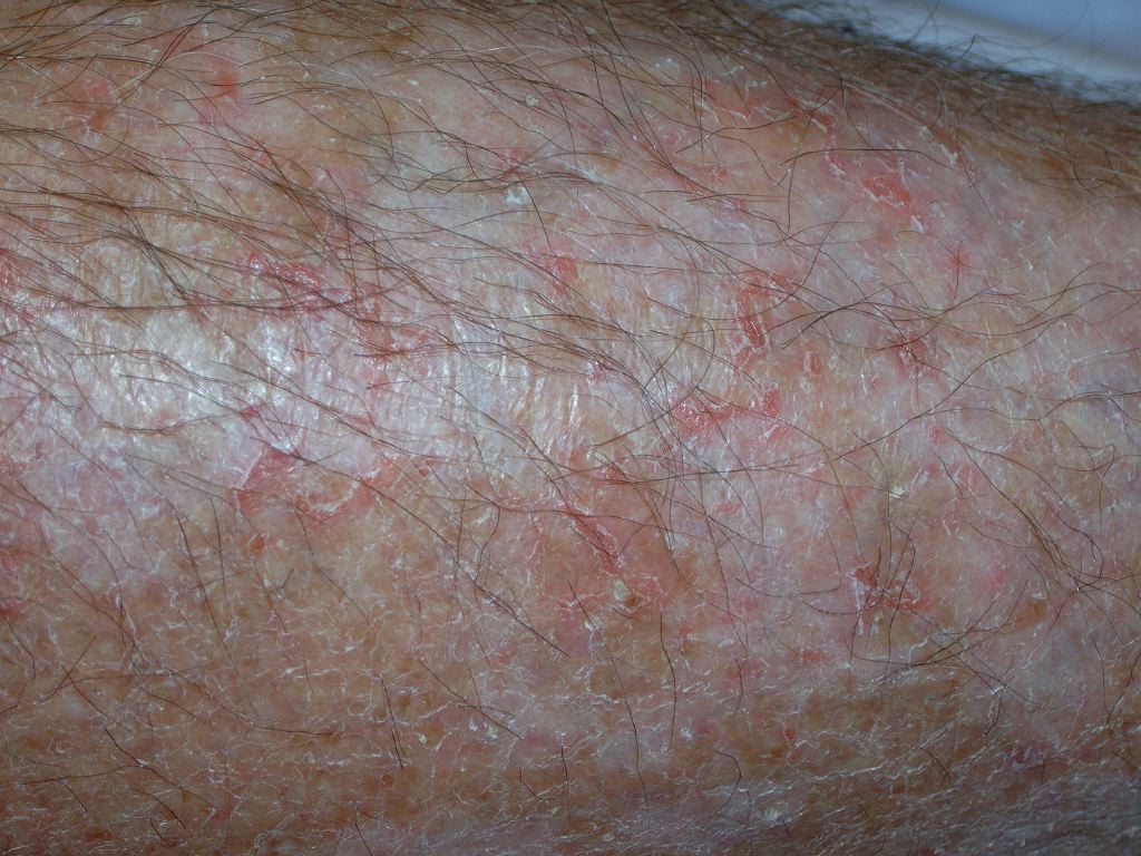
Eczema, Craquele. This image shows the hallmark characteristics of eczema: dry, scaly skin with a network of fine, erythematous fissures that create a "crazy-paving" or "cracked porcelain" appearance.
References
Specht S, Persaud Y. Asteatotic Eczema. StatPearls. 2025 Jan:(): [PubMed PMID: 31747214]
Norman RA. Xerosis and pruritus in the elderly: recognition and management. Dermatologic therapy. 2003:16(3):254-9 [PubMed PMID: 14510882]
Cassler NM, Burris AM, Nguyen JC. Asteatotic eczema in hypoesthetic skin: a case series. JAMA dermatology. 2014 Oct:150(10):1088-90. doi: 10.1001/jamadermatol.2014.394. Epub [PubMed PMID: 25029204]
Level 3 (low-level) evidenceWard S. Eczema and dry skin in older people: identification and management. British journal of community nursing. 2005 Oct:10(10):453-6 [PubMed PMID: 16234740]
Caplan RM. Superficial hemorrhagic fissures of the skin. Archives of dermatology. 1970 Apr:101(4):442-51 [PubMed PMID: 5440818]
Sparsa A, Liozon E, Boulinguez S, Bordessoule D, Vidal E, Bonnetblanc JM, Bedane C. Generalized eczema craquele as a presenting feature of systemic lymphoma: report of seven cases. Acta dermato-venereologica. 2005:85(4):333-6 [PubMed PMID: 16191855]
Level 3 (low-level) evidenceAkimoto K, Yoshikawa N, Higaki Y, Kawashima M, Imokawa G. Quantitative analysis of stratum corneum lipids in xerosis and asteatotic eczema. The Journal of dermatology. 1993 Jan:20(1):1-6 [PubMed PMID: 8482746]
Errichetti E. Dermoscopy of Inflammatory Dermatoses (Inflammoscopy): An Up-to-Date Overview. Dermatology practical & conceptual. 2019 Jul:9(3):169-180. doi: 10.5826/dpc.0903a01. Epub 2019 Jul 31 [PubMed PMID: 31384489]
Level 3 (low-level) evidenceNeena V, Asokan N, Jose R, Sarin A. Prevalence of eczema among older persons: A population-based cross-sectional study. Indian journal of dermatology, venereology and leprology. 2023 May-Jun:89(3):426-430. doi: 10.25259/IJDVL_990_19. Epub [PubMed PMID: 34491677]
Level 2 (mid-level) evidenceLim CT, Tan KJ, Kagda F, Ang KC. Implant infection caused by dermatitis: a report of two cases. Journal of orthopaedic surgery (Hong Kong). 2007 Dec:15(3):365-7 [PubMed PMID: 18162688]
Level 3 (low-level) evidenceWarin AP. Eczéma craquelé as the presenting feature of myxoedema. The British journal of dermatology. 1973 Sep:89(3):289-91 [PubMed PMID: 4743432]
Weismann K, Wadskov S, Mikkelsen HI, Knudsen L, Christensen KC, Storgaard L. Acquired zinc deficiency dermatosis in man. Archives of dermatology. 1978 Oct:114(10):1509-11 [PubMed PMID: 718188]
Level 3 (low-level) evidenceYang CS, Lott JP, Bunick CG, Bolognia JL. Eczema craquelé associated with nephrotic syndrome. JAAD case reports. 2016 May:2(3):241-3. doi: 10.1016/j.jdcr.2016.04.008. Epub 2016 Jun 24 [PubMed PMID: 27408932]
Level 3 (low-level) evidenceBarker DJ, Cotterill JA. Generalized eczéma craquelé as a presenting feature of lymphoma. The British journal of dermatology. 1977 Sep:97(3):323-6 [PubMed PMID: 921903]
Level 3 (low-level) evidenceGreist MC, Epinette WW. Cimetidine-induced xerosis and asteatotic dermatitis. Archives of dermatology. 1982 Apr:118(4):253-4 [PubMed PMID: 6461297]
Level 3 (low-level) evidenceVishwanath T, Mahajan SA, Gole PV, Kulkarni M. Asteatotic eczema following paclitaxel and carboplatin chemotherapy for lung carcinoma. Indian journal of dermatology, venereology and leprology. 2024 Aug 30:():1-3. doi: 10.25259/IJDVL_569_2024. Epub 2024 Aug 30 [PubMed PMID: 39361863]
Clabbers J, van Oosten NV, Bolling M, Vreeburg M, van Geel M, Steijlen P, Gostynski A. Alitretinoin as a Treatment Modality for Ichthyosis in Women of Childbearing Age: A Case Series and Review of the Literature. Dermatology (Basel, Switzerland). 2024:240(1):170-177. doi: 10.1159/000533934. Epub 2023 Sep 4 [PubMed PMID: 37666225]
Level 2 (mid-level) evidenceVale EPBMD, Rodrigues CHL, Kallas FE, Fucuta PDS. Eczema craquelé associated with antiviral treatment for chronic hepatitis C. Anais brasileiros de dermatologia. 2017 May-Jun:92(3):436-437. doi: 10.1590/abd1806-4841.20174464. Epub [PubMed PMID: 29186272]
Le Clainche Compagnie A, Degoutte C, Papouin B, Lamaison C, Gounot R, Ingen-Housz-Oro S. Asteatotic eczema, a cutaneous manifestation of Hodgkin lymphoma in older patients. EJHaem. 2024 Aug:5(4):883-884. doi: 10.1002/jha2.910. Epub 2024 Jun 26 [PubMed PMID: 39157615]
Garg T, Sanke S. Inflammatory dermatoses in human immunodeficiency virus. Indian journal of sexually transmitted diseases and AIDS. 2017 Jul-Dec:38(2):113-120. doi: 10.4103/ijstd.IJSTD_22_17. Epub [PubMed PMID: 30148263]
Polat M, İlhan MN. Dermatological Complaints of the Elderly Attending a Dermatology Outpatient Clinic in Turkey: A Prospective Study over a One-year Period. Acta dermatovenerologica Croatica : ADC. 2015:23(4):277-81 [PubMed PMID: 26724880]
Kimura N, Nakagami G, Takehara K, Miura Y, Nakamura T, Kawashima M, Tsunemi Y, Sanada H. Prevalence of asteatosis and asteatotic eczema among elderly residents in facilities covered by long-term care insurance. The Journal of dermatology. 2013 Sep:40(9):770-1. doi: 10.1111/1346-8138.12217. Epub 2013 Jul 16 [PubMed PMID: 23855654]
Level 3 (low-level) evidenceWang X, Li L, Shi X, Zhou P, Shen Y. Itching and its related factors in subtypes of eczema: a cross-sectional multicenter study in tertiary hospitals of China. Scientific reports. 2018 Jul 17:8(1):10754. doi: 10.1038/s41598-018-28828-6. Epub 2018 Jul 17 [PubMed PMID: 30018382]
Level 2 (mid-level) evidenceHorii I, Nakayama Y, Obata M, Tagami H. Stratum corneum hydration and amino acid content in xerotic skin. The British journal of dermatology. 1989 Nov:121(5):587-92 [PubMed PMID: 2597631]
ONKEN HD, MOYER CA. The water barrier in human epidermis. Physical and chemical nature. Archives of dermatology. 1963 May:87():584-90 [PubMed PMID: 13940146]
Schulz P, Bunselmeyer B, Bräutigam M, Luger TA. Pimecrolimus cream 1% is effective in asteatotic eczema: results of a randomized, double-blind, vehicle-controlled study in 40 patients. Journal of the European Academy of Dermatology and Venereology : JEADV. 2007 Jan:21(1):90-4 [PubMed PMID: 17207174]
Level 1 (high-level) evidenceYuan C, Wang XM, Guichard A, Tan YM, Qian CY, Yang LJ, Humbert P. N-palmitoylethanolamine and N-acetylethanolamine are effective in asteatotic eczema: results of a randomized, double-blind, controlled study in 60 patients. Clinical interventions in aging. 2014:9():1163-9. doi: 10.2147/CIA.S65448. Epub 2014 Jul 17 [PubMed PMID: 25071367]
Level 1 (high-level) evidence