 Tools
Tools
 Anatomy, Thorax: Diaphragm
Introduction
The diaphragm in the thorax is called the thoracic diaphragm and serves as an important anatomical landmark separating the thorax (chest) from the abdomen. It functions during breathing, contracting to enlarge the thoracic cavity and reduce intrathoracic pressure, allowing the lungs to expand and fill their alveoli with air. It is a dome-shaped muscle and tendon that functions as the primary respiratory muscle and is essential to the breathing process. The diaphragm is a fibromuscular sheet with a convex upper surface forming the floor of the thoracic cavity and a concave undersurface forming the roof of the abdominal cavity. The esophagus, phrenic and vagus nerves, descending aorta, and inferior vena cava pass through the diaphragm between the thoracic and abdominal cavities. The diaphragm is asymmetric with the left side slightly more inferior than the right, chiefly because of the presence of the liver located on the right. The left side may also be partially inferior due to heart pressure.[1][2]
Structure and Function
Register For Free And Read The Full Article
 Search engine and full access to all medical articles
- 10 free questions in your specialty
- Free CME/CE Activities
- Free daily question in your email
- Save favorite articles to your dashboard
- Emails offering discounts
Learn more about a Subscription to StatPearls Point-of-Care
Structure and Function
Functions of the Diaphragm
Muscle of inspiration: The diaphragm pulls its central tendon down during contraction and then increases the vertical diameter of the thorax. This action increases the negative pressure inside the thoracic cavity, drawing air in. Thus, the diaphragm is the most important muscle used in inspiration. During inhalation, the diaphragm contracts and descends into the abdominal cavity, where it appears flat. Simultaneously, the external intercostal muscles located between the ribs raise the anterior chest wall like the handles of a bucket. This results in the chest cavity becoming larger and wider, allowing air to enter from the outside. During exhalation, the rib cage and chest wall begin to sag and return to their original position. Concurrently, there is relaxation and elevation of the diaphragm. This motion forces air from the lungs.[2][3]
Muscle of abdominal straining: The contraction of the diaphragm assists the anterior abdominal muscles in raising intra-abdominal pressure during normal processes such as micturition, defecation, and parturition.
Weightlifting muscle: When a person takes and holds a deep breath, the diaphragm assists the anterior abdominal muscles in raising intra-abdominal pressure. This maneuver, also called the Valsalva maneuver, is used to accentuate heart murmurs and classify them as clinically right- or left-sided.
Thoracoabdominal pump: During respiration, the diaphragm descends, which decreases intrathoracic pressure and increases intra-abdominal pressure. This negative pressure compresses the blood in the inferior vena cava, forcing it upward into the right atrium and helping fill the heart. When the abdominal lymph vessels are also compressed, their upward passage within the thoracic duct is aided by the negative intrathoracic pressure. Furthermore, valves in the thoracic duct prevent backflow of lymph.
Embryology
Diaphragm Formation
- Septum transversum
- Pleuroperitoneal membrane
- Mesentery of esophagus
- Mesoderm of the body wall
Insertion
The diaphragm inserts into a central tendon. The top surface of the tendon is partially connected to the lower surface of the fibrous pericardium. Muscle fibers arising from the right crus traverse up on the left side and encircle the orifice of the esophagus in a sling-like loop. These fibers act as a sphincter and likely help prevent regurgitation of stomach contents into the thoracic esophagus.[4]
Origin of Diaphragm
Sternal: The sternal aspect originates as 2 fleshy slips from the back of the xiphoid process.
Costal: The costal aspect originates from the inner surfaces of the cartilages adjacent to the lower sixth ribs on each side. This section interdigitates with the transversus abdominis.
Lumbar:
- The medial lumbocostal arch is tendinous in fascia covering the psoas major. Medially, it attaches to the side of the body of vertebra L1. Laterally, it connects to the front of the transverse process of vertebra L1.
- The lateral lumbocostal arch is tendinous in fascia covering the upper part of quadratus lumborum. Medially, it attaches to the front of the transverse process of vertebra L1. Laterally, it connects to the lower border of the 12th rib.
- The right crus arises from the anterolateral surface of the bodies of the upper 3 lumbar vertebrae and also the intervening intervertebral disc.
- The left crus arises from the corresponding parts of the upper 2 lumbar vertebrae.
- The medial margin of the 2 crura forms a tendinous arc across the front of the aorta called the median arcuate ligament.
Blood Supply and Lymphatics
Major Arteries Supplying the Diaphragm
- Musculophrenic artery, a branch of the internal thoracic artery
- Superior phrenic artery, branch of the aorta
- Lower five intercostal arteries and subcostal artery
- Inferior phrenic artery
Nerves
Motor Nerve Supply
Right and left phrenic nerves (C3 through C5)
Sensory Nerve Supply
The phrenic nerve innervates the parietal pleura and peritoneum covering the central surfaces of the diaphragm. The lower 6 intercostal nerves supply the periphery of the diaphragm. When the diaphragm contracts, the large-sized myelinated phrenic afferents fire. Conversely, the smaller diameter nerves continue to discharge throughout the respiratory cycle. Activation of both nonmyelinated and myelinated phrenic sensory nerves modulates respiratory output during each breathing cycle. However, the activation of the phrenic afferents does increase significantly as the diaphragm continues to work and develops fatigue. Once the phrenic afferents are activated, they also modulate the sympathetic motor outflow. Furthermore, phrenic afferents contribute to somatosensation of the diaphragm and make one aware of breathing while awake. The exact influence of the spinal and supraspinal nerves and synapses between the nonmyelinated and myelinated phrenic nerves is not known. The use of deep muscle training contributed to a significant change in body position in the sagittal plane and an increase in breathing amplitude.[5]
Muscles
The origins of the diaphragm are along the lumbar vertebrae of the spine and the inferior border of the ribs and sternum. The superior diaphragm origin is continuous from the xiphoid process anteriorly to the lower 6 costal cartilages of the thorax laterally and the first 2 lumbar vertebrae posteriorly. The musculoskeletal fibers radiate from all angles toward the center of the body and converge into a central tendon, which is the inferior attachment or musculoskeletal point. The diaphragm has a dome-like structure with the peripheral segment attached to the chest wall and abdominal cavity. The muscle fibers from these attachments converge in a central tendon, which forms the crest of the dome. The periphery of the diaphragm is made of strong muscular fibers that have their origin from the surroundings of the inferior thoracic aperture. These muscle fibers then converge and insert into the central tendon.
Surgical Considerations
Diaphragmatic Hernia
A diaphragmatic hernia is a congenital disability that occurs when one or more of a person's abdominal organs (stomach, spleen, liver, intestines) move upward into the chest through a defect in the diaphragm. It is usually congenital but can be acquired. Congenital hernias are considered a medical emergency and require prompt surgery.[6]
Congenital diaphragmatic hernia: A congenital diaphragmatic hernia is an abnormal development of the diaphragm during fetal life. The herniation leads to the passage of one or more abdominal organs into the chest, leading to pulmonary hypoplasia, which is usually unilateral. The most common subtype of congenital diaphragmatic hernia is Bochdalek hernia. Other types include Morgagni hernia, diaphragm eventration, and central tendon defects of the diaphragm.
Acquired diaphragmatic hernia: An acquired diaphragmatic hernia happens due to penetrating or blunt injury. Falls and motor vehicle accidents are major causes of blunt injury, while stabs and gunshot wounds lead to penetrating injuries. Penetrating injuries are the more common cause than the blunt injuries as a cause of the diaphragmatic rupture. Further, there can be accidental damage to the diaphragm due to surgery. Infrequently, a diaphragmatic hernia may result without any identifiable cause and remain undiagnosed for an undetermined amount of time, until the herniation of abdominal organs into the chest starts to cause symptoms.[7]
Treatment
Both acquired and congenital diaphragmatic hernias usually require immediate surgical intervention. A surgical procedure requires returning the abdominal organs from the chest cavity to their original location in the abdominal cavity. The diaphragm should be repaired with staples or permanent sutures with or without a prosthetic patch.
Clinical Significance
The diaphragm has 3 major openings and 5 minor openings:
Major
- The vena caval trunk lies at the level of the T8 vertebra in the central tendon. It allows passage of the inferior vena cava and some branches of the right phrenic nerve.
- The esophageal hiatus lies at the level of the T10 vertebra, within a sling of muscle fibers derived from the right crus, to the left of the median plane. It allows passage of the esophagus, the right and left vagus trunks, the esophageal branches of the left gastric vessels, and the lymphatic vessels.
- The aortic hiatus lies anterior to the body of the T12 vertebra between the crura. It allows passage of the aorta, thoracic duct, and azygos vein.
Minor
- The lesser aperture of right crus (permits lesser and greater splanchnic nerves)
- The lesser aperture of left crus (permits hemiazygous vein; and lesser and greater splanchnic nerves)
- The sympathetic trunk runs posteriorly below the medial lumbocostal arches
- The foramen of Morgagni is found in the areolar tissue between the sternal and costal part of the diaphragm and contains the superior epigastric branch of the internal thoracic artery and the lymphatics of the abdominal wall
- The medial and lateral lumbocostal arches can contain areolar tissue that, when present, separates the superior and posterior surface of a kidney from the pleura
The figure below shows the openings in the diaphragm: A=vena cava, B=esophagus, C=aorta. The muscular diaphragm surrounds the central tendon.
Other Issues
The diaphragm has many other roles besides respiration. This muscle acts as a barrier between the thoracic and abdominal cavities, preventing herniation of abdominal organs into the chest cavity. Both penetrating and blunt trauma can injure the diaphragm. A tear or rupture of the diaphragm is often difficult to diagnose. Options for diagnosis include laparoscopy, thoracoscopy, or a CT scan. Irrespective of the size of the tear, the muscle must be repaired as soon as possible to prevent herniation of abdominal organs. The diaphragm is also involved in hiccups. When the muscle is irritated, it can result in sudden contractions which can be uncomfortable. Most hiccups are short-lived, but in rare cases they may last a day. If the hiccups persist, they can interfere with breathing.
Media
(Click Image to Enlarge)
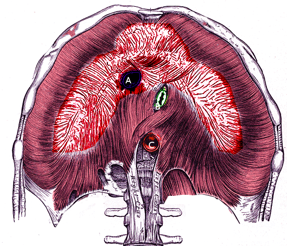
Diaphragm and its Apertures.
Contributed by S Bhimji, MD
(Click Image to Enlarge)
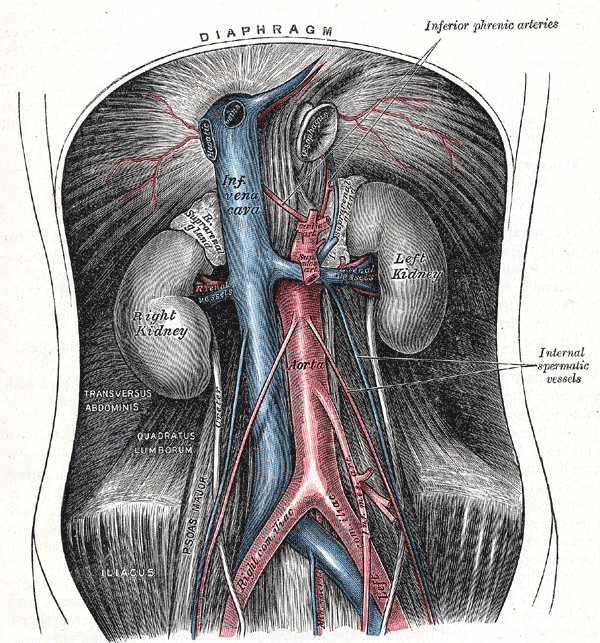
Retroperitoneal Organs. This image shows the anatomical relationships between the diaphragm, right and left suprarenal glands, right and left gonadal vessels, celiac trunk, superior and inferior mesenteric arteries, inferior phrenic arteries, internal spermatic vessels, median sacral arteries, left and right kidneys, inferior vena cava, abdominal aorta, right and left renal vessels, transversus abdominis, quadratus lumborum, right and left common Iliac vessels, psoas major, and iliacus.
Henry Vandyke Carter, Public Domain, via Wikimedia Commons
References
Oliver KA, Ashurst JV. Anatomy, Thorax, Phrenic Nerves. StatPearls. 2026 Jan:(): [PubMed PMID: 30020697]
Bordoni B, Purgol S, Bizzarri A, Modica M, Morabito B. The Influence of Breathing on the Central Nervous System. Cureus. 2018 Jun 1:10(6):e2724. doi: 10.7759/cureus.2724. Epub 2018 Jun 1 [PubMed PMID: 30083485]
McCool FD, Manzoor K, Minami T. Disorders of the Diaphragm. Clinics in chest medicine. 2018 Jun:39(2):345-360. doi: 10.1016/j.ccm.2018.01.012. Epub [PubMed PMID: 29779594]
Sefton EM, Gallardo M, Kardon G. Developmental origin and morphogenesis of the diaphragm, an essential mammalian muscle. Developmental biology. 2018 Aug 15:440(2):64-73. doi: 10.1016/j.ydbio.2018.04.010. Epub 2018 Apr 19 [PubMed PMID: 29679560]
Fayssoil A, Behin A, Ogna A, Mompoint D, Amthor H, Clair B, Laforet P, Mansart A, Prigent H, Orlikowski D, Stojkovic T, Vinit S, Carlier R, Eymard B, Lofaso F, Annane D. Diaphragm: Pathophysiology and Ultrasound Imaging in Neuromuscular Disorders. Journal of neuromuscular diseases. 2018:5(1):1-10. doi: 10.3233/JND-170276. Epub [PubMed PMID: 29278898]
Petrosyan M, Shah AA, Chahine AA, Guzzetta PC, Sandler AD, Kane TD. Congenital paraesophageal hernia: Contemporary results and outcomes of laparoscopic approach to repair in symptomatic infants and children. Journal of pediatric surgery. 2019 Jul:54(7):1346-1350. doi: 10.1016/j.jpedsurg.2018.07.008. Epub 2018 Jul 20 [PubMed PMID: 30072216]
Newbury A, Dorfman JD, Lo HS. Imaging and Management of Thoracic Trauma. Seminars in ultrasound, CT, and MR. 2018 Aug:39(4):347-354. doi: 10.1053/j.sult.2018.03.006. Epub 2018 Mar 26 [PubMed PMID: 30070227]