 Tools
Tools
 Atopic Dermatitis
Introduction
Atopic dermatitis is a specific form of eczema and the most common chronic inflammatory skin disease (see Image. Atopic Dermatitis).[1][2][3]
Atopic dermatitis usually develops in infancy and is characterized by pruritus, dry skin, eczematous lesions, and lichenification. The condition is believed to coexist with other immunoglobulin E (IgE)–associated disorders, including allergic rhinitis, asthma, and food allergies. Atopic dermatitis is associated with considerable morbidity, and its prevalence has been increasing over the past few decades.
Etiology
Register For Free And Read The Full Article
 Search engine and full access to all medical articles
- 10 free questions in your specialty
- Free CME/CE Activities
- Free daily question in your email
- Save favorite articles to your dashboard
- Emails offering discounts
Learn more about a Subscription to StatPearls Point-of-Care
Etiology
Atopic dermatitis has a complex etiology involving both genetic and environmental factors, which contribute to abnormalities in the epidermal barrier and the immune system. It is part of the atopic triad, which also includes allergic rhinitis and asthma. These conditions may develop concurrently or sequentially, in a progression known as the atopic march. Patients with the atopic triad exhibit barrier dysfunction affecting the skin and the upper and lower respiratory tracts, contributing to their clinical manifestations.
Offspring of parents with atopic disease have a greater than 50% risk of developing atopy if one parent is affected and up to an 80% risk if both parents are affected. Genetic factors include loss-of-function mutations in filaggrin (filament aggregating protein), an epidermal protein involved in maintaining the skin barrier and generating natural moisturizing factor. Filaggrin mutations are present in up to 30% of patients with atopic dermatitis and may also predispose patients to ichthyosis vulgaris, allergic rhinitis, and keratosis pilaris.
Food hypersensitivity also may cause or exacerbate atopic dermatitis in approximately 10% to 30% of patients, with about 90% of reactions or flares attributed to eggs, milk, peanuts, soy, and wheat.[4][5]
Recent studies indicate that there may be an association between smoking and adult-onset atopic dermatitis.
Epidemiology
Atopic dermatitis affects approximately 10% to 30% of children and 2% to 10% of adults in developed countries. The prevalence has increased 2- to 3-fold over the past several decades. The incidence of atopic dermatitis is higher at higher latitudes, which may be related to reduced sun exposure and lower humidity.
Atopic dermatitis is classified into 3 clinical subtypes based on the age of disease onset: early-onset, late-onset, and senile-onset. Early-onset atopic dermatitis (ie, birth to age 2) is the most common subtype, with approximately 60% of cases starting in the first year of life, and about 60% resolving by age 12. In late-onset atopic dermatitis, symptoms begin after puberty. Senile-onset atopic dermatitis is uncommon and manifests in individuals aged 60 or older.
Pathophysiology
Patients with atopic dermatitis exhibit epidermal barrier dysfunction, which increases susceptibility to xerosis, inflammation, pruritus, and other characteristic symptoms triggered by environmental irritants and allergens. Epidermal barrier dysfunction may be partly due to decreased levels of ceramides, key sphingolipids within the stratum corneum that help maintain epidermal barrier function and prevent transepidermal water loss. Dysfunction of the epidermal barrier allows irritants and allergens to penetrate the epidermis and trigger inflammation through an exaggerated T-helper 2 (Th2) immune response. Acute lesions are associated with increased levels of interleukin (IL)-4 and IL-5, whereas chronic lesions demonstrate a Th1-predominant response characterized by elevated interferon-gamma and IL-12.
Scratching further aggravates inflammation by stimulating keratinocytes to release proinflammatory cytokines, including tumor necrosis factor alpha, IL-1, and IL-6. In addition, reduced expression of antimicrobial peptides, such as human beta-defensins and cathelicidins, in the epidermis of patients with atopic dermatitis contributes to increased susceptibility to skin colonization by Staphylococcus aureus, which occurs in more than 90% of patients. Colonization with S aureus can worsen inflammation and lead to secondary infection and impetiginization.[6][7]
In atopic dermatitis, transepidermal water loss is significantly increased, although the underlying cause of epidermal barrier dysfunction is not fully understood. Filaggrin, a protein essential for maintaining epidermal integrity, is often deficient or functionally impaired in affected individuals.
History and Physical
When evaluating a patient with suspected atopic dermatitis, the clinician should obtain a detailed history, with particular attention to the onset and distribution of lesions and the severity of pruritus, including its impact on sleep. The assessment should also include a personal or family history of atopic conditions and evaluation for possible contact allergies. Identification of potential triggers is important and may include exposure to environmental allergens such as dust mites or animal dander; irritants such as soaps, fragrances, or synthetic fabrics like polyester; hot showers or excessive sweating; and food hypersensitivities.
On physical examination, the characteristic findings of atopic dermatitis vary by age group. Infants typically present with edematous papules and plaques that may show vesicles or crust on the scalp, face, and extensor extremities. Although infants may rarely develop atopic dermatitis lesions in the diaper area, they may be susceptible to other causes of diaper dermatitis, such as candidiasis and seborrheic dermatitis.
Children with atopic dermatitis classically present with less exudative patches and plaques involving the antecubital and popliteal fossae. Adults typically present with chronic lichenified lesions with accentuated skin markings, often affecting the hands. Individual lesions may also be classified by stage: acute lesions appear as edematous, erythematous papules and plaques, sometimes with vesicles or crusting; subacute lesions are characterized by erythema, scaling, and variable crusting; and chronic lesions consist of thick plaques with lichenification and scaling.
Evaluation
The diagnosis of atopic dermatitis is typically clinical and based on the characteristic presentation for the patient’s age group and disease subtype. The presence of associated findings, such as keratosis pilaris, may support the diagnosis. When performed, a skin biopsy usually demonstrates an eczematous pattern.
In pediatric cases that are recalcitrant to treatment, fluorescent enzyme immunoassays or skin prick testing may be used to detect IgE antibodies to specific allergens. However, the presence of these antibodies does not always indicate clinically relevant triggers.[8][9]
Treatment / Management
The 4 major components of atopic dermatitis treatment include daily skin care, trigger avoidance, anti-inflammatory therapy, and other complementary modalities.
Daily skin care includes twice-daily application of emollients, ideally within 3 minutes of exiting a lukewarm shower or bath, to prevent skin drying out. However, recent evidence suggests that daily emollient application may not significantly lower the risk of developing atopic dermatitis in newborns.[10] Ointments are the most occlusive vehicle and are less likely to cause burning pain or discomfort during application than other products, such as creams or lotions.(A1)
Topical corticosteroids are first-line agents for the treatment of acute flares and should be applied before emollients to enhance their efficacy. The potency should be sufficient to control the flare promptly, with consideration given to tapering to every-other-day use and then to maintenance therapy twice weekly (for example, on weekends) in commonly affected areas. Potential reversible adverse effects of topical corticosteroids include skin atrophy and telangiectasia.
Sensitive areas of the skin, such as the face and intertriginous regions (eg, the axillae and groin), may require treatment with topical nonsteroidal anti-inflammatory agents, including calcineurin inhibitors such as tacrolimus and pimecrolimus. These agents are commonly used in areas at higher risk for corticosteroid-related adverse effects. Phosphodiesterase 4 inhibitors, such as crisaborole ointment and roflumilast cream, provide additional nonsteroidal options by reducing the production of inflammatory mediators. Roflumilast is approved for use in patients aged 6 and older. Another topical nonsteroidal option is ruxolitinib cream, a Janus kinase inhibitor approved for patients aged 2 and older. This agent reduces inflammatory cytokine signaling and improves pruritus without the risk of skin atrophy associated with topical corticosteroid use.[11][12][13](A1)
When atopic dermatitis is not adequately controlled with topical therapies, systemic treatment options may be considered. These include phototherapy (eg, UVA, UVB, and narrowband UVB phototherapy) and systemic immunosuppressive agents (eg, cyclosporine, azathioprine, mycophenolate mofetil, and methotrexate). In addition, targeted biologic therapies have emerged as effective options for patients with moderate to severe disease. Dupilumab is a monoclonal antibody administered as a subcutaneous injection that blocks the IL-4 receptor, thereby inhibiting the effects of both IL-4 and IL-13. This antibody has the broadest age approval among biologic therapies, with use authorized in children as young as 6 months. Other biologic agents include lebrikizumab and tralokinumab, both IL-13 antagonists approved for use in patients aged 12 and older, with lebrikizumab requiring a minimum body weight of 40 kg. Nemolizumab, an IL-31 receptor antagonist approved for patients aged 12 and older, targets pruritus by inhibiting IL-31–mediated signaling.[14][15][16](A1)
Other complementary therapies include bleach baths (0.5 cup of bleach in a standard 40-gallon bathtub) once or twice weekly to reduce S aureus colonization. Additional strategies may include low-allergen maternal diets during breastfeeding and the use of probiotics and prebiotics in pregnant mothers and at-risk infants. These interventions have been associated with a reduction of up to 50% in the incidence of atopic dermatitis between ages 1 and 4 years compared with placebo.[17][18][19] (A1)
Numerous studies have shown that bleach baths may help relieve the symptoms of atopic dermatitis by reducing the risk of bacterial superinfection; however, other studies have found no benefit. Some patients may benefit from probiotics, which are believed to modulate the immune system and reduce the development of allergen-specific IgE responses.[20]
In patients with suspected food allergies who do not respond to initial treatment despite optimized skin care and topical corticosteroids, food allergy testing using oral food challenge or a trial elimination diet may be considered.
Differential Diagnosis
The differential diagnoses for atopic dermatitis include allergic contact dermatitis, lichen simplex, lichen planus, psoriasis, scabies, tinea infections, and seborrheic dermatitis.
Treatment Planning
Cyclosporine is approved for the treatment of moderate to severe atopic dermatitis in patients aged 16 or older. Therapy should be used with careful monitoring, and intermittent courses of up to 16 weeks are recommended for most patients. Doses should be titrated to minimize renal toxicity, and treatment should be adjusted or discontinued if hypertension or infection develops.
Patients should be educated about the signs of infection and monitored for nonmelanoma skin cancer and elevated serum lipid levels. Concomitant use with phototherapy is not recommended.
Prognosis
Many patients with atopic dermatitis experience improvement over time, although comorbid conditions such as allergic rhinitis and asthma may persist. In some cases of childhood-onset atopic dermatitis, symptoms persist for decades. Atopic dermatitis is characterized by relapsing and remitting flares, with relapses often requiring pharmacologic intervention. Individuals with ongoing exposure to irritants and allergens (eg, tobacco smoke, pet dander, chemical fumes, pollen, soaps, detergents, and wool) may experience persistent symptoms and impaired quality of life.
Persistent and recurrent pruritus is not only bothersome but also contributes to increased healthcare utilization and costs. A recognized complication of atopic dermatitis is Kaposi varicelliform eruption, which results from a primary herpes simplex virus infection (see Image. Kaposi Varicelliform Eruption in a Patient With Atopic Dermatitis). Vesicular lesions typically arise within eczematous skin and may rapidly spread to unaffected areas. Prompt treatment with acyclovir can reduce morbidity.
Complications
Complications of atopic dermatitis include Kaposi varicelliform eruption and secondary bacterial skin infections caused by Staphylococcus and Streptococcus species.
Deterrence and Patient Education
Patients should wear cotton clothing and avoid wool. Maintaining a cooler indoor temperature may help reduce sweating and pruritus. A humidifier is recommended to prevent skin dryness. Foods that trigger atopic dermatitis symptoms should be avoided.
Pearls and Other Issues
Treatment and management of atopic dermatitis should begin with patient and/or caregiver education on the chronic nature of the disease and the importance of maintenance therapy, which strengthens the epidermal barrier, reduces allergen sensitization, and may help prevent disease flares.
Enhancing Healthcare Team Outcomes
Atopic dermatitis is a chronic skin disorder that can significantly affect quality of life. Although medical therapy is central to management, nonpharmacologic strategies can also improve patient outcomes. Pharmacists and nurses play an important role in educating patients and caregivers about these measures. Patients should be advised to wear soft clothing, preferably cotton, and avoid wool. Maintaining a cooler home environment may help reduce sweating and irritation, and the use of a humidifier may help prevent skin dryness. Clothing should be washed with mild detergent, and fabric softeners and bleach should be avoided. When outdoors, patients should apply appropriate sunscreen and moisturizers. Keeping a food diary and avoiding known dietary triggers may also be beneficial. Activities that cause excessive sweating should be minimized, and patients with asthma should remain adherent to prescribed medications and avoid relevant allergens.[21][22]
Outcomes
Most patients with atopic dermatitis experience improvement with age. Approximately 30% of patients develop allergic rhinitis, and a similar proportion develop asthma. Long-term studies indicate that mild to moderate symptoms may persist for a decade or longer, and about 80% of patients require ongoing topical therapy to control symptoms. Symptom improvement is often observed after the second decade of life; however, effective management frequently requires lifestyle modifications, including avoidance of triggers such as tobacco smoke, pet dander, wool, and certain soaps. Although atopic dermatitis is not life-threatening, chronic dryness and pruritus can significantly impair quality of life, increase healthcare costs, and lead to frequent healthcare visits.[1][23]
Media
(Click Image to Enlarge)
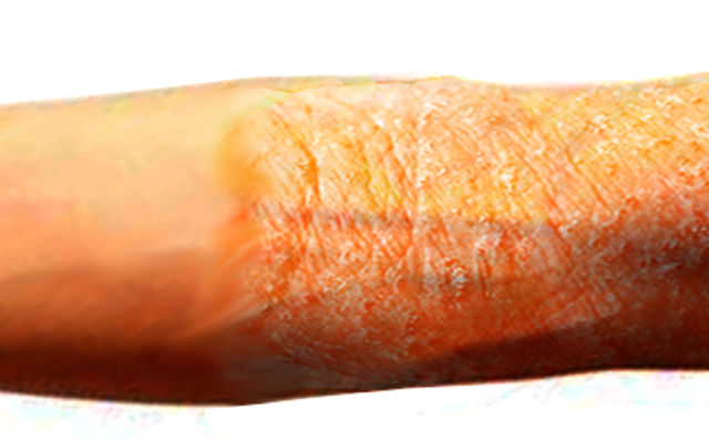
Atopic Dermatitis.
Contributed by O Chaigasame, MD
(Click Image to Enlarge)
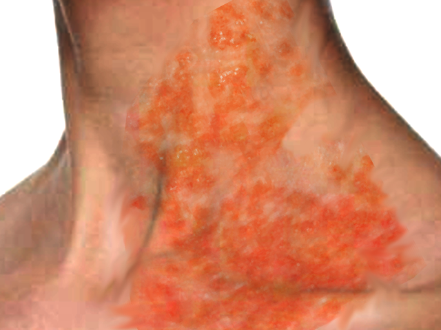
Kaposi Varicelliform Eruption in a Patient With Atopic Dermatitis. Kaposi varicelliform eruption, also known as eczema herpeticum, is a disseminated viral skin infection that occurs in patients with an underlying cutaneous disease. This condition is most commonly caused by the herpes simplex virus and presents with localized vesicular eruptions. Although uncommon, the condition can be potentially life-threatening if not treated promptly.
Contributed by O Chaigasame, MD
References
Paller A, Jaworski JC, Simpson EL, Boguniewicz M, Russell JJ, Block JK, Tofte S, Dunn JD, Feldman SR, Clark AR, Schwartz G, Eichenfield LF. Major Comorbidities of Atopic Dermatitis: Beyond Allergic Disorders. American journal of clinical dermatology. 2018 Dec:19(6):821-838. doi: 10.1007/s40257-018-0383-4. Epub [PubMed PMID: 30168085]
Giavina-Bianchi M, Giavina-Bianchi P. Systemic Treatment for Severe Atopic Dermatitis. Archivum immunologiae et therapiae experimentalis. 2019 Apr:67(2):69-78. doi: 10.1007/s00005-018-0521-y. Epub 2018 Aug 22 [PubMed PMID: 30159581]
Drucker AM, Ellis A, Jabbar-Lopez Z, Yiu ZZN, Arents BWM, Burton T, Spuls PI, Küster D, Schmitt J, Flohr C. Systemic immunomodulatory treatments for atopic dermatitis: protocol for a systematic review with network meta-analysis. BMJ open. 2018 Aug 29:8(8):e023061. doi: 10.1136/bmjopen-2018-023061. Epub 2018 Aug 29 [PubMed PMID: 30158235]
Level 1 (high-level) evidenceOliveira ADT, Sodré CS, Ferreira DC, Abad ED, Saintive S, Ribeiro M, Cavalcante FS, Piciani B, Gonçalves LS. Oral Aspects Identified in Atopic Dermatitis Patients: A Literature Review. The open dentistry journal. 2018:12():424-434. doi: 10.2174/1874210601812010424. Epub 2018 May 31 [PubMed PMID: 29988222]
Berg AK, Nørgaard K, Thyssen JP, Zachariae C, Hommel E, Rytter K, Svensson J. Skin Problems Associated with Insulin Pumps and Sensors in Adults with Type 1 Diabetes: A Cross-Sectional Study. Diabetes technology & therapeutics. 2018 Jul:20(7):475-482. doi: 10.1089/dia.2018.0088. Epub 2018 Jun 12 [PubMed PMID: 29893593]
Level 2 (mid-level) evidenceMurota H, Yamaga K, Ono E, Katayama I. Sweat in the pathogenesis of atopic dermatitis. Allergology international : official journal of the Japanese Society of Allergology. 2018 Oct:67(4):455-459. doi: 10.1016/j.alit.2018.06.003. Epub 2018 Aug 3 [PubMed PMID: 30082151]
Hulshof L, Overbeek SA, Wyllie AL, Chu MLJN, Bogaert D, de Jager W, Knippels LMJ, Sanders EAM, van Aalderen WMC, Garssen J, Van't Land B, Sprikkelman AB, Clinical Study Group. Exploring Immune Development in Infants With Moderate to Severe Atopic Dermatitis. Frontiers in immunology. 2018:9():630. doi: 10.3389/fimmu.2018.00630. Epub 2018 Mar 29 [PubMed PMID: 29966024]
Sung CT, McGowan MA, Jacob SE. Allergic Contact Dermatitis Evaluation: Strategies for the Preschooler. Current allergy and asthma reports. 2018 Aug 1:18(10):49. doi: 10.1007/s11882-018-0803-9. Epub 2018 Aug 1 [PubMed PMID: 30069607]
Batmaz SB. Laboratory and severity evaluation of pediatric atopic dermatitis and moisturizer response in different phenotypes. Allergologia et immunopathologia. 2018 Nov-Dec:46(6):571-577. doi: 10.1016/j.aller.2018.03.007. Epub 2018 Jul 4 [PubMed PMID: 29980402]
Zhong Y, Samuel M, van Bever H, Tham EH. Emollients in infancy to prevent atopic dermatitis: A systematic review and meta-analysis. Allergy. 2022 Jun:77(6):1685-1699. doi: 10.1111/all.15116. Epub 2021 Oct 12 [PubMed PMID: 34591995]
Level 1 (high-level) evidenceMeledathu S, Naidu MP, Brunner PM. Update on atopic dermatitis. The Journal of allergy and clinical immunology. 2025 Apr:155(4):1124-1132. doi: 10.1016/j.jaci.2025.01.013. Epub 2025 Jan 22 [PubMed PMID: 39855361]
Simpson EL, Augustin M, Thaçi D, Misery L, Armstrong AW, Blauvelt A, Papp KA, Szepietowski JC, Boguniewicz M, Kwatra SG, Kallender H, Sturm D, Ren H, Kircik L. Ruxolitinib Cream Monotherapy Improved Symptoms and Quality of Life in Adults and Adolescents with Mild-to-Moderate Atopic Dermatitis: Patient-Reported Outcomes from Two Phase III Studies. American journal of clinical dermatology. 2025 Jan:26(1):121-137. doi: 10.1007/s40257-024-00901-z. Epub 2024 Nov 15 [PubMed PMID: 39546129]
Level 2 (mid-level) evidenceSimpson EL, Eichenfield LF, Alonso-Llamazares J, Draelos ZD, Ferris LK, Forman SB, Gooderham M, Gonzalez ME, Hebert AA, Kircik LH, Lomaga M, Moore A, Papp KA, Prajapati VH, Hanna D, Snyder S, Krupa D, Burnett P, Almaraz E, Higham RC, Chu DH, Berk DR. Roflumilast Cream, 0.15%, for Atopic Dermatitis in Adults and Children: INTEGUMENT-1 and INTEGUMENT-2 Randomized Clinical Trials. JAMA dermatology. 2024 Nov 1:160(11):1161-1170. doi: 10.1001/jamadermatol.2024.3121. Epub [PubMed PMID: 39292443]
Level 1 (high-level) evidenceSilverberg JI, Guttman-Yassky E, Thaçi D, Irvine AD, Stein Gold L, Blauvelt A, Simpson EL, Chu CY, Liu Z, Gontijo Lima R, Pillai SG, Seneschal J, ADvocate1 and ADvocate2 Investigators. Two Phase 3 Trials of Lebrikizumab for Moderate-to-Severe Atopic Dermatitis. The New England journal of medicine. 2023 Mar 23:388(12):1080-1091. doi: 10.1056/NEJMoa2206714. Epub 2023 Mar 15 [PubMed PMID: 36920778]
Wollenberg A, Blauvelt A, Guttman-Yassky E, Worm M, Lynde C, Lacour JP, Spelman L, Katoh N, Saeki H, Poulin Y, Lesiak A, Kircik L, Cho SH, Herranz P, Cork MJ, Peris K, Steffensen LA, Bang B, Kuznetsova A, Jensen TN, Østerdal ML, Simpson EL, ECZTRA 1 and ECZTRA 2 study investigators. Tralokinumab for moderate-to-severe atopic dermatitis: results from two 52-week, randomized, double-blind, multicentre, placebo-controlled phase III trials (ECZTRA 1 and ECZTRA 2). The British journal of dermatology. 2021 Mar:184(3):437-449. doi: 10.1111/bjd.19574. Epub 2020 Dec 30 [PubMed PMID: 33000465]
Level 1 (high-level) evidenceSilverberg JI, Wollenberg A, Reich A, Thaçi D, Legat FJ, Papp KA, Stein Gold L, Bouaziz JD, Pink AE, Carrascosa JM, Rewerska B, Szepietowski JC, Krasowska D, Havlíčková B, Kalowska M, Magnolo N, Pauser S, Nami N, Sauder MB, Jain V, Padlewska K, Cheong SY, Fleuranceau Morel P, Ulianov L, Piketty C, ARCADIA 1 and ARCADIA 2 Study Investigators. Nemolizumab with concomitant topical therapy in adolescents and adults with moderate-to-severe atopic dermatitis (ARCADIA 1 and ARCADIA 2): results from two replicate, double-blind, randomised controlled phase 3 trials. Lancet (London, England). 2024 Aug 3:404(10451):445-460. doi: 10.1016/S0140-6736(24)01203-0. Epub 2024 Jul 24 [PubMed PMID: 39067461]
Level 1 (high-level) evidenceAriëns LFM, Bakker DS, van der Schaft J, Garritsen FM, Thijs JL, de Bruin-Weller MS. Dupilumab in atopic dermatitis: rationale, latest evidence and place in therapy. Therapeutic advances in chronic disease. 2018 Sep:9(9):159-170. doi: 10.1177/2040622318773686. Epub 2018 May 11 [PubMed PMID: 30181845]
Level 3 (low-level) evidenceAli Z, Ulrik CS, Agner T, Thomsen SF. Association between Atopic Dermatitis and the Metabolic Syndrome: A Systematic Review. Dermatology (Basel, Switzerland). 2018:234(3-4):79-85. doi: 10.1159/000491593. Epub 2018 Aug 15 [PubMed PMID: 30110673]
Level 1 (high-level) evidenceWollenberg A, Barbarot S, Bieber T, Christen-Zaech S, Deleuran M, Fink-Wagner A, Gieler U, Girolomoni G, Lau S, Muraro A, Czarnecka-Operacz M, Schäfer T, Schmid-Grendelmeier P, Simon D, Szalai Z, Szepietowski JC, Taïeb A, Torrelo A, Werfel T, Ring J, European Dermatology Forum (EDF), the European Academy of Dermatology and Venereology (EADV), the European Academy of Allergy and Clinical Immunology (EAACI), the European Task Force on Atopic Dermatitis (ETFAD), European Federation of Allergy and Airways Diseases Patients’ Associations (EFA), the European Society for Dermatology and Psychiatry (ESDaP), the European Society of Pediatric Dermatology (ESPD), Global Allergy and Asthma European Network (GA2LEN) and the European Union of Medical Specialists (UEMS). Consensus-based European guidelines for treatment of atopic eczema (atopic dermatitis) in adults and children: part II. Journal of the European Academy of Dermatology and Venereology : JEADV. 2018 Jun:32(6):850-878. doi: 10.1111/jdv.14888. Epub [PubMed PMID: 29878606]
Level 3 (low-level) evidenceKhan A, Adalsteinsson J, Whitaker-Worth DL. Atopic dermatitis and nutrition. Clinics in dermatology. 2022 Mar-Apr:40(2):135-144. doi: 10.1016/j.clindermatol.2021.10.006. Epub 2021 Oct 27 [PubMed PMID: 34876306]
Rastogi S, Patel KR, Singam V, Silverberg JI. Allergic contact dermatitis to personal care products and topical medications in adults with atopic dermatitis. Journal of the American Academy of Dermatology. 2018 Dec:79(6):1028-1033.e6. doi: 10.1016/j.jaad.2018.07.017. Epub 2018 Jul 25 [PubMed PMID: 30053491]
Thomas KS, Bradshaw LE, Sach TH, Cowdell F, Batchelor JM, Lawton S, Harrison EF, Haines RH, Ahmed A, Dean T, Burrows NP, Pollock I, Buckley HK, Williams HC, Llewellyn J, Crang C, Grundy JD, Guiness J, Gribbin A, Wake EV, Mitchell EJ, Brown SJ, Montgomery AA. Randomised controlled trial of silk therapeutic garments for the management of atopic eczema in children: the CLOTHES trial. Health technology assessment (Winchester, England). 2017 Apr:21(16):1-260. doi: 10.3310/hta21160. Epub [PubMed PMID: 28409557]
Level 1 (high-level) evidenceNapolitano M, Marasca C, Fabbrocini G, Patruno C. Adult atopic dermatitis: new and emerging therapies. Expert review of clinical pharmacology. 2018 Sep:11(9):867-878. doi: 10.1080/17512433.2018.1507734. Epub 2018 Aug 9 [PubMed PMID: 30073901]