 Tools
Tools
 Dermatitis Herpetiformis
Introduction
Dermatitis herpetiformis is a chronic, autoimmune blistering disease characterized by an extremely pruritic rash that predominantly affects the extensor surfaces. The characteristic vesicles are often not apparent because they are destroyed by excoriation. The disease is closely associated with gluten-sensitive enteropathy (GSE); both conditions are characterized by the development of IgA autoantibodies against transglutaminases, which, in dermatitis herpetiformis, are deposited in the superficial papillary dermis. Therapy involves strict gluten avoidance and the use of sulfonamide drugs, such as dapsone.[1][2][3][4][5]
Etiology
Register For Free And Read The Full Article
 Search engine and full access to all medical articles
- 10 free questions in your specialty
- Free CME/CE Activities
- Free daily question in your email
- Save favorite articles to your dashboard
- Emails offering discounts
Learn more about a Subscription to StatPearls Point-of-Care
Etiology
The factors that can cause dermatitis herpetiformis include the following:
Genetics
Results from studies have documented a close association between dermatitis herpetiformis and certain human leukocyte antigen (HLA) types, particularly HLA-DQ2, and, to a lesser extent, HLA-DQ8. Other non–major histocompatibility complex (MHC) genes have been linked to gluten-sensitive enteropathy (GSE) in genome-wide association studies, including myosin IXB, interleukin (IL)-12, IL-23, and CCR3, though their roles in dermatitis herpetiformis remain unclear. First-degree relatives of patients with GSE or dermatitis herpetiformis are characteristically more likely to be affected by either disorder. Thus, family screening may be indicated. Monozygotic twins have a disease concordance rate of greater than 0.9.
Environmental Factors
The principal environmental factor involved in the pathogenesis of the disease is dietary gluten and its constituent gliadin. Interestingly, tissue transglutaminase (tTG) modifies gliadin in the gastrointestinal tract mucosa into a more immunogenic form. Other environmental factors associated with dermatitis herpetiformis include iodine exposure, which can precipitate disease flares, and tobacco smoking, which can improve the condition.
Epidemiology
Incidence and Prevalence
Dermatitis herpetiformis is most common in people of northern European descent, among whom prevalence ranges from 1.2 to 39.2 per 100,000 people and incidence ranges from 0.4 to 2.6 per 100,000 people per year. Results from a recent large population-based study from the United Kingdom suggested that, although the incidence of gluten-sensitive enteropathy (GSE) increased from 1990 to 2011, the incidence of dermatitis herpetiformis fell from 1.8 per 100,000 to 0.8 per 100,000 person-years over the same period. Patients with dermatitis herpetiformis appear to have lower age-adjusted mortality than expected, possibly because of dietary modifications required by the disease.
Age
Onset is common in adult life, typically in the fourth decade, though cases have been reported in children and older people.
Sex
Results from multiple studies showed that the disease is more common in men than women, with a male-to-female ratio of 1.5 to 2:1.
Ethnicity
Dermatitis herpetiformis is a disease of people of northern European descent and is most uncommon in Asian and African populations, though a few cases have been reported. The incidence and prevalence of the disease are similar in North American populations of European descent to those seen in northern Europe, suggesting that genetic factors are important in disease susceptibility. Asian patients with dermatitis herpetiformis tend to have a distinct fibrillar pattern of IgA deposition in the skin, and GSE is very rarely associated with this pattern.
Associated Diseases
The most common association of dermatitis herpetiformis is with GSE. However, the severity of GSE in individuals with dermatitis herpetiformis varies widely and may be clinically silent or mild. Consequently, all patients with dermatitis herpetiformis should be reviewed by a gastroenterologist and investigated accordingly. Patients with dermatitis herpetiformis have an increased risk of small bowel lymphoma. The disease is also associated with other autoimmune disorders, such as autoimmune thyroid disease, and all patients should be screened for this condition. Additionally, patients with dermatitis herpetiformis have an increased risk of type 1 diabetes, Addison disease, and vitiligo.
Pathophysiology
The pathology in dermatitis herpetiformis and gluten-sensitive enteropathy (GSE) results from an IgA-dominant autoimmune response to transglutaminase molecules. In GSE, the principal target is tissue transglutaminase, whereas in dermatitis herpetiformis, increasing evidence suggests that epidermal transglutaminase 3 (TG3) may be the dominant antigen. Both proteins exhibit significant homology in their enzymatic domains and are expressed in normal epidermis, where TG3 is important for crosslinking and maintaining the cornified envelope.The series of events that result in inflammation and itch in dermatitis herpetiformis remains to be fully elucidated. Hypotheses include the release of TG3 from keratinocytes into the papillary dermis, where TG3 can bind circulating IgA antibodies, or deposition of circulating TG3 and IgA complexes in the skin.[6][7]
Histopathology
The histopathology of an intact vesicle in dermatitis herpetiformis demonstrates subepidermal blister formation with neutrophils located at the tips of the dermal papillae. Perivascular inflammatory cell infiltrate is frequently present. Because vesicles may not persist because of pruritus, clefting within the lamina lucida may not be seen.
History and Physical
History
The principal symptom of patients with dermatitis herpetiformis is pruritus. Patients report a rash, most typically over the extensor surfaces of the elbows, knees, buttocks, and scalp. Associated disorder symptoms may also be present, such as gluten-sensitive enteropathy (GSE), with bloating, diarrhea, and other gastrointestinal tract symptoms, or other autoimmune diseases, including hypothyroidism.
Presentation
The characteristic lesions of dermatitis herpetiformis are grouped erythematous papules and vesicles located over extensor sites. Because the condition is so pruritic, intact vesicles are rarely seen, and the patient may present with excoriations alone. Lesions tend to be symmetrical and heal without scarring. Patients may develop punctate purpura on the palms and soles. Mucosal changes may occur, and dental abnormalities have been reported, particularly enamel pits. Interestingly, first-degree relatives of a patient with GSE may also show enamel defects.
Evaluation
The diagnosis of dermatitis herpetiformis is made based on characteristic clinical features, histopathology, direct immunofluorescence testing, and serology. Direct immunofluorescence samples are best taken from the perilesional skin, because characteristic changes may be lost in lesional tissue. The diagnostic finding is deposition of IgA in the papillary dermis in a granular or fibrillar pattern. The latter pattern has been particularly associated with dermatitis herpetiformis in Asian patients. Complement 3 may also be found in association with IgA deposits.
Treatment / Management
The cornerstone of long-term treatment for dermatitis herpetiformis is strict adherence to a gluten-free diet. Strict gluten avoidance improves skin changes over time and is also essential in the treatment of associated gluten-sensitive enteropathy (GSE). Patients are best treated in consultation with an experienced dietitian who can provide appropriate support, because maintenance of a strict gluten-free diet can be challenging.[1][8][9][10]
Because of the slow response to a gluten-free diet, most patients with dermatitis herpetiformis require pharmacological intervention to control their disease in the short to medium term. Dapsone and related sulfonamide drugs have proven highly effective, often suppressing pruritus within days of treatment initiation. The mechanism of action of dapsone in dermatitis herpetiformis involves effects on neutrophil function and recruitment. The drug is typically used at doses of 25 to 150 mg daily. Hematological adverse effects may occur, including hemolysis, methemoglobinemia, and agranulocytosis. Consequently, patients require regular blood monitoring, particularly in the early stages of treatment. Nonhematological adverse effects include a severe drug sensitivity reaction, dapsone hypersensitivity syndrome, and peripheral neuropathy. Patients with glucose-6-phosphate dehydrogenase (G6PD) deficiency are more prone to adverse effects from dapsone, and G6PD activity should be measured before treatment. Concurrent administration of cimetidine, via inhibition of cytochrome P450, has been shown to decrease the adverse effects of dapsone.
Other sulfonamide drugs have utility in dermatitis herpetiformis, including sulfamethoxypyridazine, sulfapyridine, and sulfasalazine. These drugs may be of utility in patients who are intolerant of dapsone. Systemic corticosteroids are of little benefit, though potent topical corticosteroids may be of use in the short term to lessen pruritus.
First-Line
- Gluten-free diet
- Dapsone 25 to 150 mg daily
- Potent topical corticosteroids
Second-line
- Sulphamethoxypyridazine 0.5 to 1.5 g daily (with a gluten-free diet)
Third-line
- Sulphapyridine 250 to 750 mg daily or
- Sulphasalazine 1 to 2 g daily (with a gluten-free diet)
Differential Diagnosis
The differential diagnosis of dermatitis herpetiformis includes many pruritic and vesiculobullous disorders, such as:
- Linear IgA disease
- Pemphigoid
- Eczema
- Scabies
- Epidermolysis bullosa acquisita
Because of the often nonspecific nature of dermatitis herpetiformis lesions, skin biopsy for direct immunofluorescence should be considered when investigating patients with unexplained or treatment-refractory pruritic rashes.
Prognosis
Dermatitis herpetiformis is a chronic disease that requires patients to adopt a long-term gluten-free diet. Patients who can maintain this diet and respond to treatment seem to have excellent long-term survival and may decrease or discontinue dapsone. Recent data suggest that patients with gluten-sensitive enteropathy (GSE) who do not respond to a gluten-free diet do poorly. While the disease is chronic, remission is recognized and seems more common in patients older than 40 years.
Complications
The principal complications and comorbidities of dermatitis herpetiformis are related to gluten-sensitive enteropathy (GSE) and the associated risk of small-bowel lymphoma. GSE may lead to malabsorption, resulting in anemia, weight loss, and osteoporosis. In children, short stature may occur. Rarely, dermatitis herpetiformis may be associated with neurological diseases, such as ataxia and neuropathy.
Enhancing Healthcare Team Outcomes
Treatment of dermatitis herpetiformis is complex and requires an interprofessional team that includes a dermatologist, dietitian, gastroenterologist, nurse practitioner, and neurologist. The cornerstone of long-term treatment for dermatitis herpetiformis is strict adherence to a gluten-free diet. Strict gluten avoidance improves skin changes over time and is also essential in the treatment of associated gluten-sensitive enteropathy (GSE). Patients are best treated in consultation with an experienced dietitian who can provide appropriate support, because maintenance of a strict gluten-free diet can be challenging. For patients who adhere to the dietary restriction, the prognosis is good, but multiple relapses and remissions may still occur. For those who do not adhere to the diet, the prognosis is poor, and quality of life may be severely impaired.[11][12]
Media
(Click Image to Enlarge)
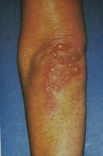
Dermatitis Herpetiformis. Intact tense bullae on the elbow.
Contributed by RJ Pye, MD
(Click Image to Enlarge)

Fig 2: Rash of dermatitis herpetiformis. Reproduced with permission from GP Core content
(Click Image to Enlarge)
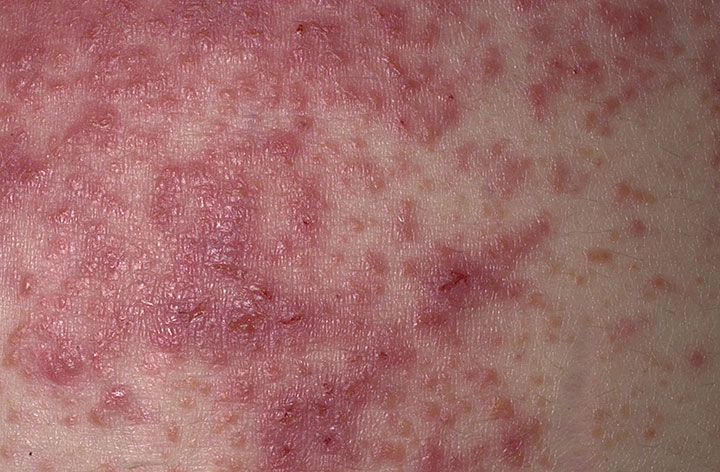
dermatitis herpetiformis Contributed by Wikimedia User: Madhero88 (CC BY-SA 3.0 https://creativecommons.org/licenses/by-sa/3.0/deed.en)
(Click Image to Enlarge)
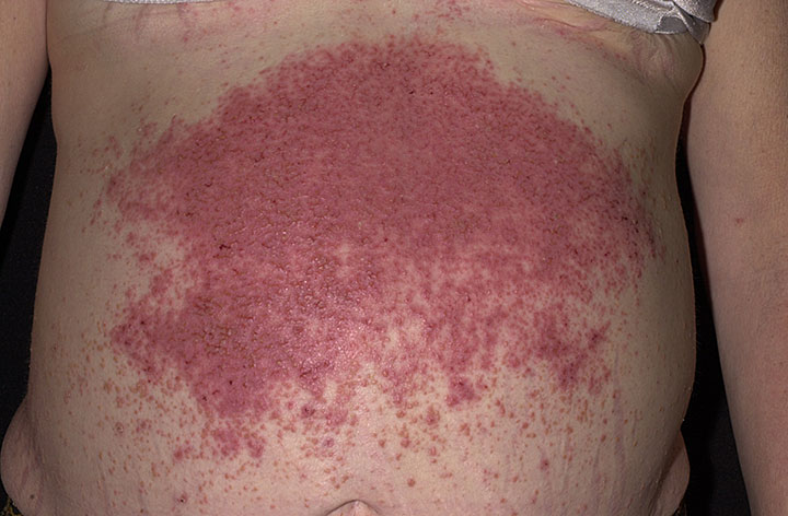
Dermatitis herpetiformis Contributed by Wikimedia User: MadHero88 ( CC BY-SA 3.0 https://creativecommons.org/licenses/by-sa/3.0/deed.pt)
References
Popp A, Mäki M. Gluten-Induced Extra-Intestinal Manifestations in Potential Celiac Disease-Celiac Trait. Nutrients. 2019 Feb 1:11(2):. doi: 10.3390/nu11020320. Epub 2019 Feb 1 [PubMed PMID: 30717318]
Adam MP, Bick S, Mirzaa GM, Pagon RA, Wallace SE, Amemiya A, Taylor AK, Lebwohl B, Snyder CL, Green PHR. Celiac Disease. GeneReviews(®). 1993:(): [PubMed PMID: 20301720]
Varpuluoma O, Jokelainen J, Försti AK, Timonen M, Huilaja L, Tasanen K. Dermatitis Herpetiformis and Celiac Disease Increase the Risk of Bullous Pemphigoid. The Journal of investigative dermatology. 2019 Mar:139(3):600-604. doi: 10.1016/j.jid.2018.10.010. Epub 2018 Oct 26 [PubMed PMID: 30612975]
Lorand L, Iismaa SE. Transglutaminase diseases: from biochemistry to the bedside. FASEB journal : official publication of the Federation of American Societies for Experimental Biology. 2019 Jan:33(1):3-12. doi: 10.1096/fj.201801544R. Epub [PubMed PMID: 30593123]
Graziano M, Rossi M. An update on the cutaneous manifestations of coeliac disease and non-coeliac gluten sensitivity. International reviews of immunology. 2018:37(6):291-300. doi: 10.1080/08830185.2018.1533008. Epub 2018 Dec 5 [PubMed PMID: 30516407]
Witte M, Zillikens D, Schmidt E. Diagnosis of Autoimmune Blistering Diseases. Frontiers in medicine. 2018:5():296. doi: 10.3389/fmed.2018.00296. Epub 2018 Nov 2 [PubMed PMID: 30450358]
Hofmann SC, Juratli HA, Eming R. Bullous autoimmune dermatoses. Journal der Deutschen Dermatologischen Gesellschaft = Journal of the German Society of Dermatology : JDDG. 2018 Nov:16(11):1339-1358. doi: 10.1111/ddg.13688. Epub [PubMed PMID: 30395395]
Rodrigo L, Beteta-Gorriti V, Alvarez N, Gómez de Castro C, de Dios A, Palacios L, Santos-Juanes J. Cutaneous and Mucosal Manifestations Associated with Celiac Disease. Nutrients. 2018 Jun 21:10(7):. doi: 10.3390/nu10070800. Epub 2018 Jun 21 [PubMed PMID: 29933630]
Reunala T, Salmi TT, Hervonen K, Kaukinen K, Collin P. Dermatitis Herpetiformis: A Common Extraintestinal Manifestation of Coeliac Disease. Nutrients. 2018 May 12:10(5):. doi: 10.3390/nu10050602. Epub 2018 May 12 [PubMed PMID: 29757210]
Losurdo G, Principi M, Iannone A, Amoruso A, Ierardi E, Di Leo A, Barone M. Extra-intestinal manifestations of non-celiac gluten sensitivity: An expanding paradigm. World journal of gastroenterology. 2018 Apr 14:24(14):1521-1530. doi: 10.3748/wjg.v24.i14.1521. Epub [PubMed PMID: 29662290]
Heikkilä K, Koskinen OA, Agarwal A, Tikkinen KAO, Mäki M, Kaukinen K. Associations of coeliac disease with coronary heart disease and cerebrovascular disease: A systematic review and meta-analysis. Nutrition, metabolism, and cardiovascular diseases : NMCD. 2015 Sep:25(9):816-831. doi: 10.1016/j.numecd.2015.05.004. Epub 2015 May 14 [PubMed PMID: 26111459]
Level 1 (high-level) evidenceWeston WL, Morelli JG, Huff JC. Misdiagnosis, treatments, and outcomes in the immunobullous diseases in children. Pediatric dermatology. 1997 Jul-Aug:14(4):264-72 [PubMed PMID: 9263305]