 Tools
Tools
 Dandy-Walker Malformation
Introduction
Dandy-Walker malformation, also known as Dandy-Walker syndrome, is a posterior fossa anomaly characterized by the following features:
- Agenesis or hypoplasia of the vermis
- Cystic enlargement of the fourth ventricle with communication to a large cystic dilated posterior fossa
- Upward displacement of the tentorium and torcula (torcular-lambdoid inversion)
- Enlargement of the posterior fossa [1][2][3]
The first description of Dandy-Walker malformation dates back to 1887 by Sutton. Dandy and Blackfan described this deformity in 1914, and Taggart and Walker expanded on it in 1942. Bender first described the condition as Dandy-Walker malformation in 1954.[4] The terms Dandy-Walker complex and Dandy-Walker spectrum are among the most frequently used radiological entities, originally proposed by Barkovich et al in 1989.[5] Dandy-Walker malformation is the most common posterior fossa malformation, and it typically occurs sporadically.[6] Most patients have hydrocephalus at the time of diagnosis.[7]
Using the development of the cerebellar vermis as the reference standard, the Dandy–Walker spectrum can be classified into:
- Dandy-Walker malformation
- Dandy-Walker variant
- Simple posterior fossa cistern widening
The Dandy-Walker variant is characterized by cerebellar vermian hypoplasia, cystic dilatation of the fourth ventricle, and a normal posterior fossa volume. However, this terminology is currently falling out of favor because it can cause confusion; rather, individual physical abnormalities should be mentioned.[8]
Many patients remain clinically asymptomatic for years, while others may present with a variety of comorbidities leading to earlier diagnosis.[9] Treatment is generally focused on alleviating hydrocephalus and posterior fossa symptoms, often including surgical interventions such as ventriculoperitoneal and cystoperitoneal shunting.[7][10]
Etiology
Register For Free And Read The Full Article
 Search engine and full access to all medical articles
- 10 free questions in your specialty
- Free CME/CE Activities
- Free daily question in your email
- Save favorite articles to your dashboard
- Emails offering discounts
Learn more about a Subscription to StatPearls Point-of-Care
Etiology
Historically, Dandy-Walker malformation was believed to be caused by atresia of the foramina of Luschka and Magendie, leading to enlargement of the fourth ventricle and vermian hypoplasia. However, recent evidence suggests that Dandy-Walker malformation results from developmental abnormalities of the rhombencephalic roof, leading to variable degrees of vermian hypoplasia and cystic enlargement. This complex malformation may be initiated by 2 different pathophysiological mechanisms: the arrest of vermian development and the failure of the fourth ventricle foramina fenestration, leading to an enlarged Blake pouch and causing compression of the vermis.[11] Alterations in the rhombic lip proliferation have been shown to result in disproportionate hypoplasia of the inferior vermis.[12]
Dandy-Walker malformation may be isolated or associated with chromosomal abnormalities, Mendelian disorders, syndromic malformations, congenital infections, and various other comorbidities.[11] Central nervous system (CNS) disorders associated with Dandy-Walker malformation include cortical malformations, holoprosencephaly, dysgenesis of the corpus callosum, and neural tube defects.[3]
Rare mutations have been described in some genes including FOXC1 (in locus 6p25.3), ZIC1, and ZIC4 (in locus 3q24), FGF17, LAMC1, and NID1.[6]
Epidemiology
Dandy-Walker malformation and related variants have a reported prevalence of 1 in 25,000 to 35,000 live births in the United States. These malformations account for approximately 1% to 4% of cases of hydrocephalus.[1][3] A female predominance has been reported, with a female-to-male ratio of approximately 3:1.[13][14]
Pathophysiology
The majority of cases are sporadic. However, some may result from chromosomal aneuploidy, Mendelian disorders, and environmental exposures, including congenital rubella and fetal alcohol exposure. This condition underscores the intricate interplay between genetic and environmental factors in brain development.[9]
Several conditions have been reported to be associated with Dandy-Walker malformation.
- Mendelian disorders
- Walker-Warburg syndrome
- Coffin-Siris syndrome
- Fraser syndrome
- Joubert-Boltshauser syndrome
- Meckel-Gruber syndrome
- Aicardi syndrome
- Neurofibromatosis type [15]
- Chromosomal aberrations [16]
- Duplication syndromes, including those involving 5q, 8p, 8q, and 17q
- Trisomies 9, 13, and 18
- Triploidy
- Deletion at 6(p24–p25)
- 9ph+ heteromorphism [16]
- Environmental agents
- Prenatal exposure to rubella, cytomegalovirus, toxoplasmosis, coumadin, and alcohol
- Maternal diabetes
- Sporadic syndromes
- Klippel-Feil syndrome
- Goldenhar syndrome
- Cornelia de Lange syndrome
Concept of the Midline as a Developmental Field (Opitz and Gilbert)
An insult to this field can result in pleiotropic sequelae on midline structures, including the CNS, heart, palate, midface, vertebrae, genitalia, and sacrococcygeal region. Dandy-Walker malformation should be considered a complex developmental disturbance of the midline of marked genetic and etiological heterogeneity.
Embryology
The cerebellar vermis develops from top to bottom at 17 to 18 weeks of gestation.[17] Due to vermian developmental arrest or compression of the vermis brought on by a failure of the fourth ventricle foramina fenestration and consequent growth of Blake pouch, the roof of the rhombencephalon does not form correctly.
History and Physical
The clinical presentation is nonspecific, subject to multiple factors, including the severity of hydrocephalus, intracranial hypertension, and associated comorbidities.[1] Most patients present with signs and symptoms of increased intracranial pressure in their first year of life.[6][7][18] Macrocephaly is the most common manifestation, affecting 90% to 100% of patients during their first months of life.[6] The syndromic form of Dandy-Walker malformation may also have malformations of the heart, face, limbs, and gastrointestinal or genitourinary system that can draw initial medical attention.[9] Almost 75% of cases have hydrocephalus by 3 months of age, and eventually, almost 90% of the patients have hydrocephalus. A case series of 42 patients with Dandy-Walker malformation showed hydrocephalus in all patients at the time of diagnosis, vermian hypoplasia in 88%, and cerebellar hypoplasia in 59%.[7]
Evaluation
Ultrasound is typically the first imaging modality used to assess the fetal brain. CNS structures evaluated using this modality should include head size and shape, choroid plexus, thalami, cerebellum, cavum septum pellucidum, lateral ventricles, nuchal fold, cisterna magna, and spine. Imaging may also demonstrate other CNS abnormalities that commonly correlate with Dandy-Walker malformation.
Measurement of the cisterna magna forms part of the ultrasound evaluation of the fetal brain. A case series reported an upper limit of normal of 10 mm and a mean size of 5 ± 3 mm at 15 or more gestational weeks. A prominent cisterna magna during prenatal fetal ultrasound evaluation may raise concern for congenital posterior fossa abnormalities. However, as an isolated finding, a prominent cisterna magna is unlikely to be clinically significant if the patient presents no other abnormalities.[19] In addition to the enlarged cisterna magna, other ultrasound findings include varying degrees of ventriculomegaly.[14]
Radiological Criteria for Diagnosis
The classical radiological triad of Dandy-Walker malformation includes hypoplasia of the vermis, enlargement of the fourth ventricle, and elevation of the torcula.[20]
However, Klein et al modified the classification proposed by Barkovich et al in 2003 to identify the radiological criteria for the diagnosis of Dandy-Walker malformation as follows:
- Large, median posterior fossa cyst widely communicating with the fourth ventricle (see Image. Occipital Meningocele With Dandy-Walker Cyst)
- Absence of the lower portion of the vermis at different degrees (lower three-fourths, lower half, and lower one-fourth)
- Hypoplasia, anterior rotation, and upward displacement of the remnant of the vermis
- Absence or flattening of the angle of the fastigium
- Large bossing of the posterior fossa with an elevation of the torcula
- Anterolateral displacement of normal or hypoplastic cerebellar hemispheres [21]
Like ultrasound, magnetic resonance imaging (MRI) is particularly useful after the 20th week of gestation and may help identify CNS malformations that are not adequately characterized by ultrasound, as well as associated abnormalities.[3][22][23]
Prenatal diagnosis by ultrasound is possible after the 18th week of gestation when the cerebellar vermis has completely developed. Diagnosis may be confirmed by MRI.[22][24] In patients with the Dandy-Walker variant, discrepancies may be observed on prenatal and postnatal imaging due to variations in vermian development.[23] MRI also helps distinguish Dandy-Walker malformation from other posterior fossa lesions (see Image. MRI of a Child's Brain with Dandy-Walker Malformation).[3]
Karyotype and postnatal imaging should be offered to all patients with prenatal imaging findings consistent with Dandy-Walker malformation to confirm the diagnosis and screen for other possible associated abnormalities.[25][23]
Measurement of the brainstem-vermian angle and the brainstem-tentorium angle substantially aids in the differential diagnosis of patients with an enlarged cisterna magna. The brainstem-vermian angle increases with the severity of the condition. Angles less than 18° are normal, whereas angles of 18° to 30° suggest Blake pouch malformation. The brainstem-tentorium angle typically ranges between 21° and 44°. Both angles greater than 45° are strongly suggestive of Dandy-Walker malformation.[26]
In contrast to the historical definition, some authors define the Dandy-Walker malformation phenotype by inferior vermian hypoplasia, enlargement of the tegmentovermian angle, displacement of the tela choroidea or choroid plexus, an obtuse fastigial recess, and an unpaired caudal lobule. They also recommend eliminating posterior fossa size and torcular location from the diagnostic criteria.[27]
Associated CNS abnormalities include occipital encephalocele, polymicrogyria, agenesis, or dysgenesis of the corpus callosum, and heterotopia.
Treatment / Management
Management of Dandy-Walker malformation is primarily focused on addressing its clinical manifestations and associated comorbidities. Most patients present with signs and symptoms from increased intracranial pressure, most commonly related to hydrocephalus. For this reason, therapy generally aims to control intracranial pressure, typically through surgery.[3]
Surgical treatment options may include placement of a ventriculoperitoneal or cystoperitoneal shunt. Selected patients may be candidates for endoscopic procedures, such as endoscopic third ventriculostomy.[28] A retrospective review of patients in the Hydrocephalus Clinical Research Network (HCRN) found no difference in treatment failure between shunting and endoscopic third ventriculostomy.[29](B2)
Differential Diagnosis
The differential diagnosis of Dandy-Walker malformation includes posterior fossa abnormalities that may be associated with hydrocephalus and may present similarly to Dandy-Walker malformation. These abnormalities include retrocerebellar arachnoid cysts, cystic hygromas, Blake pouch cysts, mega cisterna magna, and vermian hypoplasia. Additionally, several syndromes are associated with Dandy-Walker malformation, including Aase-Smith syndrome, cerebro-oculo-muscular syndrome, Coffin-Siris syndrome, Cornelia de Lange syndrome, and Aicardi syndrome.[3][9]
The non-cystic malformations include Joubert syndrome, rhombencephalosynapsis, tectocerebellar dysraphia, and neocerebellar dysgenesis.[5]
- Retrocerebellar arachnoid cysts may be large enough to cause compression of cerebellar hemispheres and the fourth ventricle.[3]
- Blake pouch cyst is a retrocerebellar fluid collection with a medial line communication to the fourth ventricle.[3]
- Mega cisterna magna refers to a fluid collection located posteroinferior to a normally developed cerebellum.[3]
Several comorbidities may also correlate with Dandy-Walker malformation, including syndromic and non-syndromic, CNS and non-CNS anomalies, chromosomal abnormalities, cardiovascular conditions, mental illness, and severe intellectual disability.[9]
Prognosis
The prognosis for patients with Dandy-Walker malformation is highly variable and dependent on several factors, including the severity of the malformation, the timeliness of diagnosis and intervention, and the presence of associated complications. In milder cases, when the cerebellar vermis is less affected and intracranial pressure is minimal, individuals may experience fewer neurological deficits and lead relatively normal lives with appropriate management and support. In contrast, in more severe cases with significant cerebellar and posterior fossa abnormalities, patients are at a higher risk of experiencing developmental delays, intellectual disabilities, motor coordination issues, and hydrocephalus[30].
Without treatment, hydrocephalus has a poor prognosis: approximately 50% of affected children die before 3 years, and only 20% to 23% survive into adulthood. Among those who survive without intervention, most exhibit motor, visual, and auditory deficits.[31]
The diameter of the fetal lateral ventricle measured by obstetric ultrasound may have substantial prognostic value. As described in a previous study, lateral ventricles measuring 11 to 15 mm are associated with a 21% risk of developmental delay. If the diameter is greater than 15 mm, the risk of developmental delay is above 50%.[32] Most case series present an overall risk for epilepsy of approximately 30%.[31] Functional outcome is subject to several factors, which include other structural brain abnormalities; extra-CNS manifestations; epilepsy; motor, visual, or hearing impairment; and other congenital abnormalities.[33][34]
Some institutions have an estimated mortality rate of 12% to 50%.[1] Treatment of hydrocephalus with shunting substantially improves mortality.[18] Fetal mortality directly correlates with the presence of extra-CNS abnormalities.[25]
Complications
Complications in Dandy-Walker malformation can encompass a broad spectrum of neurological and developmental challenges. One of the primary complications is hydrocephalus. This increased pressure can lead to symptoms such as headaches, vomiting, and cognitive impairments, making timely intervention essential. The most common complications associated with shunting include infection and shunt malfunction.[18] Patients with Dandy-Walker malformation may experience motor and coordination deficits due to cerebellar abnormalities, which can affect their daily functioning and quality of life. Cognitive and intellectual disabilities, speech and language impairments, and behavioral problems are frequently observed, adding to the complexity of managing this condition. Furthermore, the associated structural anomalies may result in additional complications, including the risk of syringomyelia or other spinal cord abnormalities.
Deterrence and Patient Education
Cerebellar vermis development varies between individuals and typically completes its formation late in the first half of pregnancy. Some fetuses may develop the vermis around 18 weeks of gestation. Therefore, the diagnosis of Dandy-Walker malformation based on imaging performed before weeks 16 to 18 of development may be premature and erroneous. Additionally, the cisterna magna has not reached its final anatomy during the first half of pregnancy. A relatively wide opening in the cerebellomedullary cistern may not indicate vermian dysgenesis at that stage of development. Therefore, the cerebellum should be re-evaluated at 20 to 22 weeks of gestation to rule out vermian abnormalities.[35] Karyotype and comprehensive fetal ultrasound should be offered for patients with features of the Dandy-Walker complex.[23] In the absence of other lesions in the posterior fossa, an isolated prominent cisterna magna may not be clinically significant.[19]
Enhancing Healthcare Team Outcomes
In managing Dandy-Walker malformation, a multidisciplinary healthcare team comprised of clinicians, advanced practitioners, nurses, pharmacists, and other healthcare professionals plays a pivotal role in enhancing patient-centered care, outcomes, patient safety, and team performance. Pediatric healthcare providers should be familiar with the broad spectrum of congenital posterior fossa abnormalities to provide an accurate diagnosis, optimal therapy, and genetic counseling.[35] Additionally, posterior fossa imaging findings change with gestational age; as such, findings suggestive of disease in earlier weeks should be reevaluated at 20 to 22 weeks of gestation.[19]
Skillful expertise, honed through continuous education and training, is essential for correctly diagnosing and treating this complex neurological condition. A well-structured strategy involving collaboration among various healthcare professionals ensures that each aspect of the patient's care plan is comprehensively addressed. Timely consultation with a pediatric neurosurgeon/neurologist should be obtained.
Ethical considerations, such as informed consent and respect for patient autonomy, guide decision-making and promote trust within the interprofessional team. Responsibilities are distributed efficiently, with clear roles and accountabilities to optimize patient care. Effective interprofessional communication fosters the exchange of critical information and seamless care coordination, helping minimize errors and improve patient safety. Care coordination, facilitated by healthcare professionals, enables a holistic approach to patient management, offering the best chance for positive outcomes in the challenging realm of Dandy-Walker malformation.
Media
(Click Image to Enlarge)
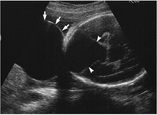
Occipital Meningocele With Dandy-Walker Cyst. A transverse image through the fetal head shows a large cystic structure outside the calvaria in the occipital region (arrows). A large cystic structure is also observed in the posterior fossa, consistent with a Dandy-Walker cyst (arrowheads). A small defect is observed in the cranium at the base of the cyst.
Departments of Radiology and Obstetrics and Gynecology, McMaster University Medical Centre. Scientific exhibit presented at: the 1998 Radiological Society of North America Scientific Assembly; 1998; Ontario, Canada.
(Click Image to Enlarge)
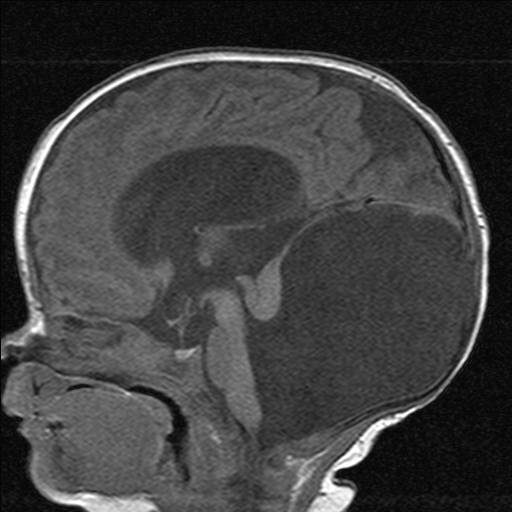
MRI of a Child's Brain with Dandy-Walker Malformation.
Contributed by S Dulebohn, MD
References
Treviño Alanís MG, González Cantú N, Montes Cruz JV, García Flores JB, Martínez Menchaca HR, Rivera Silva G. [Dandy Walker malformation]. Archivos argentinos de pediatria. 2014 Feb:112(1):103-4 [PubMed PMID: 24566791]
Level 3 (low-level) evidenceJurcă MC, Kozma K, Petcheşi CD, Bembea M, Pop OL, MuŢiu G, Coroi MC, Jurcă AD, Dobjanschi L. Anatomic variants in Dandy-Walker complex. Romanian journal of morphology and embryology = Revue roumaine de morphologie et embryologie. 2017:58(3):1051-1055 [PubMed PMID: 29250689]
Correa GG, Amaral LF, Vedolin LM. Neuroimaging of Dandy-Walker malformation: new concepts. Topics in magnetic resonance imaging : TMRI. 2011 Dec:22(6):303-12. doi: 10.1097/RMR.0b013e3182a2ca77. Epub [PubMed PMID: 24132069]
Reith W, Haussmann A. [Dandy-Walker malformation]. Der Radiologe. 2018 Jul:58(7):629-635. doi: 10.1007/s00117-018-0403-7. Epub [PubMed PMID: 29797040]
Di Nora A, Costanza G, Pizzo F, Di Mari A, Sapuppo A, Basile A, Fiumara A, Pavone P. Dandy-Walker malformation and variants: clinical features and associated anomalies in 28 affected children-a single retrospective study and a review of the literature. Acta neurologica Belgica. 2023 Jun:123(3):903-909. doi: 10.1007/s13760-022-02059-z. Epub 2022 Sep 6 [PubMed PMID: 36068432]
Level 2 (mid-level) evidenceBosemani T, Orman G, Boltshauser E, Tekes A, Huisman TA, Poretti A. Congenital abnormalities of the posterior fossa. Radiographics : a review publication of the Radiological Society of North America, Inc. 2015 Jan-Feb:35(1):200-20. doi: 10.1148/rg.351140038. Epub [PubMed PMID: 25590398]
Kumar R, Jain MK, Chhabra DK. Dandy-Walker syndrome: different modalities of treatment and outcome in 42 cases. Child's nervous system : ChNS : official journal of the International Society for Pediatric Neurosurgery. 2001 May:17(6):348-52 [PubMed PMID: 11417415]
Level 3 (low-level) evidenceAlves CAPF, Sidpra J, Manteghinejad A, Sudhakar S, Massey FV, Aldinger KA, Haldipur P, Lucato LT, Ferraciolli SF, Teixeira SR, Öztekin Ö, Bhattacharya D, Taranath A, Prabhu SP, Mirsky DM, Andronikou S, Millen KJ, Barkovich AJ, Boltshauser E, Dobyns WB, Barkovich MJ, Whitehead MT, Mankad K. Dandy-Walker Phenotype with Brainstem Involvement: 2 Distinct Subgroups with Different Prognosis. AJNR. American journal of neuroradiology. 2023 Oct:44(10):1201-1207. doi: 10.3174/ajnr.A7967. Epub 2023 Aug 17 [PubMed PMID: 37591769]
Stambolliu E, Ioakeim-Ioannidou M, Kontokostas K, Dakoutrou M, Kousoulis AA. The Most Common Comorbidities in Dandy-Walker Syndrome Patients: A Systematic Review of Case Reports. Journal of child neurology. 2017 Sep:32(10):886-902. doi: 10.1177/0883073817712589. Epub 2017 Jun 21 [PubMed PMID: 28635420]
Level 3 (low-level) evidenceOsenbach RK, Menezes AH. Diagnosis and management of the Dandy-Walker malformation: 30 years of experience. Pediatric neurosurgery. 1992:18(4):179-89 [PubMed PMID: 1472430]
Level 3 (low-level) evidenceRobinson AJ. Inferior vermian hypoplasia--preconception, misconception. Ultrasound in obstetrics & gynecology : the official journal of the International Society of Ultrasound in Obstetrics and Gynecology. 2014 Feb:43(2):123-36. doi: 10.1002/uog.13296. Epub [PubMed PMID: 24497418]
Haldipur P, Bernardo S, Aldinger KA, Sivakumar T, Millman J, Sjoboen AH, Dang D, Dubocanin D, Deng M, Timms AE, Davis BD, Plummer JT, Mankad K, Oztekin O, Manganaro L, Guimiot F, Adle-Biassette H, Russo R, Siebert JR, Kidron D, Petrilli G, Roux N, Razavi F, Glass IA, Di Gioia C, Silvestri E, Millen KJ. Evidence of disrupted rhombic lip development in the pathogenesis of Dandy-Walker malformation. Acta neuropathologica. 2021 Oct:142(4):761-776. doi: 10.1007/s00401-021-02355-7. Epub 2021 Aug 4 [PubMed PMID: 34347142]
Ocampo-Navia MI, Perez-Mendez W, Rodriguez-Alvarez MP, Chadid-Contreras J, Vergara MF. Dandy-Walker syndrome: an updated literature review. Child's nervous system : ChNS : official journal of the International Society for Pediatric Neurosurgery. 2025 May 30:41(1):194. doi: 10.1007/s00381-025-06842-0. Epub 2025 May 30 [PubMed PMID: 40445443]
Society for Maternal-Fetal Medicine (SMFM), Monteagudo A. Dandy-Walker Malformation. American journal of obstetrics and gynecology. 2020 Dec:223(6):B38-B41. doi: 10.1016/j.ajog.2020.08.184. Epub 2020 Nov 7 [PubMed PMID: 33168220]
Shah SP, Gohil JA, Kesavapisharady K, Easwer HV. Revisiting Dandy-Walker Malformation with Associated Neurofibromatosis. Asian journal of neurosurgery. 2021 Oct-Dec:16(4):850-853. doi: 10.4103/ajns.AJNS_468_20. Epub 2021 Dec 18 [PubMed PMID: 35071091]
Sun Y, Wang T, Zhang N, Zhang P, Li Y. Clinical features and genetic analysis of Dandy-Walker syndrome. BMC pregnancy and childbirth. 2023 Jan 18:23(1):40. doi: 10.1186/s12884-023-05367-1. Epub 2023 Jan 18 [PubMed PMID: 36653756]
Ecker JL, Shipp TD, Bromley B, Benacerraf B. The sonographic diagnosis of Dandy-Walker and Dandy-Walker variant: associated findings and outcomes. Prenatal diagnosis. 2000 Apr:20(4):328-32 [PubMed PMID: 10740206]
Bokhari I, Rehman L, Hassan S, Hashim MS. Dandy-Walker Malformation: A Clinical and Surgical Outcome Analysis. Journal of the College of Physicians and Surgeons--Pakistan : JCPSP. 2015 Jun:25(6):431-3 [PubMed PMID: 26100996]
Mahony BS, Callen PW, Filly RA, Hoddick WK. The fetal cisterna magna. Radiology. 1984 Dec:153(3):773-6 [PubMed PMID: 6387792]
Beaman MM, Guidugli L, Hammer M, Barrows C, Gregor A, Lee S, Deak KL, McDonald MT, Jensen C, Zaki MS, Masri AT, Hobbs CA, Gleeson JG, Cohen JL. Novel association of Dandy-Walker malformation with CAPN15 variants expands the phenotype of oculogastrointestinal neurodevelopmental syndrome. American journal of medical genetics. Part A. 2023 Nov:191(11):2757-2767. doi: 10.1002/ajmg.a.63363. Epub 2023 Aug 19 [PubMed PMID: 37596828]
Klein O, Pierre-Kahn A, Boddaert N, Parisot D, Brunelle F. Dandy-Walker malformation: prenatal diagnosis and prognosis. Child's nervous system : ChNS : official journal of the International Society for Pediatric Neurosurgery. 2003 Aug:19(7-8):484-9 [PubMed PMID: 12879343]
Kline-Fath BM, Calvo-Garcia MA. Prenatal imaging of congenital malformations of the brain. Seminars in ultrasound, CT, and MR. 2011 Jun:32(3):167-88. doi: 10.1053/j.sult.2011.02.010. Epub [PubMed PMID: 21596274]
Harper T, Fordham LA, Wolfe HM. The fetal dandy walker complex: associated anomalies, perinatal outcome and postnatal imaging. Fetal diagnosis and therapy. 2007:22(4):277-81 [PubMed PMID: 17369695]
Level 2 (mid-level) evidenceYu F, Fu L, Xu C, Wang J, Huang X, Zhang G, Zhang H. Prenatal Magnetic Resonance Imaging helps Discover Cerebellar Dysplasia or Malformations in Foetuses. Current medical imaging. 2023 Oct 31:():. doi: 10.2174/0115734056256514231020103822. Epub 2023 Oct 31 [PubMed PMID: 37921151]
Nyberg DA, Cyr DR, Mack LA, Fitzsimmons J, Hickok D, Mahony BS. The Dandy-Walker malformation prenatal sonographic diagnosis and its clinical significance. Journal of ultrasound in medicine : official journal of the American Institute of Ultrasound in Medicine. 1988 Feb:7(2):65-71 [PubMed PMID: 3279226]
Level 2 (mid-level) evidenceVolpe P, Contro E, De Musso F, Ghi T, Farina A, Tempesta A, Volpe G, Rizzo N, Pilu G. Brainstem-vermis and brainstem-tentorium angles allow accurate categorization of fetal upward rotation of cerebellar vermis. Ultrasound in obstetrics & gynecology : the official journal of the International Society of Ultrasound in Obstetrics and Gynecology. 2012 Jun:39(6):632-5. doi: 10.1002/uog.11101. Epub 2012 May 14 [PubMed PMID: 22253138]
Whitehead MT, Barkovich MJ, Sidpra J, Alves CA, Mirsky DM, Öztekin Ö, Bhattacharya D, Lucato LT, Sudhakar S, Taranath A, Andronikou S, Prabhu SP, Aldinger KA, Haldipur P, Millen KJ, Barkovich AJ, Boltshauser E, Dobyns WB, Mankad K. Refining the Neuroimaging Definition of the Dandy-Walker Phenotype. AJNR. American journal of neuroradiology. 2022 Oct:43(10):1488-1493. doi: 10.3174/ajnr.A7659. Epub 2022 Sep 22 [PubMed PMID: 36137655]
Mohanty A, Biswas A, Satish S, Praharaj SS, Sastry KV. Treatment options for Dandy-Walker malformation. Journal of neurosurgery. 2006 Nov:105(5 Suppl):348-56 [PubMed PMID: 17328256]
Level 2 (mid-level) evidenceYengo-Kahn AM, Wellons JC 3rd, Hankinson TC, Hauptman JS, Jackson EM, Jensen H, Krieger MD, Kulkarni AV, Limbrick DD Jr, McDonald PJ, Naftel RP, Pindrik JA, Pollack IF, Reeder R, Riva-Cambrin J, Rozzelle CJ, Tamber MS, Whitehead WE, Kestle JRW, Hydrocephalus Clinical Research Network. Treatment strategies for hydrocephalus related to Dandy-Walker syndrome: evaluating procedure selection and success within the Hydrocephalus Clinical Research Network. Journal of neurosurgery. Pediatrics. 2021 Jul 1:28(1):93-101. doi: 10.3171/2020.11.PEDS20806. Epub 2021 Apr 30 [PubMed PMID: 33930865]
Venkatesan C, Kline-Fath B, Horn PS, Poisson KE, Hopkin R, Nagaraj UD. Short- and Long-Term Outcomes of Prenatally Diagnosed Dandy-Walker Malformation, Vermian Hypoplasia, and Blake Pouch Cyst. Journal of child neurology. 2021 Oct:36(12):1111-1119. doi: 10.1177/08830738211049115. Epub 2021 Nov 10 [PubMed PMID: 34757866]
Chumas P, Tyagi A, Livingston J. Hydrocephalus--what's new? Archives of disease in childhood. Fetal and neonatal edition. 2001 Nov:85(3):F149-54 [PubMed PMID: 11668153]
Yashon D, Jane JA, Sugar O. The course of severe untreated infantile hydrocephalus. Prognostic significance of the cerebral mantle. Journal of neurosurgery. 1965 Nov:23(5):509-16 [PubMed PMID: 5295268]
Donders J, Canady AI, Rourke BP. Psychometric intelligence after infantile hydrocephalus. A critical review and reinterpretation. Child's nervous system : ChNS : official journal of the International Society for Pediatric Neurosurgery. 1990 May:6(3):148-54 [PubMed PMID: 2357712]
Dennis M, Fitz CR, Netley CT, Sugar J, Harwood-Nash DC, Hendrick EB, Hoffman HJ, Humphreys RP. The intelligence of hydrocephalic children. Archives of neurology. 1981 Oct:38(10):607-15 [PubMed PMID: 6975094]
Bromley B, Nadel AS, Pauker S, Estroff JA, Benacerraf BR. Closure of the cerebellar vermis: evaluation with second trimester US. Radiology. 1994 Dec:193(3):761-3 [PubMed PMID: 7972820]