 Tools
Tools
 Anatomy, Head and Neck, Thyroid Muscles
Introduction
The thyroid gland is a midline endocrine structure located in the anterior neck. It lies anterior to the trachea, and the sternocleidomastoid and infrahyoid muscles cover its anterior and lateral borders. The sternocleidomastoid is a large muscle located in the posterior triangle of the neck.
This muscle runs anterior to the common carotid arteries and is a typical muscular landmark in neck surgery. The strap muscles, also known as the infrahyoid muscles, consist of four paired muscles—the sternohyoid, sternothyroid, omohyoid, and thyrohyoid. These muscles have both a superficial layer, composed of the sternohyoid and the omohyoid, and a deep layer, consisting of the sternothyroid and the thyrohyoid.[1][2]
Structure and Function
Register For Free And Read The Full Article
 Search engine and full access to all medical articles
- 10 free questions in your specialty
- Free CME/CE Activities
- Free daily question in your email
- Save favorite articles to your dashboard
- Emails offering discounts
Learn more about a Subscription to StatPearls Point-of-Care
Structure and Function
The lateral surface of the thyroid is comprised of the sternothyroid muscle, whose attachment to the thyroid cartilage supports the superior pole of the thyroid gland. Anteriorly lies the superior belly of the omohyoid and the sternohyoid muscles. The anterior aspect of the sternocleidomastoid muscle overlaps both of these muscles. The avascular fascia joins the sternothyroid and sternohyoid muscles at the midline, and it is commonly incised during thyroid surgery.
Together, the infrahyoid muscles are important for swallowing and phonation due to their roles in the gross movement and positioning of the larynx. The sternocleidomastoid muscle arises from the clavicle and sternum and has a medial and lateral head. The muscle acts to turn the head towards the ipsilateral side and to turn the shoulder towards the contralateral side.[2]
Embryology
The muscles of the head and neck derive from 6 pairs of branchial arches. These arches form during weeks 4 to 7 of gestation. The arches consist of neural crest cells and mesoderm, with the mesodermal layer giving rise to the muscles surrounding the thyroid gland. The thyroid gland is endodermal in origin. It descends anteriorly and inferiorly, beginning at the foramen cecum in the posterior tongue. The thyroid will reach its final anatomical location by the seventh week of gestation, midline and anterior to the trachea.[2]
Blood Supply and Lymphatics
The thyroid region has an extensive blood supply. The main arterial supply to the thyroid gland is the superior and inferior thyroid arteries. These 2 arteries have a rich collateral system with numerous branches and anastomoses, both ipsilaterally and contralaterally. The superior thyroid artery is a branch of the external carotid artery. It courses under the omohyoid and sternohyoid muscles and runs lateral to the larynx.
The cricothyroid artery, a branch of the superior thyroid artery, enters the cricothyroid muscle’s superficial surface and contributes to its blood supply. The inferior thyroid artery is a branch of the thyrocervical trunk, which arises from the subclavian artery. Most of its branches supply blood to the lateral aspect of the thyroid gland and the lateral muscles of the thyroid region. Specifically, the muscular branches of the inferior lateral artery supply the infrahyoid, anterior scalene, longus colli, and inferior pharyngeal constrictor muscles. In 10% of the population, an additional artery, called the thyroid ima, may be present and contribute to the thyroid's blood supply.
The superior, middle, and inferior thyroid veins provide the venous drainage of the thyroid region. The superior thyroid vein courses along the superior thyroid artery and eventually drains into the internal jugular vein. The middle thyroid vein runs directly lateral to the internal jugular vein. The left inferior thyroid vein drains into the left brachiocephalic vein. The right inferior thyroid vein may drain into either the left or right brachiocephalic vein.
There is an abundant lymphatic supply to the thyroid region, including the prelaryngeal, paratracheal, pretracheal, and cervical nodes. The lower deep cervical nodes and the paratracheal nodes drain the isthmus and inferior thyroid. The cervical nodes and superior pretracheal nodes drain the superior thyroid. After immediate drainage to these periglandular lymph nodes, further lymphatic drainage continues to the mediastinal nodes.[2]
Nerves
The infrahyoid muscles are innervated by the deep ansa cervicalis (C1-C3), a branch of the cervical plexus. The thyrohyoid muscle also receives nerve supply from the superior ansa cervicalis, which goes alongside the hypoglossal nerve.[3] The sternocleidomastoid muscle receives motor innervation from the ipsilateral spinal accessory nerve (CN XI). The ventral rami of C2 and C3 from the cervical plexus innervate the sternocleidomastoid.
Muscles
The infrahyoid muscles and the sternocleidomastoid muscle border the thyroid gland directly. These muscles and other important muscles of note in the thyroid region are:
Infrahyoid muscles: Together, the infrahyoid muscles play an active role in swallowing by moving the larynx. The omohyoid, sternohyoid, and thyrohyoid act to depress the hyoid bone. The thyrohyoid elevates the larynx, whereas the sternothyroid depresses the larynx. The omohyoid also helps ensure proper venous return through its attachment to the carotid sheath. When the omohyoid pulls on the carotid sheath, the internal jugular vein maintains a low-pressure system, thereby increasing blood return to the superior vena cava.
Sternocleidomastoid muscle: it arises from the clavicle and sternum. It is composed of 2 heads—a lateral (clavicular) head and a medial (sternal) head. Upon contraction of one side of the muscle, it rotates the head towards the contralateral side of the body and pulls the head towards the ipsilateral shoulder. When both sternocleidomastoid muscles act together, they function to flex the cervical vertebral column. Working together, they also aid forced inspiration by elevating the thorax.
Cricothyroid muscle: The cricothyroid muscle is a relatively thin, flat muscle located on the anterior and lateral surfaces of the cricoid cartilage. It inserts at the lower lamina and inferior cornu of the thyroid cartilage. It is the only tensor muscle of the larynx that aids in phonation. When stimulated, the muscle elongates and tenses the vocal cords. It does this by gently tilting the thyroid cartilage forward. With this action, the distance between the thyroid and the vocal processes increases, resulting in elongation of the folds; the eventual result is a higher voice pitch.[4]
Platysma: the platysma muscles are paired, superficial muscles that arise from the subcutaneous layer and the neck fascia. The platysma functions to depress and draw down the lower lip. It also tenses the neck skin during jaw clenching, often seen when making facial expressions of anger. It receives innervation from the cervical branch of the facial nerve (CN XII).[5]
Suprahyoid muscles: the suprahyoid muscles comprise the digastrics, stylohyoid, mylohyoid, and geniohyoid. During swallowing, these muscles act to elevate the hyoid bone and the base of the tongue. Additionally, they depress the mandible when the hyoid bone is fixed. The mylohyoid branch of the inferior alveolar nerve supplies the anterior belly of the digastric muscle and the mylohyoid muscle. The facial nerve innervates the posterior belly of the digastric muscle and the stylohyoid muscle. Nerve fibers from C1 innervate the geniohyoid muscle.[2]
Physiologic Variants
Sternocleidomastoid innervation: As previously described, the sternocleidomastoid is typically innervated by the spinal accessory nerve and the C2 and C3 ventral rami. Multiple aberrant innervation variants of the sternocleidomastoid have been described, including branches of the external laryngeal nerve, hypoglossal nerve, accessory vagus nerve, facial nerve, and transverse cervical nerve.[6] Cricothyroid innervation: In most individuals, the cricothyroid muscle is innervated solely by the external branch of the superior laryngeal nerve. Case reports describe the cricothyroid muscle receiving a nerve supply from branches of the recurrent laryngeal nerve, which innervates the endolaryngeal muscles. This anomaly is important to consider during thyroid surgery, as changes in the patient’s voice may occur if innervation of the cricothyroid muscle is injured.[7]
Surgical Considerations
The thyroid muscles and surrounding vasculature are essential to consider during neck surgery. The infrahyoid muscles may require incision during thyroid surgery to increase exposure to the thyroid gland. There is some evidence that injury to the infrahyoid muscles during thyroid surgery may affect patients’ voices.
The primary finding regarding this injury is that patients’ ability to reach high pitches decreased postoperatively. Additionally, when dissecting the superior pole of the thyroid, it is important not to perform high ligation of the superior thyroid artery. The reason is that the external branch of the superior laryngeal nerve lies in close proximity and can be injured. Thus, the patient will present with dysphonia if the nerve has suffered an injury.[8]
Clinical Significance
The thyroid muscles are critical anatomical landmarks during thyroid surgery, and injury to these muscles can result in various pathologies:
Infrahyoid muscle paralysis: Damage to the ansa cervicalis can occur as a result of trauma to the cervical spine; this may result in infrahyoid muscle paresis or even paralysis. This injury may present in various manifestations, such as a hoarse voice, swallowing difficulties, and throat tightness. The ansa cervicalis may also be deliberately sacrificed during a radical neck dissection. This procedure may be necessary when malignancy is found in the head and neck region and can cause permanent deficits for the patient postoperatively.
Torticollis: Unilateral shortening or tightening of the sternocleidomastoid muscle may result in a pathology known as torticollis—flexion, extension, or twisting of the neck to one side of the body. The muscular contractions causing torticollis can be intermittent or sustained, acute or chronic, acquired or congenital. It is oftentimes associated with muscle spasms and can be painful. Surgical correction may be indicated in severe cases.[9][10]
Media
(Click Image to Enlarge)
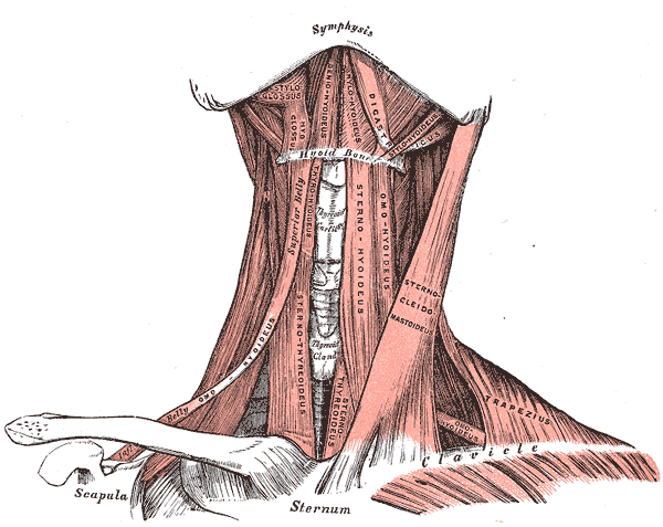
Anterior Neck Muscles and Related Structures. This illustration shows the suprahyoid, infrahyoid, styloglossus, hyoglossus, geniohyoideus, mylohyoideus, digastricus, stylohyoideus, omohyoideus, sternothyroideus, sternohyoideus, omohyoideus, sternocleidomastoideus, trapezius, and omohyoideus muscles. The mandibular symphysis, thyroid cartilage, thyroid gland, hyoid bone, clavicles, scapula, and sternum are also shown.
Henry Vandyke Carter, Public Domain, via Wikimedia Commons
References
Gervasio A, Mujahed I, Biasio A, Alessi S. Ultrasound anatomy of the neck: The infrahyoid region. Journal of ultrasound. 2010 Sep:13(3):85-9. doi: 10.1016/j.jus.2010.09.006. Epub 2010 Nov 5 [PubMed PMID: 23396844]
Allen E, Fingeret A. Anatomy, Head and Neck, Thyroid. StatPearls. 2026 Jan:(): [PubMed PMID: 29262169]
Meguid EA, Agawany AE. An anatomical study of the arterial and nerve supply of the infrahyoid muscles. Folia morphologica. 2009 Nov:68(4):233-43 [PubMed PMID: 19950073]
Pei YC, Fang TJ, Li HY, Wong AM. Cricothyroid muscle dysfunction impairs vocal fold vibration in unilateral vocal fold paralysis. The Laryngoscope. 2014 Jan:124(1):201-6. doi: 10.1002/lary.24229. Epub 2013 Jun 28 [PubMed PMID: 23712513]
Level 2 (mid-level) evidencede Almeida ART, Romiti A, Carruthers JDA. The Facial Platysma and Its Underappreciated Role in Lower Face Dynamics and Contour. Dermatologic surgery : official publication for American Society for Dermatologic Surgery [et al.]. 2017 Aug:43(8):1042-1049. doi: 10.1097/DSS.0000000000001135. Epub [PubMed PMID: 28394862]
Paraskevas G, Lazaridis N, Spyridakis I, Koutsouflianiotis K, Kitsoulis P. Aberrant innervation of the sternocleidomastoid muscle by the transverse cervical nerve: a case report. Journal of clinical and diagnostic research : JCDR. 2015 Apr:9(4):AD01-2. doi: 10.7860/JCDR/2015/11787.5757. Epub 2015 Apr 1 [PubMed PMID: 26023545]
Level 3 (low-level) evidenceMiyauchi A, Masuoka H, Nakayama A, Higashiyama T. Innervation of the cricothyroid muscle by extralaryngeal branches of the recurrent laryngeal nerve. The Laryngoscope. 2016 May:126(5):1157-62. doi: 10.1002/lary.25691. Epub 2015 Oct 28 [PubMed PMID: 26509739]
Tinckler LF. Strap muscles in thyroid surgery: to cut or not to cut? Annals of the Royal College of Surgeons of England. 1993 Sep:75(5):378-9 [PubMed PMID: 8215159]
Level 3 (low-level) evidenceKessomtini W, Chebbi W. [Congenital muscular torticollis in children]. The Pan African medical journal. 2014:18():190. doi: 10.11604/pamj.2014.18.190.4863. Epub 2014 Jul 4 [PubMed PMID: 25419317]
Level 3 (low-level) evidenceDo TT. Congenital muscular torticollis: current concepts and review of treatment. Current opinion in pediatrics. 2006 Feb:18(1):26-9 [PubMed PMID: 16470158]
Level 3 (low-level) evidence