 Tools
Tools
 Craniotomy
Introduction
A craniotomy is a surgical procedure in which a part of the skull is temporarily removed to expose the brain and perform an intracranial procedure.[1] The most common conditions that can be treated via this approach include brain tumors, aneurysms, arteriovenous malformations, subdural empyemas, subdural hematomas, and intracranial hematomas.[2] Specialized tools and equipment are used to remove the section of bone, known as the bone flap. The bone flap is temporarily removed, placed on the surgical instrument table, and then reapplied back after the brain surgery has been concluded. In some cases, depending on the indication for the procedure, the bone can be discarded, stored in an abdominal subcutaneous space, or cryopreserved under cold storage conditions.[3] If the bone flap is discarded or not replaced during the same operation, the procedure is called a craniectomy. In a decompressive craniectomy used for the treatment of malignant brain edema, the bone flap is placed back several weeks after the brain swelling has resolved (see Image. Decompressive Hemicraniectomy).[4][5] The surgical procedure to reconstruct and replace the bone flap in the skull during a second intervention is known as a cranioplasty.[6]
From a historical perspective, cranial interventions have ranged from a single burr hole trephine to a much larger craniectomy. Modern craniotomies are performed by connecting a series of burr holes. Although trephination is the oldest known cranial surgical technique, with ancient reports dating back 2300 years, our modern surgical technique for a craniotomy is the result of a procedure introduced at the end of the 19th century by the self-educated surgeon Wilhelm Wagner.[1][7][8] Although the transition from trephination to a tailored intracranial resection via craniotomy that occurred much later in history, ancient civilizations, eg, the Incas in Peru, likely had some basic familiarity with brain anatomy and surgical interventions, despite their rudimentary knowledge of pathology.[7][9]
Depending on the type of intracranial lesion, pathology that was present, and surgical approach, some craniotomy procedures can be aided by neuronavigation guidance based on magnetic resonance imaging (MRI) or computed tomographic (CT) scans to customize the procedure to the size of the mass lesion, using the smallest incision that is required. Neuronavigation is a modern, computerized technology that helps surgeons localize pathology more precisely by merging a series of craniofacial points present in the patient located in the surgical field. Neuronavigation offers improved surgical guidance, orientation, and localization that provides the surgeon with greater technical confidence and improved patient outcomes.[10]
Historical Background
The craniotomy approach has evolved, dating back to the Neolithic period. Trepanation, meaning “borer”, became synonymous with trephination because of the French instrument tres fines, meaning “3 ends”.[11] Trephination has been performed by prehistoric peoples either for magic or religious rituals to release demons and malignant spirits, or to wear the skull bone as an amulet. During the Neolithic era, therapeutic drilling was performed by pointed or sharp cutting tools made up of silica or obsidian.[12] The principle of bow drilling originated in fire-making and was used by the Egyptians around 1400 BC. A sharp rod made of hard stone or metal was swiveled rapidly between the hands and later by a cord and the string of a bow to make a circle of small holes where the bony bridges between them were connected.[13] The approach to craniotomy is attributed to Imhotep, who is believed to have described it around 2900 BCE.[12] Hippocrates first reported the therapeutic use of craniotomy for the management of fractures in the fifth century BC.[13][14] The instruments were detailed as early as 1518 in “De fractura calvae” by Berengario. Broca also related the archaeological findings of trepanation of the skull.[14] Celso advocated for trephination, a sequential process involving the external cortex, diploic space, and the internal cortex, while safeguarding the meninges.[13] William Detmold first operated on a brain abscess within the lateral ventricle in 1850.[13] Craniotomy evolved in the Renaissance, when firearms and grenades were used, in the 16th and 17th centuries. The use of angulated manual trephines, equipped with a series of perforating or cutting terminals, was introduced in the 16th century. In 1889, Wagner first performed an osteoplastic bone flap. The Gigli saw was used by Obalinski in 1897. At the beginning of the 19th century, the use of craniotomy declined primarily due to the development of infections, and trephining was limited to exceptional cases.[13] Advancements in antisepsis and general anesthesia in the 19th century led to an exponential growth in the use of trephination and craniotomy, even for nontraumatic intracranial lesions.[13][12][14]
Anatomy and Physiology
Register For Free And Read The Full Article
 Search engine and full access to all medical articles
- 10 free questions in your specialty
- Free CME/CE Activities
- Free daily question in your email
- Save favorite articles to your dashboard
- Emails offering discounts
Learn more about a Subscription to StatPearls Point-of-Care
Anatomy and Physiology
Many types of craniotomies are described in the literature, and a basic understanding of anatomy and physiology is necessary to perform a craniotomy with few complications. The type of craniotomy is named for the skull bone that is elevated. Typical skull bones selected for craniotomy include the frontal, parietal, temporal, and occipital bones. Depending on the location of the pathology to be addressed, supratentorial or infratentorial (posterior fossa) craniotomies can be utilized.
One of the craniotomies most commonly performed is the pterional craniotomy. This supratentorial craniotomy can be utilized for aneurysms of the anterior circulation, basilar tip artery aneurysms, direct surgical approaches to the cavernous sinus, frontal and temporal lobe tumors, in addition to suprasellar tumors, eg, pituitary adenomas and craniopharyngiomas.[15][16][17][18] Another type of standard supratentorial craniotomy is the temporal or subtemporal craniotomy, which can be used for temporal lobe biopsy, temporal lobectomy, temporal lobe epilepsy surgery, and access to the floor of the middle cranial fossa.[19][20]
The frontal craniotomy is used to access the anterior skull base and the frontal lobe of the brain for approaches to tumors of the third ventricle or sellar region, eg, craniopharyngiomas, planum sphenoidale meningiomas, frontal lobe tumors, and the repair of cerebrospinal fluid fistulas. Other types of common craniotomies are performed in the parietal, occipital, and retrosigmoid locations.[21]
Indications
Indication for Craniotomy
Craniotomy indications include:
- Trauma for acute epidural hematoma, acute subdural hematoma, depressed skull fractures, intracranial foreign bodies, and cerebrospinal fluid (CSF) leak repair
- Tumors (eg, meningiomas, high-grade and low-grade gliomas, epidermoid tumors, ependymomas, oligodendrogliomas, brain metastases, orbital tumors, cerebellopontine angle tumors, intrasellar lesions, and parasellar lesions)
- Vascular
- Intracerebral hematoma, malignant middle cerebral artery (MCA) territory infarction, cortical venous thrombosis (CVT) with hemorrhagic conversion, aneurysms, and vascular malformations that include arteriovenous malformations, cavernous angiomas, and arteriovenous fistulae
- Microvascular decompression of cranial nerves
- Infectious lesions (eg, brain abscess and subdural empyema)
- Parasitic mass lesions that include hydatid cyst and racemose neurocysticercosis
- Miscellaneous processes
Contraindications
The few contraindications to performing a craniotomy that have been identified include:
- High anesthetic risks: advanced age, severe medical comorbidities
- Moribund state
- Poor functional status
- High frailty index
- Severe systemic collapse (sepsis, multiorgan failure)
- Coagulation disorders
- Bleeding dyscrasias, and
- Pathologies that a single burr hole can access [35]
Absolute Contraindications
Absolute contraindications for an awake craniotomy include:
- Patient refusal
- Patient noncompliance
Relative Contraindications
Relative contraindications for an awake craniotomy include:
- Obese patients
- Obstructive sleep apnea
- Difficult airway access
- Chronic refractory cough
- Highly vascular lesions
- Posterior fossa lesions [36]
Equipment
A craniotomy is a surgical procedure that requires specific equipment. The following are the equipment requirements to perform a craniotomy:
- Suction tips
- Bipolar cautery forceps
- Scalpel handle and blades
- Needle holders
- Hemostatic clips
- Scalp retractors
- Periosteal elevator
- Bayonet forceps
- Adson forceps
- Head-fixation system
- High-speed pneumatic cranial drill (craniotome)
- Hudson brace with a perforating bit
- Round burr for the Hudson brace
- Gigli wire saw, guide, and handles
- Perforating bits, narrow burr attachment with a possible extension
- Bone curette
- Kerrison bone rongeur
- Penfield dissector
- Gerald forceps
- Hemostatic agents (bone wax, Surgicel, Gelfoam)
- Dural scissors.[37]
Personnel
An interprofessional team is required to carry out a craniotomy successfully. The following are the persons whose involvement is mandatory:
- Neurosurgeon
- Operating room head nurse
- Surgical technologist (operating room technologist)
- Anesthesiologist
- Anesthetist
- Intensive care unit nursing personnel
Preparation
Preoperatively, the patient should be in an optimal medical condition to tolerate the procedure. The patient must have an empty stomach or "nil per os" (NPO), a Latin phrase meaning "nothing through the mouth". In emergency cases, this may not be possible. Blood-thinning medications, eg, antiplatelet or antithrombotic agents, should be discontinued between 2 and 10 days preoperatively, depending on the specific medication.[38] An internal medicine or cardiology evaluation should be obtained for medical clearance and patient-specific recommendations, as well as to assess the surgical risk.
Most craniotomy procedures are performed under general anesthesia, requiring a case discussion with the anesthesiology team for any pertinent details regarding the etiology to be treated. On occasion, an awake craniotomy is performed under a local anesthetic agent, often with moderate sedation, to be able to communicate with the patient during surgery involving motor and speech areas.[39][40][41][42] Specific patient preoperative considerations include obtaining the patient's informed consent for the procedure, performing a time-out to verify the correct patient and surgical side have been confirmed, and ensuring blood products are available in case they are needed for transfusion during the procedure.[43]
Routine preoperative antibiotics are administered before the procedure begins for wound infection prophylaxis, in conjunction with any other necessary medications, eg, anticonvulsant medications or corticosteroids. The initial setup of equipment, including the neuronavigation system, surgical microscope, and neuromonitoring, should be performed. Intensive care unit availability should be discussed preoperatively since most patients will require this level of care after a craniotomy.
Anesthesia during craniotomy includes:
- General
- Awake
Awake anesthesia is comparable to general anesthesia in terms of operative and functional outcomes.[44]
Propofol-maintained and volatile-maintained anesthesia had similar brain relaxation scores; however, mean intracranial pressure (ICP) was lower, and cerebral perfusion pressure (CPP) was higher during propofol-maintained anesthesia.[45]
Technique or Treatment
Once the patient is under anesthesia, the head is positioned for the operative approach that is to be used. Avoiding pressure points on vulnerable body locations is of the utmost importance and can be achieved by adequately padding those affected areas. The location of the incision for the craniotomy is dependent on the location of the pathology within the brain that is being addressed. If the surgical craniotomy is guided by neuronavigation, anatomical points are confirmed before the incision.
For supratentorial surgery, the incision is usually made over the frontal, temporal, parietal, or occipital bones or over a combination of these bones. For surgery in the infratentorial area, the incision is usually performed through the posterior skull below the transverse sinus. After the desired incision site is selected, the hair of the skin over the area may be shaved.[37] The incision should preferably be located behind the hairline for cosmetic reasons. After the incision boundaries are confirmed, the surgical area is cleaned with the preferred antiseptic agent, followed by routine sterile draping techniques. A local anesthetic with epinephrine is typically injected into the marked skin incision to aid in hemostasis.
After the skin incision is made, the scalp muscles are dissected off the bone to expose the skull. Retractors can be placed on the edges of the incision to provide exposure to the surgical area. Alternatively, fishhook retractors or sutures can be used to secure the scalp flap away fromthe bone. The pericranium can be harvested and used as a dural substitute during closure if desired. Several burr holes are placed in the skull utilizing the craniotome or cranial drill around the surgical site.[37] Caution must be exercised to prevent the craniotome from penetrating into the brain. The holes are cleaned of any bone fragments, and the dura is separated from the undersurface of the skull with a Freer elevator or Penfield dissector. The burr holes are connected with a craniotome saw, and the bone flap is elevated after carefully separating it from the underlying dura mater. The bone flap is held in the surgical instrument table until the surgery is completed and the skull closure begins. For the intradural procedure, the dura is incised and retracted, exposing the brain.
Once the surgery on the brain is concluded, the bone is secured to the skull with plates and screws. Adequate hemostasis should be achieved for scalp closure. The overlying tissues are reapproximated, and the scalp is then sutured closed in anatomical layers. Depending on the surgeon’s preference, a subdural or subgaleal drain can be left in place to drain any accumulating blood products. A systematic review supports the use of a regional scalp block.[46]
The Dictum of Craniotomy
The general principles of craniotomy are as follows:
- Adequate surgical exposure to the underlying lesion
- The shortest distance to the lesion is chosen
- The procedure should employ minimal normal brain retraction
- Incision
- The incision should be fashioned to allow access to the lesion
- Emphasis is focused on wound healing and cosmetic aspects, eg, placement behind the hairline
- Skin incision should ideally be along Langer’s skin tension lines
- A coronal incision often provides satisfactory exposure and a more cosmetic result by maintaining forehead aesthetics
- The base of the flap should be wider than the length by no more than 1.5 times
- Safeguard the vascularity of the flap by avoiding crossed incisions
Patient positioning
Positioning of the patient should include the following:
- Direct access to the surgical site
- Promote gravity-assisted brain retraction
- Ensure effective cerebral venous blood return and airway patency
- Comply with the surgical ergonomics
Head fixation recommendations
Based upon 3-point rigid cranial fixation, consisting of a 2-point swivel arm and a contralateral single pin. The maximum allowable pin pressure is 80 lbs. However, the following should be avoided when performing head fixation:
- The course of the skin incision
- Pneumatized sinuses
- Cranial sutures
- Dural venous sinuses
- Neurovascular structures (superficial temporal artery, supraorbital artery, supraorbital nerve, occipital artery)
- Thin calvarium, eg, the temporal squamosa and the pterion
Preoperative steps
Preoperative craniotomy steps include:
- Appropriate positioning of the patient
- Adequate scalp preparation with or without hair removal
- Stringent scalp cleansing
- Marking of the appropriate skin incision
- Proper draping of the surgical site
Craniotomy types
Types of craniotomy include:
- Trephine craniotomy
- Flap craniotomy: Free bone flap or osteoplastic flap
- Keyhole craniotomy
- Stereotactic craniotomy
The Salient Craniotomy Steps
The types of skin flaps include:
- Bicoronal/Souttar flap: The skin incision starts at the superior aspect of the zygomatic arch, 1 cm anterior to the tragus, and courses over the bregma to the corresponding contralateral side. The scalp flap is reflected anteriorly down toward the supraorbital rim.
- Frontal flap: This scalp flap can be unilateral or bifrontal, and the course curves along the coronal suture to end at the mid-hairline. This incision is performed for the exposure of lesions within the anterior interhemispheric fissure, sellar, lamina terminalis, and anterior third ventricular region.
- Temporal flap: A linear or question mark incision that extends behind the zygoma, then courses just above the superior temporal line up to the hairline anteriorly. This flap provides access to the middle cranial fossa, the anterior superior brainstem, petroclival lesions, and basilar apex aneurysms, and can be combined with an anterior petrosectomy.[12]
- Parietal flap
- This flap is used for an interhemispheric approach for parafalcine, medial parietal, and splenial lesions. The transcortical route passes through the noneloquent superior parietal lobule.
- The parietal flap should spare the motor and sensory cortices by using cortical mapping, neuronavigation, or functional MRI (fMRI).
- Pterional (fronto-temporal) flap: This flap was developed by Gazi Yasargil and is centered on the pterion. It provides access to the frontal and temporal opercula, the Sylvian fissure, and the suprasellar cisterns and can be combined with a subfrontal approach to expose the anterior cranial fossa.[12]
- Fronto-temporo-parietal (question mark) flap
- Horseshoe flap: Inverted "U" shape to expose any part of the cerebral convexity.
- Orbitozygomatic (OZ)
- First described by Pellerin and Hakuba
- Can be 1 or 2-piece osteotomies. The 2-piece approach is made by removing a traditional pterional bone flap, followed by a supraorbital craniotomy.
- Used for lesions within the orbital apex, paraclinoid, and parasellar areas, the cavernous sinuses, the basal cisterns, and the upper clivus.[12]
- Occipital skin (Mitre) flap
- Vertical limb extends from inion to vertex
- The anterior limb eclipses the posterior parietal region towards the ear
- Vascularity is based on the occipital artery
- Applied for assessing lesions on the occipital lobe, posterior falx, and superior tentorial surface
- Midline suboccipital incision
- Retromastoid skin incision
- Linear and curvilinear incisions
- Trauma flap
- For traumatic lesions and malignant MCA infarct
- Based on the zygoma
- Blood supply from the superficial temporal and supraorbital vessels
- Curves around 3.5 cm posterior to the external auditory meatus
- Retrosigmoid incision
- Popularized by Woolsey and Krause as the lateral suboccipital approach
- Vertical incision 1 cm medial to the mastoid bone
- Can be a ‘Lazy S’-shaped incision, or an inverted ‘J’ Hockey stick incision
- Provides access to the cerebellopontine and cerebellomedullary cisterns
- Also used for neurovascular decompression procedures.[12]
-
Suboccipital incision
- Developed by Rand and Yasargil
- Extends from the external occipital protuberance (EOP) to the C2 level
- Allows exposure of the cerebellar hemispheres, tonsils, vermis, medulla, fourth ventricle, craniocervical junction, and foramen magnum [12]
Burr Hole and Bone Flap Techniques
The burr hole procedure should include the following:
- A handheld drill (eg, a Hudson brace or motorized drill) attached to a perforating bit
- The drill is kept perpendicular to the skull and turned rapidly
- Penetrating the outer table of the skull may be difficult initially, but is relatively easy to drill through the cancellous bone
- Cogwheel resistance is felt once the inner cortex is engaged
- Visually inspect the burr hole to confirm the breach of the inner cortex
- A sharp bone curette or a round burr for the Hudson brace is used to enlarge the hole safely
- Bone wax is applied to the edges of the burr to control bleeding [37]
Bone flap techniques should include the following:
- The flap should provide direct or the shortest access trajectory to the lesion
- The flap should be centered over the lesion when located near to the convexity
- Careful separation of the underlying dura with Penfield’s dissector
- Beveling the bone edges should be performed to avoid sinking of the bone flap
- The bone cuts along or across the dural venous sinuses should be performed last
Additionally, manual craniotomy can be physically exhausting and time-consuming. Robotic systems aid in preoperative surgical path planning and precision surgery during drilling and milling.[47] Deep learning, augmented and virtual reality (AR, VR) assisted craniotomy can supplement, augment, or even replace conventional approaches.[48][49][50][51][52]
Durotomy
Durotomy techniques include:
- Epidural tacking sutures can be applied if they are felt to be necessary.
- A dural flap based on the sinus locations should be performed.
- The dura is first opened with a sharp hook and knife.
- Further opening is made with dural scissors, with cottonoid beneath the dura over the brain.
- A suitable cuff of dura should be maintained for closure at the end of the procedure.
Wound closure includes the following:
- Dural repair should be as close to watertight as possible
- Bone flap replacement should be performed when indicated
- Even distribution of tension should be maintained along the suture line
- Tension-free closure
- Interrupted sutures better preserve the galeal vessels supplying the scalp
- The skin is closed in 2 layers
Postprocedure Admission Consideration
Routine ward admission appears safe for most patients. Approximately 2% of unplanned intensive care unit (ICU) admissions are reported. Factors for admission include:
- Lengthy operation
- Profound blood loss
- High anesthetic risks
- New neurological deficits
- Reduced level of consciousness or deficits of the lower cranial nerves
- Provisions for delayed extubation necessitate ICU admission [53][54]
Factors to take into account for ICU admission include:
- Patient-specific risk factors, eg, age, neurological status, anesthesia-associated risks, frailty index, and medical comorbidities
- Surgical factors, including the location, size, and type of the lesion, the surgical approach, the duration of surgery, the nature of the procedure (elective versus emergency), and perioperative complications
- Anticipated postoperative complications will require stringent neurological and hemodynamic assessment, and the management of hormonal or electrolyte imbalance that includes the syndrome of inappropriate antidiuretic hormone, diabetes insipidus, or cerebral salt wasting.
Univariate analysis showed that patients with diabetes, intraoperative high blood loss, the need for blood transfusion, older patients, and longer surgical procedures were associated with ICU admission. In contrast, multivariate analysis showed that only diabetes and age were predictive variables of the need for postoperative intensive care unit admission.[55] A "safe transition pathway" model should be adopted.
General Postoperative Management
Multimodal monitoring and patient-centered optimization of neurosurgical care in the perioperative period are essential.[56][57] Postoperative management recommendations include:
- Stringent neurological assessment and monitoring [58]
- Hemodynamic stability surveillance
- Seizure prophylaxis
- Adequate analgesia and sedation using opioids, paracetamol/NSAIDs, and regional anesthesia [59]
- Fluid and electrolyte monitoring
- Chest physiotherapy
- Nutritional support
- Deep venous thrombosis (DVT) prophylaxis, including low-molecular-weight heparin (LMWH) and intermittent compression devices [60][61]
The enhanced recovery after surgery (ERAS) principle incorporates the following:
- Mental state assessment
- Antimicrobial, steroidal, and antiepileptic prophylaxes
- Nutritional assessment
- Postoperative nausea and vomiting (PONV) prophylaxis
- Regional field block or scalp block
- Avoidance, minimization, and early removal of invasive monitoring
- Use of absorbable skin sutures
- Avoidance of wound drains
- Early extubation
- Early mobilization
- Early fluid deescalation
- Early initiation of oral intake
- Early postoperative imaging [56]
Complications
Complications of Head Fixation Devices
Complications of cranial fixation devices include:
- Scalp laceration
- Skull fractures
- Pin site infections and osteomyelitis (see Image. Osteomyelitis)
- Venous air embolism
- Acute epidural, subdural hematoma, and brain contusions
Specific Complications to Individual Flaps
Flap-specific complications include:
- Scalp flap necrosis
- Frontal flaps
- Cosmetic deformity
- Cerebrospinal fluid (CSF) leak
- Injury of the superior sagittal sinus
- Retraction-induced bilateral frontal lobe injury.[12]
- Temporal flaps
- Parietal flaps
- Injury to the vein of the Trolard with subsequent venous infarction
- Injury to the superior sagittal sinus and overlying cortical veins, sinus or cortical vein bleeding or thrombosis
- Injury to the motor cortex [12]
- Pterional flaps
- The frontal sinus may be violated
- The frontalis branches of the facial nerve may be injured
- Osteotomy involving the sphenoid bone may extend into the optic canal.[12]
- Orbitzygomatic flaps
- Fractures of the orbital roof and rim can lead to injury of the optic nerve
- Fractures of the sphenoid and ethmoid sinuses can cause CSF leaks
- Retrosigmoid flaps
- Injury to the lesser occipital and greater auricular nerves, causing postoperative headache and dysesthesia
- Retraction injury to the cerebellum
- Injury to the transverse, sigmoid, occipital venous sinuses, and torcula
- Damage to the cranial nerves and brainstem
- CSF leaks and pseudomeningocele formation
- The mastoid emissary vein can cause substantial bleeding and can be a source of air embolism
- Injury to the vertebral artery
- Bone-dust-induced meningitis [12]
- Suboccipital:
- Pooling of the blood within the operative bed limits surgical visibility in prone positioning
- Pressure on the eyes and face in prone positioning can cause visual impairment or loss
- The sitting position has the risk of venous air embolism and hemodynamic instability
- Injury to the transverse, sigmoid, occipital venous sinuses, and torcula
- CSF leak and pseudomeningocele formation
- Cerebellar mutism
Burr Holes, Craniotomy, and Durotomy Complications
Complications of burr holes, craniotomy, and durotomy include:
- Opening into an air sinus is repaired by removing the sinus mucosa, packing the sinus with betadine-shocked gel foam, and covering the repair with bone wax or a vascularized flap
- Bone bleeding is controlled with bone wax packing
- Dural venous sinus injury is controlled with hemostatic agent packing or by being sewn closed
- Dural lacerations are sewn closed
- Injuries to the cortical draining veins are coagulated until closed
- The drill perforator plunges into the brain, causing a cerebral contusion that is treated with hemostatic agents.[63]
The following factors increase the risk of complications:
- Prone/lateral prone positioning
- Emergency procedure
- Anesthesia fluctuation
- Increased length of the operation
- Thin scalp [64]
Postprocedure Complications of Craniotomy
Complications that may occur following craniotomy include:
- Postcraniotomy headache [2]
- Postcraniotomy emergence hypertension [65]
- Extraxial hematomas
- Seizures: Levetiracetam is superior to phenytoin for de novo seizures following craniotomy [66]
- Electrolyte abnormalities: the most common being hyponatremia and hypernatremia
- Tension pneumocephalus
- Postoperative infection: The incidence of postoperative meningitis is 2.2%, most commonly caused by gram-negative organisms, with an overall mortality rate of 5%.
- Intracranial hemorrhage (ICH)
- Ventilator-associated pneumonia (VAP) diagnosed by bronchoalveolar lavage and endotracheal tube aspiration
- Cerebral edema
- Cerebral ischemia
- Vasospasm
- Pneumocephalus
- CSF leak (studies demonstrate that dural sealants do not reduce the number of CSF leaks but minimize the risk of surgical-site infection) [67][68]
- Hydrocephalus
- Infection (eg, soft tissue infections, extradural abscess, empyema, and bone flap infection) [69][70]
- Instrument failure, eg, drill bit breakage [71]
- Incidental dropping of the craniotomy bone flap on the floor [72]
- Temporalis muscle atrophy [73]
- Myositis ossificans [74]
- Craniectomy itself can induce an inflammatory response, inhibit autophagy, and disrupt the blood-brain barrier.[75]
One study has shown that significant variables for surgical site infections include:
- American Society of Anesthesiologists (ASA) score (>2)
- The presence of other infections
- Duration of the operation >4 hours
- Sinus entry
- CSF leak (OR 7.817)
- CSF drainage
- Surgical drain placement
- Number of previous operations
- Implants present [76][77][78]
A meta-analysis has shown that prophylactic antibiotics significantly reduce the risk of meningitis after craniotomy.[79] The reported incidence of significant complications is 8.3%, while minor complications occur in up to 60% of cases. Mortality owing to major complications is reportedly 22% (0.5% in minor complications).[78]
Variables associated with significant complications include:
- Age
- An abnormal neurological examination after the end of the surgery
- Intraoperative desaturation [78]
Clinical Significance
The surgical procedure of craniotomy has become one of the essential procedures in modern healthcare for neurological disorders. Before the advent of this procedure, many patients were unable to survive conditions that are now treated on a daily basis. Disorders ranging from brain tumors, vascular pathologies, and trauma are routinely treated, which could produce devastating damage to the patient if access to the intracranial cavity were limited. The procedure itself has been modernized and will continue to evolve as new technologies are developed. Since its inception, craniotomy has revolutionized neurosurgical and neurological patient care.
Based on the specific pathology to be addressed and the physician's clinical judgment, a decision is made on whether a craniotomy is necessary for the patient. Even with the advent of new endovascular techniques for treating intracranial vascular disorders and stereotactic radiosurgery for treating intracranial tumors, craniotomy remains the primary tool for treating the majority of neurosurgical pathologies.
Scores used in assessing postoperative outcomes include:
- Preoperative ASA physical status classification
- Karnofsky performance score (KPS)
- Charlson comorbidity score
- Modified Rankin Scale
- Sex, KPS, ASA physical status classification, location, and edema (SKALE) [80]
KPS has the strongest evidence for predicting surgical outcomes. No single score strongly predicts nonsurgical outcomes after elective craniotomies. KPS and ASA physical status classification predict early (≤30-day) morbidity in tumor patients. The Charlson co-morbidity predicts mortality of elective aneurysms.[80]
Enhancing Healthcare Team Outcomes
Craniotomy is a surgical procedure that involves temporarily removing a portion of the skull to access the brain for treatment of tumors, vascular lesions, traumatic injuries, infections, or functional disorders. Modern techniques integrate neuronavigation, advanced imaging, and minimally invasive approaches to enhance precision, reduce healthy tissue disruption, and optimize patient outcomes. Perioperative management, including patient positioning, bone flap handling, dural repair, and postoperative care, is critical for safety, recovery, and functional outcomes. Historical evolution from trephination to contemporary craniotomy demonstrates continual advancement in surgical skill, technology, and evidence-based practices, enabling more effective and safer interventions.
Effective care requires coordinated skills and responsibilities across the healthcare team. In the preoperative phase, communication among neurosurgeons, emergency physicians, internists, and cardiologists ensures appropriate risk assessment and preparation. Intraoperatively, neurosurgeons, neuroanesthesiologists, and neuromonitoring technicians collaborate closely, ensuring procedural accuracy, patient safety, and timely intervention. Postoperatively, nurses, intensivists, and pharmacists coordinate care, monitor recovery, and manage medications, fostering patient-centered outcomes. Strong interprofessional communication, strategy, and shared responsibility enhance team performance, safety, and overall quality of neurosurgical care.
Nursing, Allied Health, and Interprofessional Team Interventions
Team-based practices are necessary for performing a successful craniotomy procedure. Interaction between the neurosurgeon and the anesthesiologist typically occurs even before the patient enters the operating room, through discussion of the case and the desired approach. Discussing the necessary equipment with the operating room head nurse is essential. A discussion of non-anesthetic agents to be administered by the anesthetist takes place before commencing the incision. Once the patient is under general anesthesia, effective communication between the anesthesia team and the neurosurgeon during the procedure minimizes complications and unexpected events.
Postoperative care for a patient who has undergone a craniotomy also involves an interprofessional team, including intensive care unit nursing personnel, as well as speech pathologists, physical medicine and rehabilitation physicians, practical nurses, physical therapists, respiratory therapists, and occasionally, personnel from the discharge planning team and social work team.[81]
Media
(Click Image to Enlarge)
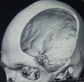
Decompressive Hemicraniectomy. Image showing decompressive hemicraniectomy.
Contributed by S Munakomi, MD
(Click Image to Enlarge)
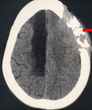
Osteomyelitis. Computed tomography demonstrating osteomyelitis.
Contributed by S Munakomi, MD
References
González-Darder JM. [History of the craniotomy]. Neurocirugia (Asturias, Spain). 2016 Sep-Oct:27(5):245-57. doi: 10.1016/j.neucir.2016.02.002. Epub 2016 Mar 19 [PubMed PMID: 27006140]
Subbarao BS, Fernández-de Thomas RJ, Das JM, Eapen BC. Postcraniotomy Headache. StatPearls. 2025 Jan:(): [PubMed PMID: 29493922]
Bhaskar IP, Zaw NN, Zheng M, Lee GY. Bone flap storage following craniectomy: a survey of practices in major Australian neurosurgical centres. ANZ journal of surgery. 2011 Mar:81(3):137-41. doi: 10.1111/j.1445-2197.2010.05584.x. Epub 2010 Dec 8 [PubMed PMID: 21342384]
Level 3 (low-level) evidenceSchizodimos T, Soulountsi V, Iasonidou C, Kapravelos N. An overview of management of intracranial hypertension in the intensive care unit. Journal of anesthesia. 2020 Oct:34(5):741-757. doi: 10.1007/s00540-020-02795-7. Epub 2020 May 21 [PubMed PMID: 32440802]
Level 3 (low-level) evidenceSahuquillo J, Dennis JA. Decompressive craniectomy for the treatment of high intracranial pressure in closed traumatic brain injury. The Cochrane database of systematic reviews. 2019 Dec 31:12(12):CD003983. doi: 10.1002/14651858.CD003983.pub3. Epub 2019 Dec 31 [PubMed PMID: 31887790]
Level 1 (high-level) evidenceAlkhaibary A, Alharbi A, Alnefaie N, Oqalaa Almubarak A, Aloraidi A, Khairy S. Cranioplasty: A Comprehensive Review of the History, Materials, Surgical Aspects, and Complications. World neurosurgery. 2020 Jul:139():445-452. doi: 10.1016/j.wneu.2020.04.211. Epub 2020 May 6 [PubMed PMID: 32387405]
Rifkinson-Mann S. Cranial surgery in ancient Peru. Neurosurgery. 1988 Oct:23(4):411-6 [PubMed PMID: 3059210]
Buchfelder M. From trephination to tailored resection: neurosurgery in Germany before World War II. Neurosurgery. 2005 Mar:56(3):605-13; discussion 605-13 [PubMed PMID: 15730586]
Andrushko VA, Verano JW. Prehistoric trepanation in the Cuzco region of Peru: a view into an ancient Andean practice. American journal of physical anthropology. 2008 Sep:137(1):4-13. doi: 10.1002/ajpa.20836. Epub [PubMed PMID: 18386793]
Enchev Y. Neuronavigation: geneology, reality, and prospects. Neurosurgical focus. 2009 Sep:27(3):E11. doi: 10.3171/2009.6.FOCUS09109. Epub [PubMed PMID: 19722813]
Hobert L, Binello E. Trepanation in Ancient China. World neurosurgery. 2017 May:101():451-456. doi: 10.1016/j.wneu.2016.10.051. Epub 2016 Oct 18 [PubMed PMID: 27769950]
Rao D, Le RT, Fiester P, Patel J, Rahmathulla G. An Illustrative Review of Common Modern Craniotomies. Journal of clinical imaging science. 2020:10():81. doi: 10.25259/JCIS_176_2020. Epub 2020 Dec 10 [PubMed PMID: 33408956]
Sperati G. Craniotomy through the ages. Acta otorhinolaryngologica Italica : organo ufficiale della Societa italiana di otorinolaringologia e chirurgia cervico-facciale. 2007 Jun:27(3):151-6 [PubMed PMID: 17883195]
Nikova A, Birbilis T. The Basic Steps of Evolution of Brain Surgery. Maedica. 2017 Dec:12(4):297-305 [PubMed PMID: 29610595]
Yasargil MG, Antic J, Laciga R, Jain KK, Hodosh RM, Smith RD. Microsurgical pterional approach to aneurysms of the basilar bifurcation. Surgical neurology. 1976 Aug:6(2):83-91 [PubMed PMID: 951657]
Yaşargil MG, Reichman MV, Kubik S. Preservation of the frontotemporal branch of the facial nerve using the interfascial temporalis flap for pterional craniotomy. Technical article. Journal of neurosurgery. 1987 Sep:67(3):463-6 [PubMed PMID: 3612281]
Hendricks BK, Cohen-Gadol AA. The Extended Pterional Craniotomy: A Contemporary and Balanced Approach. Operative neurosurgery (Hagerstown, Md.). 2020 Feb 1:18(2):225-231. doi: 10.1093/ons/opz117. Epub [PubMed PMID: 31172173]
Choque-Velasquez J, Hernesniemi J. One burr-hole craniotomy: Lateral supraorbital approach in Helsinki Neurosurgery. Surgical neurology international. 2018:9():156. doi: 10.4103/sni.sni_185_18. Epub 2018 Aug 10 [PubMed PMID: 30159200]
Choque-Velasquez J, Hernesniemi J. One burr-hole craniotomy: Subtemporal approach in helsinki neurosurgery. Surgical neurology international. 2018:9():164. doi: 10.4103/sni.sni_187_18. Epub 2018 Aug 14 [PubMed PMID: 30186665]
Zieliński G, Sajjad EA, Robak Ł, Koziarski A. Subtemporal Approach for Gross Total Resection of Retrochiasmatic Craniopharyngiomas: Our Experience on 30 Cases. World neurosurgery. 2018 Jan:109():e265-e273. doi: 10.1016/j.wneu.2017.09.159. Epub 2017 Oct 4 [PubMed PMID: 28987834]
Level 3 (low-level) evidenceZhou C, Evins AI, Boschi A, Tang Y, Li S, Przepiorka L, Sadhwani S, Stieg PE, Xu T, Bernardo A. Preoperative identification of the initial burr hole site in retrosigmoid craniotomies: A teaching and technical note. The international journal of medical robotics + computer assisted surgery : MRCAS. 2019 Jun:15(3):e1987. doi: 10.1002/rcs.1987. Epub 2019 Feb 22 [PubMed PMID: 30721556]
Stachniak JB, Layon AJ, Day AL, Gallagher TJ. Craniotomy for intracranial aneurysm and subarachnoid hemorrhage. Is course, cost, or outcome affected by age? Stroke. 1996 Feb:27(2):276-81 [PubMed PMID: 8571423]
Legnani FG, Saladino A, Casali C, Vetrano IG, Varisco M, Mattei L, Prada F, Perin A, Mangraviti A, Solero CL, DiMeco F. Craniotomy vs. craniectomy for posterior fossa tumors: a prospective study to evaluate complications after surgery. Acta neurochirurgica. 2013 Dec:155(12):2281-6 [PubMed PMID: 24078114]
Hamasaki T, Morioka M, Nakamura H, Yano S, Hirai T, Kuratsu J. A 3-dimensional computed tomographic procedure for planning retrosigmoid craniotomy. Neurosurgery. 2009 May:64(5 Suppl 2):241-5; discussion 245-6. doi: 10.1227/01.NEU.0000336763.90656.2B. Epub [PubMed PMID: 19404104]
Broggi G, Broggi M, Ferroli P, Franzini A. Surgical technique for trigeminal microvascular decompression. Acta neurochirurgica. 2012 Jun:154(6):1089-95. doi: 10.1007/s00701-012-1324-2. Epub 2012 Apr 25 [PubMed PMID: 22531963]
Alvis-Miranda H, Castellar-Leones SM, Moscote-Salazar LR. Decompressive Craniectomy and Traumatic Brain Injury: A Review. Bulletin of emergency and trauma. 2013 Apr:1(2):60-8 [PubMed PMID: 27162826]
Schaller K, Cabrilo I. Anterior temporal lobectomy. Acta neurochirurgica. 2016 Jan:158(1):161-6. doi: 10.1007/s00701-015-2640-0. Epub 2015 Nov 23 [PubMed PMID: 26596998]
Duckworth EA, Vale FL. Trephine epilepsy surgery: the inferior temporal gyrus approach. Neurosurgery. 2008 Jul:63(1 Suppl 1):ONS156-60; discussion ONS160-1. doi: 10.1227/01.neu.0000335030.85402.5f. Epub [PubMed PMID: 18728594]
Yang PF, Zhang HJ, Pei JS, Lin Q, Mei Z, Chen ZQ, Jia YZ, Zhong ZH, Zheng ZY. Keyhole epilepsy surgery: corticoamygdalohippocampectomy for mesial temporal sclerosis. Neurosurgical review. 2016 Jan:39(1):99-108; discussion 108. doi: 10.1007/s10143-015-0657-8. Epub 2015 Aug 16 [PubMed PMID: 26277790]
Smyth MD, Tenenbaum MJ, Kaufman CB, Kane AA. The "clamshell" craniotomy technique in treating sagittal craniosynostosis in older children. Journal of neurosurgery. 2006 Oct:105(4 Suppl):245-51 [PubMed PMID: 17328272]
Akai T, Shiraga S, Sasagawa Y, Iizuka H, Yamashita M, Kawakami S. Troubleshooting distraction osteogenesis for craniosynostosis. Pediatric neurosurgery. 2013:49(6):380-3. doi: 10.1159/000369029. Epub 2014 Dec 9 [PubMed PMID: 25500456]
Alford J, Derderian CA, Smartt JM Jr. Surgical Treatment of Nonsyndromic Unicoronal Craniosynostosis. The Journal of craniofacial surgery. 2018 Jul:29(5):1199-1207. doi: 10.1097/SCS.0000000000004509. Epub [PubMed PMID: 29570518]
Ciporen JN, Lucke-Wold B, Gillham HE, Cua D, Kim J, Akins P. Paramedian Forehead Flap for Repair of Refractory High-Flow Anterior Skull Base CSF Leak. Turkish neurosurgery. 2024:34(2):351-357. doi: 10.5137/1019-5149.JTN.20662-17.1. Epub [PubMed PMID: 28944942]
Sivanaser V, Manninen P. Preoperative assessment of adult patients for intracranial surgery. Anesthesiology research and practice. 2010:2010():. pii: 241307. doi: 10.1155/2010/241307. Epub 2010 Mar 31 [PubMed PMID: 20700431]
Alali J, Dua A. Anesthesia for Awake Craniotomy. StatPearls. 2025 Jan:(): [PubMed PMID: 34283419]
Donovan DJ, Moquin RR, Ecklund JM. Cranial burr holes and emergency craniotomy: review of indications and technique. Military medicine. 2006 Jan:171(1):12-9 [PubMed PMID: 16532867]
Greuter L, Ullmann M, Mariani L, Guzman R, Soleman J. Effect of preoperative antiplatelet or anticoagulation therapy on hemorrhagic complications in patients with traumatic brain injury undergoing craniotomy or craniectomy. Neurosurgical focus. 2019 Nov 1:47(5):E3. doi: 10.3171/2019.8.FOCUS19546. Epub [PubMed PMID: 31675713]
Bilotta F, Rosa G. 'Anesthesia' for awake neurosurgery. Current opinion in anaesthesiology. 2009 Oct:22(5):560-5. doi: 10.1097/ACO.0b013e3283302339. Epub [PubMed PMID: 19623055]
Level 3 (low-level) evidenceFang S, Li Y, Wang Y, Zhang Z, Jiang T. Awake craniotomy for gliomas involving motor-related areas: classification and function recovery. Journal of neuro-oncology. 2020 Jun:148(2):317-325. doi: 10.1007/s11060-020-03520-w. Epub 2020 Apr 29 [PubMed PMID: 32350781]
O'Neill M, Henderson M, Duffy OM, Kernohan WG. The emerging contribution of speech and language therapists in awake craniotomy: a national survey of their roles, practices and perceptions. International journal of language & communication disorders. 2020 Jan:55(1):149-162. doi: 10.1111/1460-6984.12510. Epub 2019 Nov 28 [PubMed PMID: 31778003]
Level 3 (low-level) evidenceÖzlü O. Anaesthesiologist's Approach to Awake Craniotomy. Turkish journal of anaesthesiology and reanimation. 2018 Aug:46(4):250-256. doi: 10.5152/TJAR.2018.56255. Epub 2018 Aug 1 [PubMed PMID: 30140530]
Oszvald Á, Vatter H, Byhahn C, Seifert V, Güresir E. "Team time-out" and surgical safety-experiences in 12,390 neurosurgical patients. Neurosurgical focus. 2012 Nov:33(5):E6. doi: 10.3171/2012.8.FOCUS12261. Epub [PubMed PMID: 23116101]
Lu VM, Phan K, Rovin RA. Comparison of operative outcomes of eloquent glioma resection performed under awake versus general anesthesia: A systematic review and meta-analysis. Clinical neurology and neurosurgery. 2018 Jun:169():121-127. doi: 10.1016/j.clineuro.2018.04.011. Epub 2018 Apr 9 [PubMed PMID: 29655013]
Level 1 (high-level) evidenceChui J, Mariappan R, Mehta J, Manninen P, Venkatraghavan L. Comparison of propofol and volatile agents for maintenance of anesthesia during elective craniotomy procedures: systematic review and meta-analysis. Canadian journal of anaesthesia = Journal canadien d'anesthesie. 2014 Apr:61(4):347-56. doi: 10.1007/s12630-014-0118-9. Epub 2014 Jan 31 [PubMed PMID: 24482247]
Level 1 (high-level) evidenceGuilfoyle MR, Helmy A, Duane D, Hutchinson PJA. Regional scalp block for postcraniotomy analgesia: a systematic review and meta-analysis. Anesthesia and analgesia. 2013 May:116(5):1093-1102. doi: 10.1213/ANE.0b013e3182863c22. Epub 2013 Mar 11 [PubMed PMID: 23477962]
Level 1 (high-level) evidenceZhichao L, Ren W, Ren H, Ma X, Wu D. Precise path planning for robot-assisted craniotomy: a CT-driven virtual center method. Biomedical physics & engineering express. 2025 Oct 15:11(6):. doi: 10.1088/2057-1976/ae0e26. Epub 2025 Oct 15 [PubMed PMID: 41033327]
Kumar R, Dhanakshirur RR, Singh R, Suri A, Kalra PK, Arora C. A deep learning approach for objective evaluation of microscopic neuro-drilling craniotomy skills. Computers in biology and medicine. 2025 Sep:196(Pt A):110650. doi: 10.1016/j.compbiomed.2025.110650. Epub 2025 Jul 11 [PubMed PMID: 40644889]
Fu Z, Wang J, Shen W, Wu Y, Zhang J, Liu Y, Wang C, Shen Y, Zhu Y, Zhang W, Lv C, Peng L. Multimodal nomogram integrating deep learning radiomics and hemodynamic parameters for early prediction of post-craniotomy intracranial hypertension. Scientific reports. 2025 Jul 2:15(1):23595. doi: 10.1038/s41598-025-09115-7. Epub 2025 Jul 2 [PubMed PMID: 40603533]
Neyazi B, Amini A, Swiatek VM, Stein KP, Rashidi A, Sandalcioglu IE. Reshaping neurosurgical training: a novel simulation-based concept for structured skill acquisition and curriculum integration. Neurosurgical review. 2025 Jun 20:48(1):517. doi: 10.1007/s10143-025-03666-z. Epub 2025 Jun 20 [PubMed PMID: 40540088]
Davis P, Napole A, Reddy C, Palepu SK, Wu AQ, Mannam SS, Gajjar AA, Jabarkheel R, Ajmera S, Ahmad H, Scott KW, Sioutas G, Shekhtman O, Kandregula S, Rahmani R, Catapano JS, Burkhardt JK, Srinivasan VM. Systematic evaluation of AI-based text-to-image models for generating medical illustrations in neurosurgery: a multi-stage comparative study. Clinical neurology and neurosurgery. 2025 Oct:257():109039. doi: 10.1016/j.clineuro.2025.109039. Epub 2025 Jul 2 [PubMed PMID: 40614319]
Level 1 (high-level) evidenceMofatteh M, Mashayekhi MS, Arfaie S, Chen Y, Mirza AB, Fares J, Bandyopadhyay S, Henich E, Liao X, Bernstein M. Augmented and virtual reality usage in awake craniotomy: a systematic review. Neurosurgical review. 2022 Dec 19:46(1):19. doi: 10.1007/s10143-022-01929-7. Epub 2022 Dec 19 [PubMed PMID: 36529827]
Level 1 (high-level) evidenceBadenes R, Prisco L, Maruenda A, Taccone FS. Criteria for Intensive Care admission and monitoring after elective craniotomy. Current opinion in anaesthesiology. 2017 Oct:30(5):540-545. doi: 10.1097/ACO.0000000000000503. Epub [PubMed PMID: 28682828]
Level 3 (low-level) evidenceBui JQ, Mendis RL, van Gelder JM, Sheridan MM, Wright KM, Jaeger M. Is postoperative intensive care unit admission a prerequisite for elective craniotomy? Journal of neurosurgery. 2011 Dec:115(6):1236-41. doi: 10.3171/2011.8.JNS11105. Epub 2011 Sep 2 [PubMed PMID: 21888476]
Level 2 (mid-level) evidenceHanak BW, Walcott BP, Nahed BV, Muzikansky A, Mian MK, Kimberly WT, Curry WT. Postoperative intensive care unit requirements after elective craniotomy. World neurosurgery. 2014 Jan:81(1):165-72. doi: 10.1016/j.wneu.2012.11.068. Epub 2012 Nov 24 [PubMed PMID: 23182731]
Stumpo V, Staartjes VE, Quddusi A, Corniola MV, Tessitore E, Schröder ML, Anderer EG, Stienen MN, Serra C, Regli L. Enhanced Recovery After Surgery strategies for elective craniotomy: a systematic review. Journal of neurosurgery. 2021 Dec 1:135(6):1857-1881. doi: 10.3171/2020.10.JNS203160. Epub 2021 May 7 [PubMed PMID: 33962374]
Level 1 (high-level) evidenceSiegemund M, Steiner LA. Postoperative care of the neurosurgical patient. Current opinion in anaesthesiology. 2015 Oct:28(5):487-93. doi: 10.1097/ACO.0000000000000229. Epub [PubMed PMID: 26263123]
Level 3 (low-level) evidenceWagner A, Quiring A, Do A, Heim M, Schneider G, Meyer B. Postoperative Monitoring After Elective Intracranial Surgery in a Postanesthesia Care Unit is Safe, Efficient, and Cost-Effective. Neurocritical care. 2025 Dec:43(3):927-933. doi: 10.1007/s12028-025-02323-z. Epub 2025 Jul 21 [PubMed PMID: 40691410]
Kulikov A, Tere V, Sergi PG, Bilotta F. Prevention and treatment of postoperative pain in pediatric patients undergone craniotomy: Systematic review of clinical evidence. Clinical neurology and neurosurgery. 2021 Jun:205():106627. doi: 10.1016/j.clineuro.2021.106627. Epub 2021 Apr 1 [PubMed PMID: 33857811]
Level 1 (high-level) evidenceCollen JF, Jackson JL, Shorr AF, Moores LK. Prevention of venous thromboembolism in neurosurgery: a metaanalysis. Chest. 2008 Aug:134(2):237-249. doi: 10.1378/chest.08-0023. Epub 2008 Jul 18 [PubMed PMID: 18641095]
Level 1 (high-level) evidenceBanerjee O, Rodrigues R, Adkins L, Busl KM. Venous Thromboembolism Prophylaxis in the Neurocritically Ill Population. Journal of clinical medicine. 2025 Jun 22:14(13):. doi: 10.3390/jcm14134434. Epub 2025 Jun 22 [PubMed PMID: 40648808]
Vaca EE, Purnell CA, Gosain AK, Alghoul MS. Postoperative temporal hollowing: Is there a surgical approach that prevents this complication? A systematic review and anatomic illustration. Journal of plastic, reconstructive & aesthetic surgery : JPRAS. 2017 Mar:70(3):401-415. doi: 10.1016/j.bjps.2016.10.008. Epub 2016 Nov 5 [PubMed PMID: 27894915]
Level 1 (high-level) evidenceBeniwal M, Shukla D. Management of Perforator Plunge in the Transverse Sinus. Pediatric neurosurgery. 2016:51(5):273-5. doi: 10.1159/000445901. Epub 2016 May 20 [PubMed PMID: 27193189]
Cheng G, Hao S, Ye Z, Wang B, Huangpu B, Zhang P, Wang H, Hao Q. Potential risk analysis and experience summarization of unstable factors of cranial fixation devices in neurosurgical operations: three-case reports and systematic review. Chinese neurosurgical journal. 2021 Apr 28:7(1):25. doi: 10.1186/s41016-021-00244-2. Epub 2021 Apr 28 [PubMed PMID: 33910652]
Level 3 (low-level) evidenceGauhar F, Kamal N, Kelly PD. Post-craniotomy emergence hypertension: A scoping review. Journal of clinical neuroscience : official journal of the Neurosurgical Society of Australasia. 2025 Nov:141():111629. doi: 10.1016/j.jocn.2025.111629. Epub 2025 Sep 17 [PubMed PMID: 40967176]
Level 2 (mid-level) evidenceLee CH, Koo HW, Han SR, Choi CY, Sohn MJ, Lee CH. Phenytoin versus levetiracetam as prophylaxis for postcraniotomy seizure in patients with no history of seizures: systematic review and meta-analysis. Journal of neurosurgery. 2019 Jun 1:130(6):2063-2070. doi: 10.3171/2018.4.JNS1891. Epub 2018 Jul 1 [PubMed PMID: 30004278]
Level 1 (high-level) evidenceKinaci A, Algra A, Heuts S, O'Donnell D, van der Zwan A, van Doormaal T. Effectiveness of Dural Sealants in Prevention of Cerebrospinal Fluid Leakage After Craniotomy: A Systematic Review. World neurosurgery. 2018 Oct:118():368-376.e1. doi: 10.1016/j.wneu.2018.06.196. Epub 2018 Jun 30 [PubMed PMID: 29969744]
Bauman MMJ, Webb KL, Michaelcheck CE, Carlstrom LP, Morshed RA, Van Gompel JJ, Link MJ, Peris Celda M. Use of calcium phosphate bone cement in retrosigmoid craniotomies to reduce rates of pseudomeningocele and craniotomy-site CSF leakage: a cohort study and meta-analysis. Journal of neurosurgery. 2025 Oct 1:143(4):1027-1036. doi: 10.3171/2025.1.JNS241076. Epub 2025 Jun 6 [PubMed PMID: 40479821]
Level 1 (high-level) evidenceChughtai KA, Nemer OP, Kessler AT, Bhatt AA. Post-operative complications of craniotomy and craniectomy. Emergency radiology. 2019 Feb:26(1):99-107. doi: 10.1007/s10140-018-1647-2. Epub 2018 Sep 25 [PubMed PMID: 30255407]
Rangankar V, Singh A, Khaladkar S. Pictorial review of the post-operative cranium. SA journal of radiology. 2023:27(1):2684. doi: 10.4102/sajr.v27i1.2684. Epub 2023 Jul 21 [PubMed PMID: 38059119]
Aladdam M, Kahraman MA, Aksu ME, Gürbüz MS. Drill bit breakage with tip dislodgment deep into the brain parenchyma: A unique craniotomy complicatıon. Surgical neurology international. 2025:16():333. doi: 10.25259/SNI_416_2025. Epub 2025 Aug 8 [PubMed PMID: 41030391]
Marzouq A, Ahmad MKB, Bokhari R, Baeesa SS. Reuse or replacement of incidentally dropped craniotomy bone flap: An institutional perspective and literature review. Surgical neurology international. 2025:16():309. doi: 10.25259/SNI_517_2025. Epub 2025 Jul 25 [PubMed PMID: 40837297]
Level 3 (low-level) evidenceJackson M, Karuparti S, Nunna R, Willson TD, Rivera A, Carr SB. Predictors of temporalis muscle atrophy and head asymmetry following frontotemporal craniotomy: A retrospective analysis of clinical factors and volumetric comparison. Surgical neurology international. 2025:16():159. doi: 10.25259/SNI_996_2024. Epub 2025 May 2 [PubMed PMID: 40469335]
Level 2 (mid-level) evidenceAntoniou A, Tatsis D, Louizakis A, Domvri K, Paraskevopoulos K. Myositis Ossificans of the Temporalis Muscle Following Neurosurgical Intervention: A Report of a Rare Case and Literature Review. Cureus. 2025 Jul:17(7):e87512. doi: 10.7759/cureus.87512. Epub 2025 Jul 8 [PubMed PMID: 40777678]
Level 3 (low-level) evidenceTarudji AW, McDonald BZ, Curtis E, Gee C, Kievit FM. Structural Defects Associated with Craniectomy Induce Neuroinflammation and Blood-Brain Barrier Permeability. Neurotrauma reports. 2025:6(1):586-599. doi: 10.1177/08977151251362176. Epub 2025 Jul 26 [PubMed PMID: 40933834]
Schipmann S, Akalin E, Doods J, Ewelt C, Stummer W, Suero Molina E. When the Infection Hits the Wound: Matched Case-Control Study in a Neurosurgical Patient Collective Including Systematic Literature Review and Risk Factors Analysis. World neurosurgery. 2016 Nov:95():178-189. doi: 10.1016/j.wneu.2016.07.093. Epub 2016 Aug 6 [PubMed PMID: 27506410]
Level 2 (mid-level) evidenceFang C, Zhu T, Zhang P, Xia L, Sun C. Risk factors of neurosurgical site infection after craniotomy: A systematic review and meta-analysis. American journal of infection control. 2017 Nov 1:45(11):e123-e134. doi: 10.1016/j.ajic.2017.06.009. Epub 2017 Jul 24 [PubMed PMID: 28751035]
Level 1 (high-level) evidenceAlexandre V, Guyonaud C, Frasca D, Dahyot-Fizelier C. Major complications after scheduled craniotomy: A justification for systematic postoperative intensive care admission? European journal of anaesthesiology. 2020 Feb:37(2):147-149. doi: 10.1097/EJA.0000000000001045. Epub [PubMed PMID: 31913939]
Level 1 (high-level) evidenceAlotaibi AF, Hulou MM, Vestal M, Alkholifi F, Asgarzadeh M, Cote DJ, Bi WL, Dunn IF, Mekary RA, Smith TR. The Efficacy of Antibacterial Prophylaxis Against the Development of Meningitis After Craniotomy: A Meta-Analysis. World neurosurgery. 2016 Jun:90():597-603.e1. doi: 10.1016/j.wneu.2016.02.048. Epub 2016 Feb 26 [PubMed PMID: 26921699]
Level 1 (high-level) evidenceReponen E, Tuominen H, Korja M. Evidence for the use of preoperative risk assessment scores in elective cranial neurosurgery: a systematic review of the literature. Anesthesia and analgesia. 2014 Aug:119(2):420-432. doi: 10.1213/ANE.0000000000000234. Epub [PubMed PMID: 25046789]
Level 1 (high-level) evidenceLivesay S, Moser H. Evidence-based nursing review of craniectomy care. Stroke. 2014 Nov:45(11):e217-9. doi: 10.1161/STROKEAHA.114.006355. Epub 2014 Oct 14 [PubMed PMID: 25316281]