 Tools
Tools
 Anatomy, Bony Pelvis and Lower Limb: Calf Common Peroneal Nerve (Common Fibular Nerve)
Introduction
The common peroneal nerve, or the common fibular nerve, is a major nerve that innervates the lower extremity (see Image. The Sacral and Coccygeal Nerves). As 1 of the 2 major branches of the sciatic nerve, the common peroneal nerve receives fibers from the posterior divisions of L4 through S2. Specifically, the common peroneal nerve separates from the sciatic nerve proximal to the popliteal fossa and courses along the posterolateral aspect of the leg, deep to the long head of the biceps femoris and through the popliteal fossa, before passing behind the proximal fibular head. Just inferior and lateral to the fibular head, the common peroneal nerve divides into 2 nerves at the fibular neck: the superficial and deep fibular nerves. The superficial fibular nerve courses anterolaterally between the fibularis longus muscle and extensor digitorum longus muscle within the lateral compartment.
The superficial fibular nerve terminates within the lateral compartment before reaching the ankle and foot. The deep fibular nerve courses anteriorly and runs adjacent to the anterior tibial artery between the extensor digitorum longus muscle and the tibialis anterior muscle. As the deep fibular nerve travels distally, the nerve runs within the anterior leg compartment between the extensor hallucis longus muscle and the tibialis anterior muscle. As the nerve approaches the foot anterior to the talus, the deep fibular nerve divides into medial and lateral branches. The medial branch travels alongside the dorsalis pedis artery and terminates between the first 2 metatarsals. The lateral branch travels alongside the lateral tarsal artery and terminates near the fifth metatarsal.[1]
Structure and Function
Register For Free And Read The Full Article
 Search engine and full access to all medical articles
- 10 free questions in your specialty
- Free CME/CE Activities
- Free daily question in your email
- Save favorite articles to your dashboard
- Emails offering discounts
Learn more about a Subscription to StatPearls Point-of-Care
Structure and Function
The common fibular nerve is ultimately responsible for innervating the muscles that compose the lower extremity's anterior and lateral muscular compartments. The anterior muscular compartment of the lower extremity consists of the tibialis anterior muscle, extensor hallucis longus muscle, and extensor digitorum longus muscle. This compartment is responsible primarily for dorsiflexion of the foot.[2] The lateral muscular compartment of the lower extremity consists of the fibularis longus muscle and fibularis brevis muscle. This compartment is responsible primarily for eversion of the foot.
In addition to carrying motor fibers, the common fibular nerve carries afferent cutaneous sensory fibers from the lower extremities that travel proximally to the spinal cord. The superficial fibular nerve carries most anterior sensation in the leg; however, the deep fibular nerve also carries some anterior sensory fibers. Specifically, the superficial fibular nerve carries sensory fibers from the anterolateral aspect of the lower extremity, extending from midway down the leg to most of the dorsal aspect of the foot and toes. The deep fibular nerve carries sensory fibers between the first and second toes. These nerves also carry some posterolateral sensation, although this contribution is minimal. The common fibular nerve provides sensation inferolateral to the knee via the lateral sural cutaneous nerve, a small cutaneous branch of the nerve. The superficial fibular nerve is responsible for some lateral leg sensation just below where the sensation from the lateral sural cutaneous nerve ends.[1]
Physiologic Variants
As with most parts of human anatomy, the course and surrounding anatomy of the common fibular nerve have several variants that should be noted, especially for surgeons who may have to decompress the nerve. These variants may also increase or decrease the likelihood of nerve compression.[3] Results from a study comparing cadaveric anatomy with surgically decompressed nerves found a few major variants. The first variant includes fibrous tissue forming a bandlike structure beneath the superficial head of the fibularis longus. The next variant also includes fibrous tissue forming a bandlike structure; however, this tissue is located on the superficial surface of the deep head of the fibularis longus. The last variant noted includes 2 muscles and their uncommon junction and origin. Usually, the soleus and fibularis longus muscles originate separately from the fibular head; however, in some individuals, they originate together at the fibular head and separate as they move distally.[4]
Clinical Significance
The common fibular nerve is a more frequently discussed subject than other neuromuscular complaints due to its high incidence of injury and classic clinical presentation. Because of its location, the common fibular nerve is fairly vulnerable to damage. The nerve travels across the lateral aspect of the knee over the neck of the fibula, where its only protection is skin, subcutaneous fat, and fascia. Consequently, various traumas to the lateral knee often damage the common fibular nerve. For example, if a football player is tackled from the side, the result may be a fibular neck fracture and a common fibular nerve injury.[5] The patient would most likely present with knee pain, loss of dorsiflexion at the foot, ankle eversion, and loss of sensation along the lateral leg and dorsum of the foot. Because of the patient’s inability to dorsiflex the foot, foot drop is also likely. This condition occurs during the swing phase of the gait cycle. Because dorsiflexion is lost, the toes drag on the ground rather than clear the ground during normal gait. The patient often compensates for foot dragging by exaggerating hip abduction to increase foot clearance.[6][7]
Another frequent mechanism of injury to the common fibular nerve is nerve compression as the nerve crosses the fibular head and neck or courses distal to these structures. This may occur after a splint or cast is placed incorrectly or too tightly or in the setting of compartment syndrome after trauma or a burn. These examples would present similarly: the compressed nerve would initially cause paresthesias, followed by loss of sensation and weakness, ultimately resulting in loss of dorsiflexion of the foot and ankle eversion if left untreated. Clinicians should note that these cases may also involve vascular compromise; therefore, distal pulses should be examined.[2]
Clinical presentations may also mimic an injury to the common fibular nerve despite resulting from proximal injuries at the level of the sciatic nerve. One common example is piriformis syndrome. In some cases or anatomical variants, the sciatic nerve, which carries fibers for the common fibular nerve, may become entrapped within the piriformis muscle. A tight piriformis may cause symptoms similar to those of classic common fibular nerve injury: loss of dorsiflexion, loss of foot eversion, and loss of sensation in the anterolateral aspect of the leg and dorsum of the foot. However, in addition to these symptoms, the patient might also experience symptoms proximal to the knee, similar to the classic presentation of sciatica. Therefore, differentiating among sciatica, piriformis syndrome, and isolated injury to the common fibular nerve is essential for developing a proper treatment plan.[8]
Media
(Click Image to Enlarge)
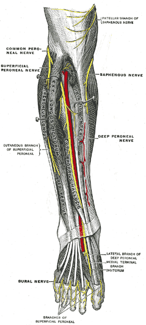
Deep Fibular Nerve and Related Structures. The deep fibular nerve courses through the anterior compartment of the leg in close association with the anterior tibial artery. Distal branching includes lateral and medial terminal branches on the dorsum of the foot. Adjacent nerves, such as the superficial fibular, saphenous, and sural nerves, are also illustrated.
Henry Vandyke Carter, Public Domain, via Wikimedia Commons
References
Khan IA, Mahabadi N, D'Abarno A, Varacallo MA. Anatomy, Bony Pelvis and Lower Limb: Leg Lateral Compartment. StatPearls. 2026 Jan:(): [PubMed PMID: 30137811]
Chow AL, Levidy MF, Luthringer M, Vasoya D, Ignatiuk A. Clinical Outcomes After Neurolysis for the Treatment of Peroneal Nerve Palsy: A Systematic Review and Meta-Analysis. Annals of plastic surgery. 2021 Sep 1:87(3):316-323. doi: 10.1097/SAP.0000000000002833. Epub [PubMed PMID: 34397520]
Level 2 (mid-level) evidenceTomaszewski KA, Graves MJ, Vikse J, Pękala PA, Sanna B, Henry BM, Tubbs RS, Walocha JA. Superficial fibular nerve variations of fascial piercing: A meta-analysis and clinical consideration. Clinical anatomy (New York, N.Y.). 2017 Jan:30(1):120-125. doi: 10.1002/ca.22741. Epub 2016 Jul 7 [PubMed PMID: 27271092]
Level 1 (high-level) evidenceDellon AL, Ebmer J, Swier P. Anatomic variations related to decompression of the common peroneal nerve at the fibular head. Annals of plastic surgery. 2002 Jan:48(1):30-4 [PubMed PMID: 11773727]
Walters BB, Constant D, Anand P. Fibula Fractures. StatPearls. 2026 Jan:(): [PubMed PMID: 32310599]
Lezak B, Massel DH, Varacallo MA. Peroneal Nerve Injury. StatPearls. 2026 Jan:(): [PubMed PMID: 31751049]
Nori SL, Stretanski MF. Foot Drop. StatPearls. 2026 Jan:(): [PubMed PMID: 32119280]
van Zantvoort APM, Setz MJM, Hoogeveen AR, Scheltinga MRM. Common Peroneal Nerve Entrapment in the Differential Diagnosis of Chronic Exertional Compartment Syndrome of the Lateral Lower Leg: A Report of 5 Cases. Orthopaedic journal of sports medicine. 2018 Aug:6(8):2325967118787761. doi: 10.1177/2325967118787761. Epub 2018 Aug 17 [PubMed PMID: 30148178]
Level 3 (low-level) evidence