 Tools
Tools
 Chronic Cholecystitis
Introduction
Chronic cholecystitis is a prolonged, subacute condition caused by mechanical or functional dysfunction of gallbladder emptying, leading to chronic inflammation. This condition may present as chronic, intermittent epigastric or right upper quadrant pain that occurs after eating, but it more often presents during an acute exacerbation. Chronic cholecystitis is usually caused by recurrent episodes of biliary colic, which occurs when gallstones intermittently obstruct the flow of bile through the cystic duct. Pain lasting for more than 6 hours is typically diagnostic of acute cholecystitis. Most cases of chronic cholecystitis are associated with gallstones, though acalculous chronic cholecystitis can be found in certain patient populations.
Etiology
Register For Free And Read The Full Article
 Search engine and full access to all medical articles
- 10 free questions in your specialty
- Free CME/CE Activities
- Free daily question in your email
- Save favorite articles to your dashboard
- Emails offering discounts
Learn more about a Subscription to StatPearls Point-of-Care
Etiology
Under normal conditions, bile produced in the liver flows through the hepatic ducts into the common hepatic duct. Bile may then enter the gallbladder through the cystic duct for storage or continue through the common bile duct into the duodenum through the ampulla of Vater. After food ingestion, particularly after high-fat meals, cholecystokinin stimulates gallbladder contraction. The gallbladder releases bile through the cystic duct and common bile duct into the duodenum, where bile facilitates fat emulsification and absorption.[1] Chronic cholecystitis occurs when intermittent obstruction of the cystic duct develops, usually because of gallstones. The condition is more commonly seen in patients with recurrent biliary colic or recurrent episodes of acute cholecystitis that have not been treated with cholecystectomy. Some patients have mild chronic symptoms and may not seek medical care until symptoms worsen.
Epidemiology
The epidemiology of chronic cholecystitis generally parallels that of cholelithiasis. Gallstone disease is very common.[2] Approximately 10% to 20% of the global population will develop gallstones during their lifetime, and about 80% of gallstones are asymptomatic.[3] Each year, more than 500,000 cholecystectomies are performed in the US for gallbladder disease.
The incidence of gallstone formation increases with age. Furthermore, obesity increases the likelihood of gallstones, especially in women, due to increased biliary cholesterol secretion. Risk also increases with drastic weight loss or fasting because of biliary stasis.[4][5] Notably, gallstones are associated with hormones. Results from studies showed that estrogen increases bile cholesterol and decreases gallbladder contractility. Women of reproductive age or who use estrogen-containing contraceptives have a 2-fold increase in gallstone formation compared with men. Additionally, people with chronic illnesses such as diabetes mellitus also have an increased risk of gallstone formation and reduced gallbladder wall contractility due to neuropathy.[6]
Pathophysiology
Occlusion of the cystic duct or malfunction of gallbladder emptying mechanics is the underlying pathology of this disease. More than 90% of cases of chronic cholecystitis are associated with gallstones.[2] Gallstones cause intermittent obstruction of bile flow through the cystic duct, leading to inflammation and edema of the gallbladder wall. Occlusion of the common bile duct, such as by neoplasms or strictures, can also lead to bile stasis, which can cause gallstone formation.[7]
Researchers have proposed that lithogenic bile leads to increased free radical–mediated damage from hydrophobic bile salts. This process, together with reduced mucosal protection due to lower prostaglandin E2 levels, results in a continuous inflammatory state. When cholecystokinin receptors in the smooth muscle are affected, gallbladder contraction is impaired, leading to stasis and worsening of the permissive environment in which lithogenic bile promotes inflammation.[8]
Histopathology
The gallbladder wall may be thickened to variable degrees, and adhesions may involve the serosal surface. In some cases, extensive fibrosis may cause the gallbladder to appear shrunken. Smooth muscle hypertrophy may be present and more pronounced in prolonged chronic conditions. Additionally, calcium bilirubinate or cholesterol stones are most common and can range in size from sand-like particles or sludge to stones that completely fill the gallbladder lumen. These stones may be multiple or single. Interestingly, normal-appearing bile can also be present. Finally, various bacterial species can be found in 11% to 30% of cases.
Rokitansky-Aschoff sinuses are present or accentuated in 90% of chronic cholecystitis specimens. Rokitansky-Aschoff sinuses represent herniations of intraluminal sinuses from increased pressure, possibly associated with the ducts of Luschka. Furthermore, the mucosa may exhibit varying degrees of inflammation. T lymphocytes are the predominant cells, followed by plasma cells and histiocytes. Moreover, metaplastic changes may be present. Notably, hypertrophy of the muscularis mucosa is usually present, with varying degrees of mural fibrosis and elastosis. A variant in which calcium deposition and hyaline fibrosis lead to diffuse thinning of the gallbladder wall is called hyalinizing cholecystitis. The brittle consistency and hyperdense appearance on advanced imaging also give hyalinizing cholecystitis the name porcelain gallbladder (see Image. Porcelain Gallbladder).[9]
History and Physical
Patients with symptomatic chronic cholecystitis usually present with dull right upper quadrant abdominal pain or epigastric pain that may radiate around the waist to the midback or right scapular tip. High-fat foods may exacerbate the pain, but the classic prolonged postprandial pain of acute cholecystitis is less common. Nausea and occasional vomiting also accompany concerns about increased bloating and flatulence.
The symptoms often occur in the evening or at night, and are usually present for weeks to months in contrast to the abrupt, severe presentation of acute cholecystitis. Symptoms may gradually worsen, or episodes may increase in frequency. Furthermore, fever and tachycardia are rare. Older adults with cholecystitis may present with nonspecific symptoms, and they are at risk of progression to acute or complicated disease, such as cholecystoduodenal fistula formation. Clinicians need a high index of clinical suspicion to diagnose this condition in this population.
Evaluation
Laboratory testing is neither specific nor sensitive for diagnosing chronic cholecystitis. Leukocytosis and abnormal liver function test results may be absent in these patients, unlike in acute disease. However, basic laboratory testing, including a metabolic panel, liver function tests, and a complete blood count, should be performed. Cardiac testing, including electrocardiography and troponin testing, should be considered in the appropriate clinical setting.
The diagnostic test of choice when chronic cholecystitis is suspected clinically is right upper quadrant ultrasonography. This noninvasive study, which is readily available in most facilities, can accurately evaluate for a thickened gallbladder wall or signs of inflammation. Right upper quadrant ultrasonography also aids in evaluating gallstones or sludge. Computed tomography with intravenous contrast usually reveals cholelithiasis, increased bile attenuation, and gallbladder wall thickening. The gallbladder itself may appear distended or contracted; however, pericholecystic inflammation and fluid collection are usually absent in chronic disease.[10] A distended gallbladder and increased enhancement of adjacent hepatic tissue favor findings of acute cholecystitis, whereas hyperenhancement of the gallbladder wall is more commonly seen in chronic disease.[11] Given the overlap in findings between acute and chronic cholecystitis, ultrasonography and CT are generally sufficient to confirm the diagnosis.[12]
An MRI study is a useful alternative in patients who are unable to undergo a CT scan due to radiation concerns or renal disease.[13] The diagnostic test of choice to confirm chronic cholecystitis is hepatobiliary scintigraphy (HIDA) with cholecystokinin. The most common scintigraphic findings are delayed gallbladder visualization (between 1 and 4 hours) and delayed increase in biliary-to-bowel transit time.[14] The tracer is injected intravenously and concentrates in the gallbladder. Cholecystokinin is then administered, and the percentage of gallbladder emptying (ejection fraction) is calculated. An ejection fraction less than 35% at the 15-min cutoff is considered evidence of a dyskinetic gallbladder and is suggestive of chronic cholecystitis.
Treatment / Management
The preferred treatment for chronic cholecystitis is elective laparoscopic cholecystectomy. Laparoscopic cholecystectomy has a low morbidity rate and can be performed as an outpatient surgical procedure. An open cholecystectomy is rarely required but can be used when laparoscopic cholecystectomy cannot be performed. Endoscopic retrograde cholangiopancreatography is usually performed when choledocholithiasis is suspected. Patients with suspected choledocholithiasis usually undergo endoscopic retrograde cholangiopancreatography before elective cholecystectomy.
Patients who are not surgical candidates or who prefer not to undergo a surgical procedure can be closely observed and treated conservatively. A low-fat diet can help reduce the frequency of symptoms. In patients with symptomatic cholelithiasis, Results from studies showed that the use of ursodeoxycholic acid or ursodiol reduced the rates of biliary colic and acute cholecystitis.[15] However, the literature on its role in chronic cholecystitis is limited. The treatment of asymptomatic patients with incidentally detected chronic cholecystitis depends on patient characteristics. Asymptomatic patients with no radiographic or clinical concerns for malignant neoplasm can also be closely monitored with follow-up imaging. Patients who are high-risk surgical candidates and progress to acute cholecystitis may have additional options. Patients can be evaluated for percutaneous cholecystostomy tubes when critically ill or unable to tolerate surgical intervention.[16](B3)
Endoscopic alternatives, such as cystic duct stent placement or transduodenal gallbladder drainage, offer additional options for patients who are unable to undergo a surgical procedure.[17][18] These procedures remain less widely available than percutaneous cholecystostomy. Results from recent studies showed promise in patients who are poor surgical candidates, but long-term outcomes require further investigation.[19](B3)
Differential Diagnosis
Other common medical conditions can mimic the presentation of chronic cholecystitis. Common clinical features of these disorders are as follows:
- Acute cholecystitis: Continuous, severe right-sided abdominal pain lasting at least 6 hours, with fever, nausea, and vomiting in an ill-appearing patient, is suggestive of acute cholecystitis.[20]
- Gallbladder cancer: Chronic abdominal symptoms associated with weight loss or other constitutional symptoms should raise suspicion for a neoplasm. Imaging and histology are helpful in making a definitive diagnosis.[21]
- Peptic ulcer disease: The presence of epigastric abdominal pain and early satiety should raise suspicion for peptic ulcer disease. Alarm symptoms include weight loss, anemia, melena, or dysphagia. Please see StatPearls' companion reference, "Peptic Ulcer Disease," for further information.
- Gastroesophageal reflux disease: A burning sensation in the epigastrium or retrosternal region may be associated with regurgitation of food material.
- Gastric cancer: The presence of alarm symptoms of peptic ulcer disease, persistent vomiting, evidence of a malignant neoplasm, or other risk factors should raise concern for gastric cancer.[22]
- Myocardial infarction: In cases of inferior wall or right ventricular ischemia, the presenting symptoms may be epigastric pain with nausea and vomiting. Other cardiac symptoms, such as dizziness or shortness of breath, or risk factors for coronary ischemia should prompt further evaluation.[23]
- Mesenteric ischemia: Acute mesenteric ischemia presents with severe acute abdominal pain, and the chronic variant typically presents with postprandial pain. Advanced age, risk factors for atherosclerosis, blood in the stool, and weight loss are concerning features.[24]
- Mesenteric vasculitis: Ongoing abdominal symptoms unexplained by standard evaluation and other features consistent with systemic vasculitis may suggest this relatively underrecognized but dangerous condition. Please see StatPearls' companion reference, "Mesenteric Vasculitis," for further information.
Prognosis
Most uncomplicated cases of chronic cholecystitis have an excellent outcome. In many cases, supportive treatments can help with symptoms. Most recognized cases are treated with elective cholecystectomy to prevent future complications. Surgical treatment is considered safe for patients who are appropriate surgical candidates. The outcomes of surgical treatment for chronic cholecystitis are not as well studied as those for acute cholecystitis, but outcomes are likely as good as, if not better than, those for acute cholecystitis. In general, laparoscopic cholecystectomy has a morbidity rate as low as 1.6% and a mortality rate as low as 0.08%.[25]
Complications
Disease progression or worsening infection can lead to acute cholecystitis. Gallstones also occasionally erode through the gallbladder wall into the duodenum and become impacted in the terminal ileum, presenting as a cholecystoduodenal fistula with gallstone ileus. Rarely, the patient may develop emphysematous cholecystitis due to gas-forming organisms such as Clostridium spp, Escherichia coli, and Klebsiella species. Emphysematous cholecystitis is most common in patients with diabetes mellitus and carries a high mortality rate.
The relationship between chronic cholecystitis and gallbladder cancer is controversial. Although results from studies showed that chronic inflammation was associated with an increased risk of cancer,[26] the data are limited. Additionally, xanthogranulomatous cholecystitis is a variant of chronic cholecystitis in which ongoing inflammation leads to extensive thickening and fibrosis that extend locally beyond the gallbladder wall. In this severe variant, complications such as abscesses and fistulas are more common. Xanthogranulomatous cholecystitis is considered a premalignant condition. A porcelain gallbladder tends to be asymptomatic in most cases. The association with malignant neoplasm is again controversial, but the consensus is that a porcelain gallbladder carries a slightly increased risk of cancer.[27]
Consultations
The diagnosis is usually made in the primary care or inpatient setting. Most patients are referred to general surgery for consideration of elective cholecystectomy. A gastroenterology consultation can also be considered when gallstone obstruction of the biliary system is suspected. However, surgeons with the appropriate expertise may be comfortable performing minimally invasive common bile duct explorations to relieve obstructions at the same time as cholecystectomy. A gastroenterology consultation may also help evaluate nonsurgical treatment options.
Enhancing Healthcare Team Outcomes
The diagnosis and treatment of chronic cholecystitis require an interdisciplinary team approach. Primary care clinicians and emergency department clinicians must maintain a high index of suspicion. Referral to the surgical team, followed by shared decision-making between the surgeon and the patient, should be initiated when there is concern for chronic cholecystitis. Gastroenterologists may offer additional treatment options for patients deemed poor surgical candidates. Appropriate postprocedural or postoperative follow-up is also essential. Counseling on dietary habits, with support from a dietitian, and lifestyle changes are crucial for patients undergoing conservative treatment.
Media
(Click Image to Enlarge)
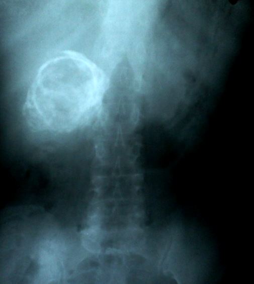
Porcelain Gallbladder. Abdominal radiography demonstrates a calcified gallbladder wall, a finding consistent with porcelain gallbladder in chronic cholecystitis.
Contributed by S Munakomi, MD
References
Ahmed M. Functional, Diagnostic and Therapeutic Aspects of Bile. Clinical and experimental gastroenterology. 2022:15():105-120. doi: 10.2147/CEG.S360563. Epub 2022 Jul 20 [PubMed PMID: 35898963]
Gallaher JR, Charles A. Acute Cholecystitis: A Review. JAMA. 2022 Mar 8:327(10):965-975. doi: 10.1001/jama.2022.2350. Epub [PubMed PMID: 35258527]
Stinton LM, Shaffer EA. Epidemiology of gallbladder disease: cholelithiasis and cancer. Gut and liver. 2012 Apr:6(2):172-87. doi: 10.5009/gnl.2012.6.2.172. Epub 2012 Apr 17 [PubMed PMID: 22570746]
Shamim Seth U, Iqbal F, Akhtar MB, Naveed M, Akhtar R, Uddin M, Shaheen A. A Cross-Sectional Analysis of Body Mass Index With Cholesterol Gallstones in Patients With Cholelithiasis in Tertiary Healthcare Settings. Cureus. 2025 Nov:17(11):e96214. doi: 10.7759/cureus.96214. Epub 2025 Nov 6 [PubMed PMID: 41356989]
Level 2 (mid-level) evidencePak M, Lindseth G. Risk Factors for Cholelithiasis. Gastroenterology nursing : the official journal of the Society of Gastroenterology Nurses and Associates. 2016 Jul-Aug:39(4):297-309. doi: 10.1097/SGA.0000000000000235. Epub [PubMed PMID: 27467059]
Andercou O, Olteanu G, Mihaileanu F, Stancu B, Dorin M. Risk factors for acute cholecystitis and for intraoperative complications. Annali italiani di chirurgia. 2017:88():318-325 [PubMed PMID: 29068324]
Wang L, Sun W, Chang Y, Yi Z. Differential proteomics analysis of bile between gangrenous cholecystitis and chronic cholecystitis. Medical hypotheses. 2018 Dec:121():131-136. doi: 10.1016/j.mehy.2018.07.004. Epub 2018 Jul 5 [PubMed PMID: 30396466]
Guarino MP, Cong P, Cicala M, Alloni R, Carotti S, Behar J. Ursodeoxycholic acid improves muscle contractility and inflammation in symptomatic gallbladders with cholesterol gallstones. Gut. 2007 Jun:56(6):815-20 [PubMed PMID: 17185355]
Level 1 (high-level) evidenceBenkhadoura M, Elshaikhy A, Eldruki S, Elfaedy O. Routine histopathological examination of gallbladder specimens after cholecystectomy: Is it time to change the current practice? Turkish journal of surgery. 2019 Jun:35(2):86-90. doi: 10.5578/turkjsurg.4126. Epub 2018 Sep 11 [PubMed PMID: 32550311]
Smith EA, Dillman JR, Elsayes KM, Menias CO, Bude RO. Cross-sectional imaging of acute and chronic gallbladder inflammatory disease. AJR. American journal of roentgenology. 2009 Jan:192(1):188-96. doi: 10.2214/AJR.07.3803. Epub [PubMed PMID: 19098200]
Level 2 (mid-level) evidenceYeo DM, Jung SE. Differentiation of acute cholecystitis from chronic cholecystitis: Determination of useful multidetector computed tomography findings. Medicine. 2018 Aug:97(33):e11851. doi: 10.1097/MD.0000000000011851. Epub [PubMed PMID: 30113479]
Childs DD, Lalwani N, Craven T, Arif H, Morgan M, Anderson M, Fulcher A. A meta-analysis of the performance of ultrasound, hepatobiliary scintigraphy, CT and MRI in the diagnosis of acute cholecystitis. Abdominal radiology (New York). 2024 Feb:49(2):384-398. doi: 10.1007/s00261-023-04059-w. Epub 2023 Nov 20 [PubMed PMID: 37982832]
Level 1 (high-level) evidenceKaura SH, Haghighi M, Matza BW, Hajdu CH, Rosenkrantz AB. Comparison of CT and MRI findings in the differentiation of acute from chronic cholecystitis. Clinical imaging. 2013 Jul-Aug:37(4):687-91. doi: 10.1016/j.clinimag.2013.02.009. Epub 2013 Mar 28 [PubMed PMID: 23541278]
Level 3 (low-level) evidenceChamarthy M, Freeman LM. Hepatobiliary scan findings in chronic cholecystitis. Clinical nuclear medicine. 2010 Apr:35(4):244-51. doi: 10.1097/RLU.0b013e3181d18ef5. Epub [PubMed PMID: 20305411]
Guarino MP, Cocca S, Altomare A, Emerenziani S, Cicala M. Ursodeoxycholic acid therapy in gallbladder disease, a story not yet completed. World journal of gastroenterology. 2013 Aug 21:19(31):5029-34. doi: 10.3748/wjg.v19.i31.5029. Epub [PubMed PMID: 23964136]
Koushesh P, Ayaz T, Tullius T. Percutaneous Cholecystostomy: Procedural Guidance and Future Directions for Clinical Management. Seminars in interventional radiology. 2024 Oct:41(5):460-465. doi: 10.1055/s-0044-1791724. Epub 2024 Dec 10 [PubMed PMID: 39664225]
Level 3 (low-level) evidenceStorm AC, Vargas EJ, Chin JY, Chandrasekhara V, Abu Dayyeh BK, Levy MJ, Martin JA, Topazian MD, Andrews JC, Schiller HJ, Kamath PS, Petersen BT. Transpapillary gallbladder stent placement for long-term therapy of acute cholecystitis. Gastrointestinal endoscopy. 2021 Oct:94(4):742-748.e1. doi: 10.1016/j.gie.2021.03.025. Epub 2021 Mar 30 [PubMed PMID: 33798540]
Troncone E, Amendola R, Moscardelli A, De Cristofaro E, De Vico P, Paoluzi OA, Monteleone G, Perez-Miranda M, Del Vecchio Blanco G. Endoscopic Gallbladder Drainage: A Comprehensive Review on Indications, Techniques, and Future Perspectives. Medicina (Kaunas, Lithuania). 2024 Apr 14:60(4):. doi: 10.3390/medicina60040633. Epub 2024 Apr 14 [PubMed PMID: 38674279]
Level 3 (low-level) evidenceInoue T, Yoshida M, Suzuki Y, Kitano R, Urakabe K, Haneda K, Okumura F, Naitoh I. Comparison of the long-term outcomes of EUS-guided gallbladder drainage and endoscopic transpapillary gallbladder drainage for calculous cholecystitis in poor surgical candidates: a multicenter propensity score-matched analysis. Gastrointestinal endoscopy. 2023 Sep:98(3):362-370. doi: 10.1016/j.gie.2023.04.002. Epub 2023 Apr 12 [PubMed PMID: 37059367]
Huffman JL, Schenker S. Acute acalculous cholecystitis: a review. Clinical gastroenterology and hepatology : the official clinical practice journal of the American Gastroenterological Association. 2010 Jan:8(1):15-22. doi: 10.1016/j.cgh.2009.08.034. Epub 2009 Sep 10 [PubMed PMID: 19747982]
Adsay NV, Basturk O. Dysplasia and Early Carcinoma of the Gallbladder and Bile Ducts: Terminology, Classification, and Significance. Gastroenterology clinics of North America. 2024 Mar:53(1):85-108. doi: 10.1016/j.gtc.2023.10.001. Epub 2023 Nov 17 [PubMed PMID: 38280752]
Ajani JA, Lee J, Sano T, Janjigian YY, Fan D, Song S. Gastric adenocarcinoma. Nature reviews. Disease primers. 2017 Jun 1:3():17036. doi: 10.1038/nrdp.2017.36. Epub 2017 Jun 1 [PubMed PMID: 28569272]
Albulushi A, Giannopoulos A, Kafkas N, Dragasis S, Pavlides G, Chatzizisis YS. Acute right ventricular myocardial infarction. Expert review of cardiovascular therapy. 2018 Jul:16(7):455-464. doi: 10.1080/14779072.2018.1489234. Epub 2018 Jun 27 [PubMed PMID: 29902098]
Gnanapandithan K, Feuerstadt P. Review Article: Mesenteric Ischemia. Current gastroenterology reports. 2020 Mar 17:22(4):17. doi: 10.1007/s11894-020-0754-x. Epub 2020 Mar 17 [PubMed PMID: 32185509]
Pucher PH, Brunt LM, Davies N, Linsk A, Munshi A, Rodriguez HA, Fingerhut A, Fanelli RD, Asbun H, Aggarwal R, SAGES Safe Cholecystectomy Task Force. Outcome trends and safety measures after 30 years of laparoscopic cholecystectomy: a systematic review and pooled data analysis. Surgical endoscopy. 2018 May:32(5):2175-2183. doi: 10.1007/s00464-017-5974-2. Epub 2018 Mar 19 [PubMed PMID: 29556977]
Level 1 (high-level) evidenceGoetze TO. Gallbladder carcinoma: Prognostic factors and therapeutic options. World journal of gastroenterology. 2015 Nov 21:21(43):12211-7. doi: 10.3748/wjg.v21.i43.12211. Epub [PubMed PMID: 26604631]
Patel S, Roa JC, Tapia O, Dursun N, Bagci P, Basturk O, Cakir A, Losada H, Sarmiento J, Adsay V. Hyalinizing cholecystitis and associated carcinomas: clinicopathologic analysis of a distinctive variant of cholecystitis with porcelain-like features and accompanying diagnostically challenging carcinomas. The American journal of surgical pathology. 2011 Aug:35(8):1104-13. doi: 10.1097/PAS.0b013e31822179cc. Epub [PubMed PMID: 21716080]