 Tools
Tools
 Chordoma
Introduction
Chordomas were first described in 1846 by Virchow and were not determined to be derived from notochordal remnants until later. While histologically considered a low-grade malignancy, chordomas are locally aggressive with a high rate of recurrence even after surgical resection.[1][2] The World Health Organization (WHO) classifies chordomas into 3 groups: conventional, dedifferentiated, and poorly differentiated.[3][1]
Chordomas predominantly arise in the axial skeleton between the skull base and the coccyx in individuals 40 to 60 years of age.[1] The clinical presentation of a patient with a chordoma depends on the chordoma's location, size, and whether surrounding structures are involved. Surgery is the primary treatment for chordomas, with the goal being a gross total resection. Conventional external beam radiation therapy, particle beam radiation, and systemic therapy are also utilized, especially when a complete resection is not possible or metastatic disease occurs. The overall 5-year survival is approximately 50%.[4][5][6]
Etiology
Register For Free And Read The Full Article
 Search engine and full access to all medical articles
- 10 free questions in your specialty
- Free CME/CE Activities
- Free daily question in your email
- Save favorite articles to your dashboard
- Emails offering discounts
Learn more about a Subscription to StatPearls Point-of-Care
Etiology
Chordomas demonstrate notochordal differentiation. The notochord is an embryological structure that provides structural support and plays an important role in the arrangement of the adjacent mesoderm and ectoderm. Ultimately, the notochord disappears, but remnants are retained in the nucleus pulposus in the vertebral discs. Notochordal remnants have also been found within the vertebral bodies. Given that chordomas have been reported outside the neuroaxial structures, they may not specifically arise from notochordal remnants but may instead reflect notochordal differentiation.[1]
Genes implicated in chordoma formation include the brachyury (TBXT) gene, the mechanistic target of rapamycin (mTOR) signaling pathway, phosphatase and tensin homolog (PTEN) deficiency, INI-1, and platelet-derived growth factor receptor beta (PDGFR-beta).[7] While most chordomas are sporadic, a few familial cases are associated with a mutation in the brachyury gene.[8] Chordomas have also been reported in pediatric patients with tuberous sclerosis (TSC1 or TSC2 mutations).[9]
Epidemiology
Chordoma is a rare malignancy affecting 1 person per million per year and accounting for only 1% to 4% of malignant bone tumors.[10][11] Chordomas typically occur in patients aged 40 to 60 years, with less than 5% diagnosed in pediatric patients.[12][5][13] Males are affected more often than females. Chordomas occur in the axial skeleton from the base of the skull to the coccyx in >95% of cases in almost equal proportions.[1] Chordomas have been reported in other locations in the body, including the lungs, soft tissues of the upper extremities, buttocks, tibia, and mediastinum.[14][15][16][17]
A nationwide Dutch pathology registry study reported an increase in pathology-confirmed chordoma incidence from 0.593 per million inhabitants in 1991 to 1995 to 1.111 per million inhabitants in 2015 to 2019. The same study showed increasing centralization of diagnostic review in specialized bone tumor centers, supporting referral to experienced interprofessional centers for diagnosis and treatment planning.[18]
Pathophysiology
Chordoma pathophysiology is driven by persistence of a notochordal transcriptional program, with TBXT/brachyury serving as both a defining diagnostic marker and a central biologic dependency in many tumors. Molecular alterations in poorly differentiated chordoma may involve cell-cycle regulation, PI3K/AKT/mTOR signaling, receptor tyrosine kinase pathways, and loss of SMARCB1/INI1. Most tumors are sporadic, although familial TBXT duplication and other germline cancer-predisposition variants have been reported. Poorly differentiated and dedifferentiated chordomas have more aggressive biology than conventional chordoma.[2][9][19]
Histopathology
Conventional Chordomas
Conventional chordomas contain lobules separated by fibrous septa that grow in an infiltrative pattern. The lobules contain nests of epithelioid cells interspersed in a myxoid matrix. Large cells with bubbly clear cytoplasm (physaliphorous cells) are also often seen. Mitotic activity is usually low, although necrosis and atypia may be present, particularly in more aggressive tumors.[3][1]
A subtype of conventional chordoma, chondroid chordoma, resembles conventional chordoma except for areas with a hyaline cartilage matrix rather than a myxoid matrix. The proportion of hyaline cartilage is variable.[1] Immunohistochemically, chordomas stain positive for keratins (CK 8,18,19), EMA (epithelial membrane antigen), and S100. Nuclear expression of brachyury seen in chordomas is highly sensitive and specific for the diagnosis.[1][3][9]
Dedifferentiated Chordomas
A dedifferentiated chordoma is biphasic. The 2 distinct components represent a conventional chordoma and a high-grade sarcoma. These areas can be intermixed or can demonstrate an abrupt transition. The sarcomatous portion may have features of a high-grade pleomorphic sarcoma or a high-grade osteosarcoma.[1]
Poorly Differentiated Chordomas
Poorly differentiated chordomas contain sheets of poorly differentiated epithelioid cells that contain intracytoplasmic vacuoles, giving the cells the appearance of signet rings.[1] Mitotic figures and necrosis are commonly present. Poorly differentiated chordomas contain focal areas of rhabdoid morphology.[3] A myxoid matrix and physaliphorous cells seen in conventional chordomas are absent. Immunohistochemically, in addition to being positive for keratins, EMA, and brachyury, poorly differentiated chordomas also show loss of SMARCB1/IMI1.[3]
Histopathologic Differentiation of Chordoid Mimics
A recent large tissue microarray study of 14,976 cancers across 135 tumor types and subtypes confirmed that nuclear brachyury expression is highly specific for chordoma, supporting its use in distinguishing chordoma from chondrosarcoma, myoepithelial tumors, metastatic carcinoma, and other chordoid mimics.[19]
History and Physical
The specific location of the chordoma will affect the history and physical findings reported by a patient with a chordoma. For instance, skull base and clival chordomas typically present with headaches or cranial nerve dysfunction that include facial numbness, cranial nerve VI palsy, and palpebral ptosis. Vision loss and dysphagia have also been reported.[20][6]
However, cervical chordomas typically present with nonspecific neck, shoulder, or arm pain with occasional dysphagia due to mass effect. Cervical chordomas can also extend intracranially, causing lower cranial nerve dysfunction and compression of the spinal cord or exiting cervical nerves, resulting in myelopathy or radiculopathy, respectively. Thoracic and lumbar chordomas also present with nonspecific localized pain and may cause a pathologic fracture, radiculopathy, or myelopathy. Sacral chordomas commonly present with pain, although more than 25% of patients will have motor deficits or cauda equina syndrome.[21]
Evaluation
Imaging Studies
Chordomas are evaluated initially with imaging studies. Plain radiographs may demonstrate a locally destructive lytic lesion. However, the complexity of the 3-dimensional structure of the skull base and sacrum may make the interpretation of radiographic imaging studies difficult. When a mass lesion is suspected, advanced imaging should be obtained, eg, computed tomography (CT) or magnetic resonance imaging (MRI).
CT imaging can demonstrate the osteolytic nature of chordomas (see Image. Chordoma on CT). The extent of bone destruction and the presence of an associated soft-tissue mass can be visualized on CT. Internal punctate calcifications may also be seen in the mass.[11] Sacrococcygeal chordomas are located in the midline of the sacrum, often with a large mixed lytic/sclerotic mass extending into the presacral area. Chordomas in the mobile spine are centered in the vertebral body and can extend into adjacent vertebrae. Pathological fractures may be present.[11] Skull base chordomas commonly originate at the spheno-occipital synchondrosis of the clivus.[22]
MRI is the best radiographic modality for evaluating the extent of a chordoma and its association with surrounding structures (see Image. Chordoma on MRI). The appearance of chordomas is variable on T1-weighted imaging and can demonstrate low to intermediate signal intensity. Due to the high fluid content of the vacuolated cellular component and the myxoid matrix present in chordomas, conventional chordomas universally have high signal intensity on T2-weighted images.[22] Areas of high signal are seen on both T1 and T2 images, representing intratumoral hemorrhage. T1-weighted imaging with gadolinium contrast administration demonstrates mild-to-moderate, heterogeneous contrast enhancement of the tumor in a honeycomb pattern.[11][22]
Tissue Diagnosis
The diagnosis is made through either an image-guided needle biopsy or open surgical biopsy. Biopsy should be planned with the definitive surgical team so that the biopsy tract can be removed during definitive resection when feasible. For suspected sacral or mobile-spine chordoma, image-guided core biopsy through a safe posterior tract is generally preferred to avoid contamination of uninvolved compartments.
Once the diagnosis of chordoma is determined, the National Comprehensive Cancer Network Guidelines (Version 2.2025) recommend a screening MRI of the entire spinal axis and CT scans of the chest, abdomen, and pelvis to evaluate for the presence of metastatic disease. Consideration should also be given to obtaining either a fluorodeoxyglucose-positron emission tomography (FDG-PET)/CT or a bone scan. Metastatic disease is present in about 10% of patients, and the most common sites of metastases are bone, brain, liver, lung, and lymph nodes.[5][23][24]
Treatment / Management
The European Society for Medical Oncology consensus guidelines for the treatment of chordomas recommend an R0 (microscopic margin of 1mm or greater) resection when possible and when patients are willing to accept the potential sequelae. When an R0 resection is not possible, an R1 resection (negative microscopic margin of less than 1 mm) should be the goal. When the location and/or size of the chordoma precludes an en bloc resection, surgery aimed at decompressing vital anatomical structures and reducing the tumor volume should be performed, followed by radiation therapy.[25] Inoperable cases can be treated primarily with radiation therapy. Because chordomas are not radiosensitive, the recommended radiation dose is at least 70 Gy for conventional fractionated or proton therapy.[25](B3)
The National Comprehensive Cancer Network (NCCN) guidelines for chordomas also recommend a wide excision, with or without radiation, if the chordoma is in the sacrococcygeal area or the mobile spine. For skull base or clival chordomas, the primary treatment is intralesional excision with or without adjuvant radiation followed by a postoperative MRI with contrast to evaluate for the volume of residual disease. Adjuvant radiation therapy or repeat surgical excision may be indicated based on the postoperative MRI findings. Unresectable chordomas can be treated definitively with radiation therapy. The NCCN guideline recommendations for surveillance of chordomas are an annual MRI, with and without contrast, for up to 10 years after diagnosis to monitor for local recurrence, and chest imaging every 6 months for 5 years, then annually, to monitor for metastatic disease.
For inoperable patients or those declining highly morbid sacrectomy, definitive high-dose radiation may be considered in selected sacral chordomas after interprofessional review. A recent systematic review of 52 patients with nonoperatively managed sacral chordoma found that nonoperative strategies may provide local disease control with functional preservation in selected cases, although recurrence remains a risk and the evidence is limited to small retrospective series and case reports.[26](A1)
| Pause and Reflect |
A 55-year-old patient presents with several months of progressive sacral pain and new bowel and bladder symptoms. Imaging reveals a midline destructive sacral mass suspicious for chordoma.
|
Differential Diagnosis
The differential diagnoses of chordomas include several of the following benign and malignant entities:
- Benign notochordal cell tumors (BNCT): Benign intraosseous tumor with notochordal differentiation that is often found incidentally on imaging or at autopsy.[1] On imaging, BNCTs are intraosseous lesions with intact trabecular architecture, without bone destruction or an associated soft-tissue mass.[11] On a contrast MRI, BNCTs do not enhance. Histologically, BNCTs consist of sheets of adipocyte-like cells with vacuolated cytoplasm and rounded, eccentric nuclei that stain positive for brachyury, cytokeratins, S-100, and EMA. Unlike chordoma, BNCTs lack cellular atypia, necrosis, mitotic figures, a lobular growth pattern, or a myxoid matrix.[27][28][11]
- Ecchordosis physaliphora: An extraskeletal hamartomatous lesion arising from notochordal remnants along the dura, commonly adjacent to the clivus. Ecchordosis physaliphoras are usually small (<2 cm) and asymptomatic, and are found incidentally on imaging studies or at autopsy. Histologically, ecchordosis physaliphora have a similar appearance to BNCTs.
- Chondrosarcoma: A malignant cartilaginous tumor which can appear radiographically similar to a chordoma with an infiltrative growth pattern. While chordomas are usually located in the midline, chondrosarcomas are more often located eccentrically. Histologically, chondrosarcomas do not stain positive for EMA or brachyury, which are characteristic of chordomas. Chondrosarcomas can also have IDH1 or IDH2 mutations.[1]
Clival/spheno-occipital lesions that should also be considered include:
- Meningioma: Arising from the arachnoid cells of the meninges, meningiomas are the second most common central nervous system neoplasm in adults.[29]
- Pituitary macroadenoma: These lesions are benign tumors arising from the pituitary gland.
- Olfactory neuroblastoma: This lesion, also known as an esthesioneuroblastoma, is a rare, locally invasive tumor arising from nerve cells that provide the sense of smell in the upper nasal cavity.
Vertebral/sacrococcygeal lesions that should be considered include:
- Spinal metastases
- Osteomyelitis
- Diffuse large B-cell lymphoma (the third most common malignant tumor in the sacrum)
- Giant cell tumor (GCTs)
- Benign locally aggressive tumor that is the second most common primary sacral tumor
- Most commonly occur in young adults and are not midline in location [30]
- Osteosarcomas (malignant osseous tumor usually arising from the posterior elements of the vertebra) [30]
- Osteoblastomas (benign but locally aggressive bone tumor usually in patients younger than 20) [30]
Surgical Oncology
The mainstay of treatment of chordomas is surgical resection. Ideally, en bloc resection is performed due to the risk of seeding the surrounding tissues with tumor cells. However, because of the midline location along the neuroaxial spine and the association with surrounding structures, an en bloc resection is not always possible. En bloc resection has been demonstrated to decrease local recurrence rates and improve overall survival.[6][4][21]
For skull base chordomas, the goal of surgery is to maximize the tumor resection while maintaining neurologic function and quality of life. Surgery should be planned with input from radiation oncology, including what spinal implants may be utilized if stabilization of the spine is necessary to avoid interference with adjuvant radiation administration. The surgical approach depends on the location of the tumor and, on occasion, multiple approaches may be needed. Midline anterior approaches include endoscopic, endonasal, and transoral, transverse trajectories to the clivus for resection of midline tumors.[31] Lateral approaches, including transpetrous and transtemporal, are utilized to access tumors with lateral extensions.[2][25][4] Complications of surgery include cerebrospinal fluid leak, meningitis, cerebral infarction, surgical site infection, and hemorrhage.[32][33]
En bloc resection is often more feasible in chordomas of the mobile spine and sacrum than those located in the skull base. Depending on the size of the soft-tissue mass and its relationship to surrounding structures, an interprofessional team, including orthopedic surgery, general surgery, urology, and vascular surgery, may be needed. The following nerve roots may need to be sacrificed, especially with a sacral resection:
- Sacrum: When only the S4 or S5 nerve roots are sacrificed, over 90% of patients will maintain normal ambulation and 100% have normal bowel and bladder function. With a more cranial resection, if only one of the S3 nerve roots is sacrificed, 70% of patients maintain normal bowel and bladder function.[34] When both S3 nerve roots are sacrificed, 40% of patients have normal bladder function and 50% have normal bowel function. If only both S1 and 1 S2 nerve roots can be spared, only 25% have normal bladder function and 12.5% have normal bowel function. If unilateral S2-S5 are preserved, normal bowel and bladder function are seen in 82.6% and 75% of patients, respectively.[35] A complication rate of up to 45% is reported, including infection, wound dehiscence, CSF fistula, muscle necrosis, and sacral stress fracture.[36] No consensus exists for whether reconstruction or stabilization is indicated after sacrectomy. If the resection involves a significant portion of the sacral bone contributing to the sacroiliac joint, spinopelvic instability becomes more likely, and spinopelvic reconstruction may be indicated.
- Mobile spine: En bloc resection of chordomas of the mobile spine should be pursued by the surgeon when feasible. The proximity of these tumors to neurovascular structures can make en bloc resection impossible or be accompanied by unacceptable morbidity. Resection of thoracic nerve roots results in minimal morbidity, while significant functional deficits accompany lumbar or cervical nerve root resection.[25]
Recent long-term functional data after definitive surgery for sacrococcygeal chordoma reinforce the importance of preoperative counseling about nerve-root sacrifice. In a 66-patient series with median 5.8-year follow-up, pain-free status increased from 22.7% preoperatively to 54.5% postoperatively, but bowel or bladder dysfunction remained strongly associated with the level of nerve-root sacrifice; preservation of the S3 roots was critical to minimizing bowel or bladder dysfunction.[37]
Radiation Oncology
Radiation Therapy
Radiation therapy is recommended as part of the management of chordomas by the NCCN guidelines (version 2.2025), although specific indications, type, or timing of the radiation are not defined.
The European Society for Medical Oncology consensus guidelines recommend the following:
- Skull base/cervical spine: Adjuvant radiotherapy after gross total resection or when surgical resection is not feasible, radiation therapy alone, to a dose of at least 74 Gy.
- Sacrum: In the setting of gross total resection, 70 Gy is recommended. A higher dose, greater than 74 Gy, is recommended when there is macroscopic residual disease. In patients who decline surgery, definitive particle beam therapy should be utilized.
- Mobile spine: same recommendations as for the skull base/cervical spine. Dural plaque intraoperative brachytherapy can be used to increase the localized dose.[25]
Chordoma is a radioresistant tumor; however, some studies have demonstrated improved local control with doses greater than 70 Gy.[38][39][40][41] Overall, the efficacy of neoadjuvant and adjuvant radiotherapy remains a subject of investigation.[42]
Particle-beam radiotherapy and conventional radiotherapy
In comparing radiation types, multiple published series support the use of particle-beam radiotherapy over conventional radiotherapy. Particle beam radiotherapy, proton or carbon ion therapy, focuses the energy with a steep falloff at the end of the penetration range, thereby reducing radiation exposure to surrounding tissues.[38][43][39] Particle beam radiotherapy has been shown in multiple studies to have improved outcomes and decreased toxicity compared to conventional photon therapy.[40][33][39] Studies have shown a trend towards improved overall survival among patients receiving particle-beam radiation and surgery for cranial chordomas.[33][6][44] Stereotactic radiosurgery has also been shown to be beneficial in skull base chordomas.[45] Particle-beam radiotherapy may also decrease the risk of local recurrence in chordomas of the mobile spine and sacrum, in both gross-total and subtotal resection settings.[46]
Carbon ion radiotherapy and hypofractionated proton radiation
Carbon ion radiotherapy is an important particle therapy option for selected patients with chordoma because it combines a conformal dose distribution with higher linear energy transfer and relative biological effectiveness, which may be advantageous for radioresistant tumors. The best available data remain largely nonrandomized and center-specific. A 2023 evidence-based review concluded that carbon ion radiotherapy provides benchmark disease-control outcomes for skull base chordoma with acceptable toxicity, while emphasizing the need for further research on patient selection, comparative benefit, and cost-effectiveness.[43] A 2024 systematic review and meta-analysis of 14 studies, including 1,145 patients with skull base chordoma, found no significant difference between proton therapy and carbon ion radiotherapy in 5-year local control or overall survival, supporting individualized modality selection.[39]
Hypofractionated proton versus carbon ion beam radiotherapy for sacrococcygeal chordoma was evaluated in the ISAC open-label, randomized, stratified phase II trial. The study enrolled 82 patients with inoperable or incompletely resected sacrococcygeal chordoma, with 41 patients assigned to proton therapy and 41 to carbon ion therapy, using 64 Gy relative biological effectiveness in 16 fractions. Two-year and 4-year overall survival were 96% and 81%, respectively, and 2-year and 4-year local progression-free survival were 84% and 70%, respectively. No significant difference was observed between the proton and carbon ion arms with this hypofractionated regimen.[47]
Radiation Treatment Planning and Monitoring
Radiation treatment planning should use preoperative and postoperative MRI fused with planning CT when available, with careful delineation of gross residual disease, operative bed, biopsy tract when relevant, and organs at risk. Critical structures may include the brainstem, optic apparatus, cochlea, temporal lobes, spinal cord, bowel, bladder, rectum, sacral plexus, skin flaps, and reconstructed tissues. Because durable local control often requires dose escalation near critical normal structures, proton or carbon ion therapy should be considered when available, especially for skull base, mobile spine, and sacral tumors that require highly conformal, high-dose radiation.
Radiation toxicity should be monitored longitudinally because complications may appear months to years after treatment. Skull base patients require surveillance for cranial neuropathy, optic pathway injury, hearing loss, pituitary dysfunction, temporal lobe necrosis, and neurocognitive effects. Sacral and mobile-spine patients require monitoring for wound complications, neuropathic pain, bowel or bladder dysfunction, insufficiency fracture, plexopathy, and soft-tissue fibrosis.
Pertinent Studies and Ongoing Trials
Current trials investigating the role of radiation in chordoma treatment include:
- Sacral Chordoma: Surgery versus Definitive Radiation Therapy and Primary Localized Disease, SACRO Study, NCT 02986516
- Proton Beam Therapy for Chordoma Patients, NCT00496119
- Proton Radiation for Chordomas and Chondrosarcomas, NCT01449149
- Hypofractionated Proton Therapy in Chordomas and Chondrosarcomas of the Skull Base, NCT05861245
- High Dose Intensity Modulated Proton Radiation Treatment +/- Surgical Resection of Sarcomas of the Spine, Sacrum and Base of Skull, NCT01346124
- Randomized Carbon Ions vs Standard Radiotherapy for Radioresistant Tumors (ETOILE), NCT02838602
- Comparing Carbon Ion Therapy, Surgery, and Proton Therapy for Management of Pelvic Sarcomas Involving the Bone, NCT05033288
Toxicity and Adverse Effect Management
Radiation toxicity associated with the treatment of skull base chordoma includes mucositis and temporal lobe necrosis, optic neuropathy, hearing loss, and mild cognitive dysfunction.[48][39] Complications from radiation of the sacrococcygeal region include wound complications and sacral stress fractures.[41]
Medical Oncology
Conventional cytotoxic chemotherapy has limited activity in chordoma, and no systemic therapy is established as curative or standard first-line treatment for localized disease. For unresectable, recurrent, or metastatic chordoma, systemic therapy is best pursued in a clinical trial when available.
The only chemotherapeutic agent tested in a phase 2 trial of chordoma patients was irinotecan, a topoisomerase I inhibitor, which achieved a partial response in 1 patient. Immunotherapies based on the genetic profile of chordomas are under investigation. Drugs targeting the platelet-derived growth factor pathway, vascular endothelial growth factor receptor pathway, epidermal growth factor receptor pathway, mTOR pathway, and nuclear export pathway have been investigated without significant benefit.[49] Current research includes further evaluation of the efficacy of kinase inhibitors and cyclin-dependent kinases in ongoing clinical trials, including the multikinase inhibitors regorafenib and anlotinib, the tyrosine kinase inhibitor sorafenib, the EGFR inhibitor monoclonal antibody cetuximab, the CDK 4/6 inhibitor palbociclib, and the PD-1 inhibitor nivolumab.[49][2]
Current clinical trials include:
- Cetuximab for the Treatment of Advanced Unresectable or Metastatic Chordoma, NCT05041127
- Pembrolizumab and Pemetrexed for Progressive Chordoma, NCT06794645
- A Study of BL-B01D1 in Patients With Locally Advanced or Metastatic Chordoma, NCT06787664
- Nivolumab With or Without Stereotactic Radiosurgery in Treating Patients With Recurrent, Advanced, Metastatic Chordoma, NCT02989636
- Apatinib Combined With Camrelizumab in Treating Participants With Advanced Chordoma, NCT06140732
- Study to Evaluate the Efficacy of Afatinib in Skull Base Chordoma, NCT05519917
- Trial of Palbociclib in Second Line of Advanced Sarcomas With CDK4 Overexpression (PalboSarc), NCT03242382
- Tazemetostat+Nivo/Ipi in INI1-Neg/SMARCA4-Def Tumors, NCT05407441
- A Dose Escalation/Expansion Study of ERAS-601 in Patients With Advanced or Metastatic Solid Tumors, NCT04670679
- Nivolumab and Ipilimumab in Treating Patients with Rare Tumors, NCT02834013
Staging
No chordoma-specific staging system is used uniformly across all anatomic sites. Clinical risk stratification should document primary site, tumor size, local extent, relationship to neurovascular structures, histologic subtype, resectability, margin status after surgery, recurrence status, and presence or absence of metastatic disease. Current NCCN Bone Cancer guidelines provide a framework for evaluation, management, and surveillance of chordoma and other malignant bone tumors.[50]
Prognosis
The prognosis for chordoma depends on tumor location, histologic subtype, extent of resection, margin status, recurrence, metastatic disease, and access to specialized interprofessional care. Chordomas have a high risk of local recurrence and may metastasize late. Survival estimates vary across cohorts; recent population-based studies have reported 5-year overall survival ranging from approximately 39% to 74%, reflecting differences in disease site, treatment era, surgical resectability, and cohort selection. Complete resection with negative margins, when safely achievable, is associated with improved local control and survival.[5][51]
Complications
Surgical complications for chordomas include:
- For cranial chordoma resections, the surgical complications include cerebrospinal fluid leak, meningitis, cerebral infarction, surgical site infection, and hemorrhage.[32][33]
- For sacral chordoma resections, a complication rate of up to 45% is reported, including infection, wound dehiscence, CSF fistula, muscle necrosis, and sacral stress fracture.[36]
- En bloc resection of chordomas of the mobile spine should be pursued by the surgeon when feasible. The proximity of these tumors to neurovascular structures can make en bloc resection impossible or can be accompanied by unacceptable morbidity. Resection of thoracic nerve roots results in minimal morbidity, while lumbar or cervical nerve root resection is accompanied by significant functional neurological deficits.
Radiation therapy for chordomas is also associated with complications. Radiation toxicity associated with treatment of skull base chordoma includes mucositis and temporal lobe necrosis, optic neuropathy, hearing loss, and mild cognitive dysfunction.[48][39] Complications from radiation therapy of the sacrococcygeal region include wound complications and sacral stress fractures.[41]
Postoperative and Rehabilitation Care
Postoperative care should be individualized by tumor site, surgical approach, reconstruction, neurologic deficit, and planned adjuvant radiation. Following skull base surgery, care focuses on cranial nerve assessment, cerebrospinal fluid leak monitoring, swallowing and airway evaluation, endocrine assessment when the sellar region is involved, and early coordination with radiation oncology. Following mobile-spine or sacral resection, rehabilitation should address gait training, wound care, pain control, bowel and bladder dysfunction, sexual function, sensory deficits, and durable orthotic or reconstructive needs. Sacral chordoma patients should receive preoperative and postoperative counseling regarding expected bowel, bladder, sensory, and motor outcomes, particularly when S2 or S3 roots are at risk.
Consultations
Patients with suspected chordoma should be referred early to an interprofessional center with expertise in sarcoma, skull base surgery, spine oncology, musculoskeletal radiology, pathology, and high-dose conformal radiation therapy. Depending on tumor location, consultations may include neurosurgery, orthopedic oncology, otolaryngology or skull base surgery, radiation oncology, medical oncology, plastic surgery, vascular surgery, urology, colorectal surgery, rehabilitation medicine, physical therapy, occupational therapy, pain or palliative care, genetic counseling, and oncology nursing. Centralized pathology review and coordinated treatment planning are important because chordoma is rare, anatomically complex, and associated with a high risk of local recurrence.
Deterrence and Patient Education
Chordomas cannot currently be prevented because they arise from biologic processes related to notochordal differentiation rather than modifiable environmental or lifestyle risk factors. Consequently, deterrence focuses on timely recognition of persistent or progressive symptoms that warrant diagnostic evaluation. Clinicians should educate patients to seek medical assessment for unexplained headaches, cranial nerve deficits, persistent neck or back pain, radicular symptoms, bowel or bladder dysfunction, or other neurologic changes that may indicate a lesion involving the skull base, mobile spine, or sacrum. Early evaluation with appropriate MRI and CT can facilitate diagnosis before extensive local invasion or neurologic compromise occurs, potentially expanding treatment options and improving local disease control.
Patient education should emphasize that maximal safe surgical resection remains the cornerstone of treatment and is frequently combined with high-dose radiation therapy, particularly when complete tumor removal is not feasible or residual disease is present. Patients should understand the rationale for treatment sequencing, the potential benefits and limitations of surgery and radiation, and the possibility of treatment-related complications, including neurologic deficits, cerebrospinal fluid leak, wound complications, bowel or bladder dysfunction, and radiation-associated toxicities. Preoperative counseling is especially important for patients undergoing sacral resection, as the extent of nerve-root preservation has a major impact on postoperative bowel, bladder, motor, and sensory function.
Because chordomas have a high risk of local recurrence despite aggressive therapy, clinicians should reinforce the importance of lifelong surveillance. Patients should be educated regarding recommended follow-up imaging, including periodic MRI of the primary site and chest imaging to detect metastatic disease in accordance with current guideline recommendations. They should also be encouraged to promptly report new or worsening pain, neurologic symptoms, or functional decline, as early identification of recurrence may allow additional surgical, radiation, or systemic treatment interventions.
Optimal patient education is best delivered through an interprofessional team that includes neurosurgeons or orthopedic oncologists, radiation oncologists, medical oncologists, radiologists, pathologists, rehabilitation specialists, nurses, physical and occupational therapists, pain management specialists, and primary care clinicians. This collaborative approach promotes informed shared decision-making, coordinated postoperative rehabilitation, management of long-term treatment effects, adherence to surveillance recommendations, and timely referral for clinical trials evaluating emerging targeted therapies and immunotherapeutic strategies for recurrent, unresectable, or metastatic chordoma.
Pearls and Other Issues
The following key factors should be kept in mind when managing chordomas:
- If chordoma is considered in the differential diagnosis, careful planning is required for any biopsy, as the biopsy tract must be included in the ultimate resection to reduce the risk of local recurrence.
- Despite the low-grade status of chordomas, they have a high recurrence rate and significant mortality. The 5-year survival is approximately 50% overall but improves to 65% with a complete en bloc resection with negative margins. Surgical resection with positive margins has approximately a 50% 5-year survival, and if the chordoma is inoperable, 5-year survival is approximately 40%.
- Macroscopically, chordomas are soft, gelatinous tumors and may show evidence of a prior hemorrhage. Microscopically, they consist of fibrous septa separating lobules of physaliphorous (bubbly) cells with an extensive myxoid stroma and rare mitotic figures.
- Chordomas are classified into 3 major WHO types: conventional chordoma, dedifferentiated chordoma, and poorly differentiated chordoma. Chondroid chordoma is a histologic variant of conventional chordoma that may mimic chondrosarcoma. Conventional (classic) chordomas are the most common variety and may show areas of dedifferentiation. Poorly differentiated chordomas are more common in young adult and pediatric patients as well as skull base chordomas. Poorly differentiated chordomas show a loss of the gene INI-1. Dedifferentiated chordomas typically are the fastest-growing and most aggressive chordomas and can also have a loss of INI-1 and are more common in pediatric patients. Chondroid chordoma describes chordomas that are difficult to distinguish from chondrosarcoma on histology. Typically, chordomas express the gene brachyury, whereas chondrosarcomas do not express the brachyury gene.
- Chordomas have been reported to dedifferentiate into high-grade spindle cell tumors, which portend a worse prognosis.
Enhancing Healthcare Team Outcomes
Chordoma is a rare, locally aggressive malignant bone tumor that demonstrates notochordal differentiation and most commonly arises in the skull base, mobile spine, or sacrum. Although histologically low grade in many cases, chordomas have a high propensity for local recurrence and can produce substantial neurologic morbidity through invasion of adjacent structures. Clinical manifestations vary by tumor location and may include headaches, cranial nerve deficits, dysphagia, persistent spinal pain, radiculopathy, myelopathy, or bowel and bladder dysfunction. MRI is the preferred modality for defining tumor extent, while CT characterizes osseous involvement. Diagnosis requires carefully planned image-guided or open biopsy coordinated with the definitive surgical team. Management is centered on maximal safe resection combined with high-dose radiation therapy when indicated, with particle-beam therapy offering advantages for selected patients because of improved dose conformity. Long-term surveillance is essential because of the substantial risk of local recurrence and metastatic disease.
Optimal outcomes depend on coordinated interprofessional care throughout diagnosis, treatment, rehabilitation, and surveillance. Surgeons, radiation oncologists, medical oncologists, radiologists, and pathologists collaborate to establish the diagnosis, determine resectability, optimize treatment sequencing, and individualize radiation planning. Primary care clinicians and advanced practitioners facilitate early recognition of concerning symptoms, coordinate timely referral to specialized sarcoma centers, manage comorbidities, and reinforce adherence to surveillance recommendations. Nurses provide perioperative care, patient education, symptom assessment, and monitoring for treatment-related complications, while pharmacists evaluate medication safety, optimize supportive care, and monitor potential adverse effects of systemic therapies when used. Rehabilitation specialists, pain management clinicians, and social workers address functional recovery, quality of life, and psychosocial needs. Ongoing communication, shared decision-making, and coordinated follow-up reduce treatment-related morbidity, promote early detection of recurrence, and improve patient-centered outcomes through a systems-based, multidisciplinary approach.
Media
(Click Image to Enlarge)
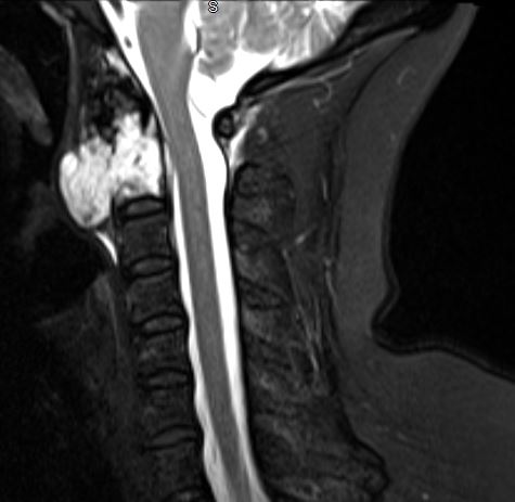
Chordoma on MRI. A sagittal T2 fat-saturation MRI image showing a chordoma arising from the C2 vertebral body and growing exophytically.
Contributed by S Tenny, MD, and C Gillis, MD
(Click Image to Enlarge)
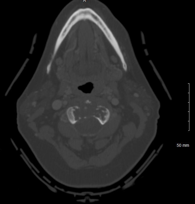
Chordoma on CT. An axial CT of the cervical spine with contrast demonstrates lytic destruction of the C2 vertebral body due to a chordoma.
Contributed by S Tenny, MD, and C Gillis, MD
References
Ulici V, Hart J. Chordoma. Archives of pathology & laboratory medicine. 2022 Mar 1:146(3):386-395. doi: 10.5858/arpa.2020-0258-RA. Epub [PubMed PMID: 34319396]
Desai R, Pelargos PE, Dunn IF. Chordoma: Genetics and Contemporary Management. International journal of molecular sciences. 2024 May 28:25(11):. doi: 10.3390/ijms25115877. Epub 2024 May 28 [PubMed PMID: 38892063]
Choi JH, Ro JY. The 2020 WHO Classification of Tumors of Bone: An Updated Review. Advances in anatomic pathology. 2021 May 1:28(3):119-138. doi: 10.1097/PAP.0000000000000293. Epub [PubMed PMID: 33480599]
Level 3 (low-level) evidenceLee S, Teferi N, Vivanco-Suarez J, Chowdhury A, Glennon S, Kato K, Matern T, Eschbacher KL, Petronek M, Hitchon P. Surgical management of skull base and spinal chordomas: A case series with comprehensive review of the literature. North American Spine Society journal. 2024 Dec:20():100569. doi: 10.1016/j.xnsj.2024.100569. Epub 2024 Nov 4 [PubMed PMID: 39687058]
Level 2 (mid-level) evidenceUllah A, Kenol GS, Lee KT, Yasinzai AQK, Waheed A, Asif B, Khan I, Sharif H, Khan J, Heneidi S, Karki NR, Tareen TK. Chordoma: demographics and survival analysis with a focus on racial disparities and the role of surgery, a U.S. population-based study. Clinical & translational oncology : official publication of the Federation of Spanish Oncology Societies and of the National Cancer Institute of Mexico. 2024 Jan:26(1):109-118. doi: 10.1007/s12094-023-03227-0. Epub 2023 Jun 12 [PubMed PMID: 37306806]
Zhou Y, Hu B, Wu Z, Cheng H, Dai M, Zhang B. The clinical outcomes for chordomas in the cranial base and spine: A single center experience. Medicine. 2019 Jun:98(23):e15980. doi: 10.1097/MD.0000000000015980. Epub [PubMed PMID: 31169734]
Level 2 (mid-level) evidenceGulluoglu S, Turksoy O, Kuskucu A, Ture U, Bayrak OF. The molecular aspects of chordoma. Neurosurgical review. 2016 Apr:39(2):185-96; discussion 196. doi: 10.1007/s10143-015-0663-x. Epub 2015 Sep 12 [PubMed PMID: 26363792]
Yang XR, Ng D, Alcorta DA, Liebsch NJ, Sheridan E, Li S, Goldstein AM, Parry DM, Kelley MJ. T (brachyury) gene duplication confers major susceptibility to familial chordoma. Nature genetics. 2009 Nov:41(11):1176-8. doi: 10.1038/ng.454. Epub 2009 Oct 4 [PubMed PMID: 19801981]
Raygada M, John L, Liu A, Schultz J, Thomas BJ, Bernstein D, Miettinen M, Raffeld M, Xi L, Tyagi M, Aldape K, Glod J, Reilly KM, Widemann BC, Wedekind MF. Germline findings in cancer predisposing genes from a small cohort of chordoma patients. Journal of cancer research and clinical oncology. 2024 May 3:150(5):227. doi: 10.1007/s00432-024-05706-5. Epub 2024 May 3 [PubMed PMID: 38700789]
Stiller CA, Trama A, Serraino D, Rossi S, Navarro C, Chirlaque MD, Casali PG, RARECARE Working Group. Descriptive epidemiology of sarcomas in Europe: report from the RARECARE project. European journal of cancer (Oxford, England : 1990). 2013 Feb:49(3):684-95. doi: 10.1016/j.ejca.2012.09.011. Epub 2012 Oct 15 [PubMed PMID: 23079473]
Murphey MD, Minn MJ, Contreras AL, Koeller KK, Shih RY, Inwards CY, Yamaguchi T. Imaging of spinal chordoma and benign notochordal cell tumor (BNCT) with radiologic pathologic correlation. Skeletal radiology. 2023 Mar:52(3):349-363. doi: 10.1007/s00256-022-04158-7. Epub 2022 Sep 5 [PubMed PMID: 36063190]
McMaster ML, Goldstein AM, Parry DM. Clinical features distinguish childhood chordoma associated with tuberous sclerosis complex (TSC) from chordoma in the general paediatric population. Journal of medical genetics. 2011 Jul:48(7):444-9. doi: 10.1136/jmg.2010.085092. Epub 2011 Jan 25 [PubMed PMID: 21266383]
Dahl NA, Luebbert T, Loi M, Neuberger I, Handler MH, Kleinschmidt-DeMasters BK, Mulcahy Levy JM. Chordoma Occurs in Young Children With Tuberous Sclerosis. Journal of neuropathology and experimental neurology. 2017 Jun 1:76(6):418-423. doi: 10.1093/jnen/nlx032. Epub [PubMed PMID: 28498973]
Shigeta N, Isaka T, Ono K, Tanaka M, Yokose T, Adachi H, Usuba W, Ito H. Primary lung chordoma: a case report. Diagnostic pathology. 2024 Jul 3:19(1):91. doi: 10.1186/s13000-024-01522-0. Epub 2024 Jul 3 [PubMed PMID: 38961474]
Level 3 (low-level) evidenceTirabosco R, Mangham DC, Rosenberg AE, Vujovic S, Bousdras K, Pizzolitto S, De Maglio G, den Bakker MA, Di Francesco L, Kalil RK, Athanasou NA, O'Donnell P, McCarthy EF, Flanagan AM. Brachyury expression in extra-axial skeletal and soft tissue chordomas: a marker that distinguishes chordoma from mixed tumor/myoepithelioma/parachordoma in soft tissue. The American journal of surgical pathology. 2008 Apr:32(4):572-80. doi: 10.1097/PAS.0b013e31815b693a. Epub [PubMed PMID: 18301055]
Suster S, Moran CA. Chordomas of the mediastinum: clinicopathologic, immunohistochemical, and ultrastructural study of six cases presenting as posterior mediastinal masses. Human pathology. 1995 Dec:26(12):1354-62 [PubMed PMID: 8522309]
Level 3 (low-level) evidenceLauer SR, Edgar MA, Gardner JM, Sebastian A, Weiss SW. Soft tissue chordomas: a clinicopathologic analysis of 11 cases. The American journal of surgical pathology. 2013 May:37(5):719-26. doi: 10.1097/PAS.0b013e31827813e7. Epub [PubMed PMID: 23588366]
Level 3 (low-level) evidenceLipplaa A, van der Wal RJP, Krol ADG, Peul WC, Bovée JVMG, Gelderblom H. Incidence and centralization of chordoma in the Netherlands: A nationwide study between 1991 and 2020. Cancer epidemiology. 2024 Apr:89():102527. doi: 10.1016/j.canep.2024.102527. Epub 2024 Jan 25 [PubMed PMID: 38277716]
Caneve P, Schraps N, Möller K, Büyücek S, Lutz F, Chirico V, Viehweger F, Reiswich V, von Bargen C, Kind S, Menz A, Kluth M, Hube-Magg C, Bernreuther C, Sauter G, Marx AH, Simon R, Krech T, Steurer S, Fraune C, Minner S, Gorbokon N, Lennartz M, Burandt E, Rico SD, Freytag M, Luebke AM. Brachyury expression is highly specific for chordoma: A tissue microarray study involving 14,976 cancers from 135 different tumor types and subtypes. Annals of diagnostic pathology. 2025 Jun:76():152448. doi: 10.1016/j.anndiagpath.2025.152448. Epub 2025 Feb 6 [PubMed PMID: 39929118]
Boari N, Gagliardi F, Cavalli A, Gemma M, Ferrari L, Riva P, Mortini P. Skull base chordomas: clinical outcome in a consecutive series of 45 patients with long-term follow-up and evaluation of clinical and biological prognostic factors. Journal of neurosurgery. 2016 Aug:125(2):450-60. doi: 10.3171/2015.6.JNS142370. Epub 2016 Jan 8 [PubMed PMID: 26745472]
Level 2 (mid-level) evidenceVarga PP, Szövérfi Z, Fisher CG, Boriani S, Gokaslan ZL, Dekutoski MB, Chou D, Quraishi NA, Reynolds JJ, Luzzati A, Williams R, Fehlings MG, Germscheid NM, Lazary A, Rhines LD. Surgical treatment of sacral chordoma: prognostic variables for local recurrence and overall survival. European spine journal : official publication of the European Spine Society, the European Spinal Deformity Society, and the European Section of the Cervical Spine Research Society. 2015 May:24(5):1092-101. doi: 10.1007/s00586-014-3728-6. Epub 2014 Dec 23 [PubMed PMID: 25533857]
Soule E, Baig S, Fiester P, Holtzman A, Rutenberg M, Tavanaiepour D, Rao D. Current Management and Image Review of Skull Base Chordoma: What the Radiologist Needs to Know. Journal of clinical imaging science. 2021:11():46. doi: 10.25259/JCIS_139_2021. Epub 2021 Aug 30 [PubMed PMID: 34513210]
Patel S, Nunna RS, Nie J, Ansari D, Chaudhry NS, Mehta AI. Incidence, Management, and Outcomes of Adult Spinal Chordoma Patients in the United States. Global spine journal. 2023 Mar:13(2):334-343. doi: 10.1177/2192568221995155. Epub 2021 Feb 15 [PubMed PMID: 33583227]
Zuckerman SL, Bilsky MH, Laufer I. Chordomas of the Skull Base, Mobile Spine, and Sacrum: An Epidemiologic Investigation of Presentation, Treatment, and Survival. World neurosurgery. 2018 May:113():e618-e627. doi: 10.1016/j.wneu.2018.02.109. Epub 2018 Feb 25 [PubMed PMID: 29486315]
Stacchiotti S, Sommer J, Chordoma Global Consensus Group. Building a global consensus approach to chordoma: a position paper from the medical and patient community. The Lancet. Oncology. 2015 Feb:16(2):e71-83. doi: 10.1016/S1470-2045(14)71190-8. Epub [PubMed PMID: 25638683]
Level 3 (low-level) evidenceBae S, Ehioghae M, Japa JP, Alshomali A, Phung A, Lee L, Hyde J, Lee J, Yoon K, Mesfin A. Nonoperative management of sacral chordomas: A systematic review of the literature. Journal of orthopaedics. 2026 Apr:74():186-194. doi: 10.1016/j.jor.2026.01.007. Epub 2026 Jan 8 [PubMed PMID: 41583620]
Level 1 (high-level) evidencePena-Burgos EM, Torena Lerchundi N, Fuentes-Sánchez J, Tapia-Viñe M, Fernández-Baíllo N, Pozo-Kreilinger JJ. Notochordal cell derived lesions: a 55-year casuistic analysis of 50 cases with radiologic-pathologic correlation in a tertiary referral hospital, and literature review. European spine journal : official publication of the European Spine Society, the European Spinal Deformity Society, and the European Section of the Cervical Spine Research Society. 2024 Sep:33(9):3315-3323. doi: 10.1007/s00586-024-08419-y. Epub 2024 Jul 24 [PubMed PMID: 39048841]
Level 3 (low-level) evidenceYamaguchi T, Iwata J, Sugihara S, McCarthy EF Jr, Karita M, Murakami H, Kawahara N, Tsuchiya H, Tomita K. Distinguishing benign notochordal cell tumors from vertebral chordoma. Skeletal radiology. 2008 Apr:37(4):291-9. doi: 10.1007/s00256-007-0435-y. Epub 2008 Jan 8 [PubMed PMID: 18188556]
Walsh KM. Epidemiology of meningiomas. Handbook of clinical neurology. 2020:169():3-15. doi: 10.1016/B978-0-12-804280-9.00001-9. Epub [PubMed PMID: 32553297]
Adin ME, Woolf G, Hegde R, Elsamadicy AA, Mendel E, Zucconi WB, Pucar D, Aygün N. Sacral tumors: a comprehensive review of imaging, diagnostic challenges, and tumor mimics. Skeletal radiology. 2025 Aug:54(8):1581-1606. doi: 10.1007/s00256-024-04862-6. Epub 2025 Jan 16 [PubMed PMID: 39821683]
Gersey ZC, Abou-Al-Shaar H, Cabral DTF, Alattar AA, Abdallah HM, McDowell MM, Wang EW, Snyderman CH, Choby G, Gardner PA, Zenonos GA. A refined treatment strategy for skull base chordoma: A protocol and management algorithm. Neurosurgical review. 2025 Oct 23:48(1):729. doi: 10.1007/s10143-025-03869-4. Epub 2025 Oct 23 [PubMed PMID: 41125900]
Wang L, Wu Z, Tian K, Wang K, Li D, Ma J, Jia G, Zhang L, Zhang J. Clinical features and surgical outcomes of patients with skull base chordoma: a retrospective analysis of 238 patients. Journal of neurosurgery. 2017 Dec:127(6):1257-1267. doi: 10.3171/2016.9.JNS16559. Epub 2017 Jan 6 [PubMed PMID: 28059654]
Level 2 (mid-level) evidenceHulou MM, Garcia CR, Slone SA, Dugan A, Lei F, Huang B, Pittman T, Villano JL. Comprehensive Review of Cranial Chordomas Using National Databases in the USA. Clinical oncology (Royal College of Radiologists (Great Britain)). 2019 Sep:31(9):e149-e159. doi: 10.1016/j.clon.2019.06.004. Epub 2019 Jul 11 [PubMed PMID: 31303332]
Court C, Briand S, Mir O, Le Péchoux C, Lazure T, Missenard G, Bouthors C. Management of chordoma of the sacrum and mobile spine. Orthopaedics & traumatology, surgery & research : OTSR. 2022 Feb:108(1S):103169. doi: 10.1016/j.otsr.2021.103169. Epub 2021 Dec 7 [PubMed PMID: 34890865]
Zoccali C, Skoch J, Patel AS, Walter CM, Maykowski P, Baaj AA. Residual neurological function after sacral root resection during en-bloc sacrectomy: a systematic review. European spine journal : official publication of the European Spine Society, the European Spinal Deformity Society, and the European Section of the Cervical Spine Research Society. 2016 Dec:25(12):3925-3931 [PubMed PMID: 26914097]
Level 1 (high-level) evidenceRuggieri P, Angelini A, Ussia G, Montalti M, Mercuri M. Surgical margins and local control in resection of sacral chordomas. Clinical orthopaedics and related research. 2010 Nov:468(11):2939-47. doi: 10.1007/s11999-010-1472-8. Epub [PubMed PMID: 20635173]
Xia Y, Papali P, Al-Mistarehi AH, Ahmed AK, Hansen LJ, Meyer C, Gross J, Khan M, Winkle M, Bettegowda C, Witham T, Bydon A, Theodore N, Wolinsky JP, Gokaslan Z, Lo SL, Sciubba DM, Colakoglu S, Lee SH, Redmond KJ, Lubelski D. Long-term functional outcomes and their relationship to nerve root sacrifice after definitive surgery for sacrococcygeal chordoma. Journal of neurosurgery. Spine. 2025 Oct 1:43(4):481-488. doi: 10.3171/2025.3.SPINE241408. Epub 2025 Jul 11 [PubMed PMID: 40644718]
Iannalfi A, Riva G, Ciccone L, Orlandi E. The role of particle radiotherapy in the treatment of skull base tumors. Frontiers in oncology. 2023:13():1161752. doi: 10.3389/fonc.2023.1161752. Epub 2023 Jun 7 [PubMed PMID: 37350949]
Rodrigues ACLF, Tos SM, Shaaban A, Mantziaris G, Trifiletti DM, Sheehan J. Proton beam and carbon ion radiotherapy in skull base chordoma: a systematic review, meta-analysis and meta-regression with trial sequential analysis. Neurosurgical review. 2024 Dec 7:47(1):893. doi: 10.1007/s10143-024-03117-1. Epub 2024 Dec 7 [PubMed PMID: 39644344]
Level 1 (high-level) evidenceMercado CE, Holtzman AL, Rotondo R, Rutenberg MS, Mendenhall WM. Proton therapy for skull base tumors: A review of clinical outcomes for chordomas and chondrosarcomas. Head & neck. 2019 Feb:41(2):536-541. doi: 10.1002/hed.25479. Epub 2018 Dec 7 [PubMed PMID: 30537295]
Level 2 (mid-level) evidenceChhabra AM, Rice SR, Holtzman A, Choi JI, Hasan S, Press RH, Chang J, Halasz L, Tsai HK, Wang CJ, Kabolizadeh P, Gondi V, Hartsell WF, Vora SA, Vargas CE, Simone CB 2nd. Clinical outcomes and toxicities of 100 patients treated with proton therapy for chordoma on the proton collaborative group prospective registry. Radiotherapy and oncology : journal of the European Society for Therapeutic Radiology and Oncology. 2023 Jun:183():109551. doi: 10.1016/j.radonc.2023.109551. Epub 2023 Feb 20 [PubMed PMID: 36813169]
Level 2 (mid-level) evidenceBorkowska AM, Pieńkowski A, Chmiel P, Skóra T, Michalik R, Rutkowski P, Spałek MJ. Long-Term Outcomes of Patients Diagnosed With Sacral Chordoma in a Retrospective Multicenter Study. Cancer control : journal of the Moffitt Cancer Center. 2025 Jan-Dec:32():10732748251323730. doi: 10.1177/10732748251323730. Epub 2025 Mar 11 [PubMed PMID: 40067816]
Level 2 (mid-level) evidenceHoltzman AL, Seidensaal K, Iannalfi A, Kim KH, Koto M, Yang WC, Shiau CY, Mahajan A, Ahmed SK, Trifiletti DM, Peterson JL, Koffler DM, Vallow LA, Hoppe BS, Rutenberg MS. Carbon Ion Radiotherapy: An Evidence-Based Review and Summary Recommendations of Clinical Outcomes for Skull-Base Chordomas and Chondrosarcomas. Cancers. 2023 Oct 17:15(20):. doi: 10.3390/cancers15205021. Epub 2023 Oct 17 [PubMed PMID: 37894388]
Level 2 (mid-level) evidenceGendreau J, Jimenez A, Lozinsky S, Zenonos G, Gardner P, Raza S, Dea N, Gokaslan Z, Choby G, Van Gompel J, Redmond K, Gallia G, Bettegowda C, Rowan N, Kuo CC, Mukherjee D. Radiotherapy After Gross Total Resection of Skull Base Chordoma: A Surveillance, Epidemiology, and End Results Database Analysis of Survival Outcomes. World neurosurgery. 2023 Apr:172():e68-e76. doi: 10.1016/j.wneu.2022.12.012. Epub 2022 Dec 9 [PubMed PMID: 36509323]
Bin-Alamer O, Mallela AN, Palmisciano P, Gersey ZC, Elarjani T, Labib MA, Zenonos GA, Dehdashti AR, Sheehan JP, Couldwell WT, Lunsford LD, Abou-Al-Shaar H. Adjuvant stereotactic radiosurgery with or without postoperative fractionated radiation therapy in adults with skull base chordomas: a systematic review. Neurosurgical focus. 2022 Nov:53(5):E5. doi: 10.3171/2022.8.FOCUS22239. Epub [PubMed PMID: 36321281]
Level 1 (high-level) evidenceGhaith AK, Nguyen R, El-Hajj VG, Montaser A, De Biase G, Ravindran K, Perez-Vega C, Lee SJ, Dominari A, Battistin U, Suárez-Meade P, Moussalem C, Patel NP, Kalani MA, Clarke MJ, Rose PS, Bydon M, Abode-Iyamah K, Akinduro OO. Proton versus photon adjuvant radiotherapy: a multicenter comparative evaluation of recurrence following spinal chordoma resection. Neurosurgical focus. 2024 May:56(5):E9. doi: 10.3171/2024.2.FOCUS23927. Epub [PubMed PMID: 38691864]
Level 2 (mid-level) evidenceSeidensaal K, Froehlke A, Lentz-Hommertgen A, Lehner B, Geisbuesch A, Meis J, Liermann J, Kudak A, Stein K, Uhl M, Tessonnier T, Mairani A, Debus J, Herfarth K. Hypofractionated proton and carbon ion beam radiotherapy for sacrococcygeal chordoma (ISAC): An open label, randomized, stratified, phase II trial. Radiotherapy and oncology : journal of the European Society for Therapeutic Radiology and Oncology. 2024 Sep:198():110418. doi: 10.1016/j.radonc.2024.110418. Epub 2024 Jun 27 [PubMed PMID: 38944346]
Level 1 (high-level) evidenceMattke M, Ohlinger M, Bougatf N, Harrabi S, Wolf R, Seidensaal K, Welzel T, Röder F, Gerum S, Ellerbrock M, Jäkel O, Haberer T, Herfarth K, Uhl M, Debus J. Proton and carbon ion beam treatment with active raster scanning method in 147 patients with skull base chordoma at the Heidelberg Ion Beam Therapy Center-a single-center experience. Strahlentherapie und Onkologie : Organ der Deutschen Rontgengesellschaft ... [et al]. 2023 Feb:199(2):160-168. doi: 10.1007/s00066-022-02002-4. Epub 2022 Sep 23 [PubMed PMID: 36149438]
Biczo A, Sahgal A, Verlaan JJ, Shreyaskumar P, Szoverfi Z, Schultheiss M, Rhines L, Reynolds J, Laufer I, Gasbarrini A, Dea N, Gokaslan Z, Fisher C, Bettegowda C, Boriani S, Hornicek F, Goodwin R, Lazary A. Latest Developments in Targeted Biological Therapies in the Management of Chordoma and Chondrosarcoma. Global spine journal. 2025 Jan:15(1_suppl):120S-131S. doi: 10.1177/21925682241227917. Epub [PubMed PMID: 39801117]
Biermann JS, Hirbe A, Ahlawat S, Bernthal NM, Binitie O, Boles S, Brigman B, Callan AK, Cipriano C, Cranmer LD, Davis J, Donnelly E, Ferguson M, Graham A, Groundland J, Hess M, Hiniker SM, Hoover-Regan ML, Hornick JL, Jonard B, Kuechle JB, Lindskog D, Mayerson JL, McGarry SV, Morris CD, Olson D, Rose PS, Santana VM, Satcher RL, Schwartz H, Shulman RM, Thorpe SW, Wilky BA, Wustrack RL, Yoon J, Hang LE, Jones F, Sansone N, Lyons M. Bone Cancer, Version 2.2025, NCCN Clinical Practice Guidelines In Oncology. Journal of the National Comprehensive Cancer Network : JNCCN. 2025 Apr:23(4):. pii: e250017. doi: 10.6004/jnccn.2025.0017. Epub [PubMed PMID: 40203873]
Level 1 (high-level) evidenceShakil H, Malhotra AK, Essa A, Landry AP, Suppiah S, Sahgal A, Dea N, Zadeh G, Fehlings MG, Witiw CD, Wilson JR. Chordoma incidence, treatment, and survival in the 21st century: a population-based Ontario cohort study. Journal of neurosurgery. 2025 Mar 1:142(3):702-711. doi: 10.3171/2024.6.JNS24426. Epub 2024 Oct 18 [PubMed PMID: 39423422]