 Tools
Tools
 Chiari Malformation Type 1
Introduction
At the end of the 19th century, pathologists Julius Arnold (1835-1915) and Hans Chiari (1851-1916) were the first to describe the condition as a complex clinical and pathological condition involving cerebellar and brainstem deformities in children. Chiari malformations are defined as a spectrum of hindbrain abnormalities involving the cerebellum, brainstem, skull base, and cervical spinal cord. Different subtypes of Chiari malformations have been identified, based on the degree and contents of herniated tissue (see Table. Chiari Malformation Types).[1]
Among the various types, the Chiari type 1 malformation is the most common subtype. Chiari 1 malformations are characterized by the caudal descent of the cerebellar tonsils through the foramen magnum of at least 5 millimeters.[1][2][3] (see Image. Chiari Malformation Types 1 and 2). Clinically, Chiari type 1 malformations may be asymptomatic or manifest from childhood to adulthood with tussive headaches, sleep disordered breathing, scoliosis, and focal neurological deficits. On the other hand, Chiari types 2, 3, and 4 are congenital and present much earlier in life than Chiari type 1 malformations.
Table. Chiari Malformation Types
| Chiari Type | Features |
| Chiari 0 |
|
| Chiari 0.5 |
|
| Chiari 1 |
|
| Chiari 1.5 |
|
| Chiari 2 (Arnold-Chiari malformation) |
|
| Chiari 3 |
|
| Chiari 4 |
|
Etiology
Register For Free And Read The Full Article
 Search engine and full access to all medical articles
- 10 free questions in your specialty
- Free CME/CE Activities
- Free daily question in your email
- Save favorite articles to your dashboard
- Emails offering discounts
Learn more about a Subscription to StatPearls Point-of-Care
Etiology
Several theories have been proposed regarding the etiology of Chiari type 1 malformations, including the molecular genetic theory,[4][5] hydrodynamic pulsion theory, and crowding theory, which suggests that restricted growth in the posterior fossa causes compression of neural tissue and downward herniation of the cerebellar tonsils.[6][7] The tethered cord theory has been proposed, suggesting that caudal traction of the spinal cord results in downward displacement of the cerebellar tonsils;[8] however, other studies have shown that caudal traction of the spinal cord only transmits downward force to the caudal-most pair of dentate ligaments, which is unlikely to result in a Chiari malformation.[9][10][11]
Chiari malformations resulting from hydrocephalus, idiopathic intracranial hypertension, pseudotumor cerebri, intracranial hypotension, and intracranial mass lesions are frequently referred to as acquired Chiari malformations.[12] Furthermore, iatrogenic Chiari 1, due to lumboperitoneal shunting for the treatment of idiopathic intracranial hypertension, has also been reported.[13]
Genetic alterations in chromosomes 2, 9, 14, and 15 have been associated with Chiari malformations.[14][15][16] For instance, a mutation in the NKX2-1 gene (14q13.3), which encodes the protein Nkx-2.1, a member of the homeobox protein family involved in the formation of body structures such as the forebrain during early embryonic development, has recently been identified in a girl diagnosed with a Chiari 1 malformation.[17] Another potential mutation can involve EPAS1 (2p21), which encodes hypoxia-inducible factor 2-alpha (HIF-2-alpha), the major regulator of erythropoietin and implicated in endochondral and intramembranous ossification.[18]
These genetic disorders likely impair the development of the para-axial mesoderm, leading to herniation of the cerebellar tonsils and a small posterior fossa, with a crowded foramen magnum. This is consistent with a proposed association with other mesodermal connective tissue disorders, eg, Ehlers-Danlos syndrome, although the relationship between this condition and Chiari 1 malformation remains unclear.[19] Researchers from the Italian neurological Carlo Besta Institute have recently dissected different pathways (eg, disorders of the RAS/MAPK pathway) that can be found altered in Chiari 1 malformation and defined genetic syndromes.[20] Despite these data, the genetic basis of Chiari 1 malformations requires more evidence.[21] A recent exome sequencing identified the chromodomain genes CHD3 and CHD8 harboring numerous variants associated with CM1.[22] Multiple etiologic subtypes are likely, and Chiari 1 malformation can be heritable.
Epidemiology
Chiari 1 malformation is the most common Chiari malformation. This malformation type is estimated to occur in approximately 1 in 1000 live births, with a slight female-to-male predominance of 1.3 to 1.[23][24] The incidence is likely underestimated since many forms are asymptomatic and manifest clinically only in adulthood. Indeed, neuroimaging data suggest that the clinical condition may be more frequent, with an estimated prevalence of around 1% in the pediatric population.[25][26][27][28][29][30] Other studies have reported up to 3.6% of children undergoing brain and spinal MRIs to have a Chiari malformation.[31] Many children are diagnosed during workup for other conditions, with rates of symptomatic cases ranging from roughly 30% to 60%.[32][31][30]
Pathophysiology
In Chiari 1 malformation, the primary pathophysiology is tonsillar invagination and direct compression of neurological structures within the foramen magnum and upper spinal cord, with obstruction of cerebrospinal fluid (CSF) and, potentially, the development of syringomyelia. Regarding the location of syringomyelia development, the most commonly affected region is the cervical spinal cord, followed by combined cervical and thoracic syringomyelia(reference). Regarding the pathophysiology of syrinx formation, one theory posits that arterial pulsation induces pressure dissociation between the subarachnoid spaces of the cranium and spinal cord. Other theories suggest that syrinx may represent the effect of the anatomical defect of the cerebellar tonsils or the consequence of the altered CSF flow.[33][34]
Hydrocephalus may occur in approximately 10% of patients and usually results from obstruction at the fourth ventricular outlet. Skeletal associations may include platybasia or basilar invagination, Sprengel deformity, and atlantooccipital assimilation. Syndromic associations include Marfan, Ehlers-Danlos, Klippel-Feil, Gorham-Stout disease, Crouzon, and Pfeiffer syndromes.[35][36] Many of these conditions involve alterations of the base of the skull, resulting in a Chiari malformation.
History and Physical
Patients with Chiari 1 malformation are frequently asymptomatic. However, in symptomatic patients, the most common symptoms are pain or headache in the occipital or cervical region (ie, neck pain), reported in up to 88% of patients.[37][38][39] A history of headache or pain may be difficult to elicit in very young children whose symptoms are more likely to manifest as excessive crying, nighttime waking, irritability, reaching for the head and neck, and failure to thrive.[40][41] Younger patients may also present with a shorter duration of symptoms compared to older children and adolescents.[37]
Valsalva maneuvers often exacerbate the headache associated with Chiari type 1 malformation. This should be differentiated from chronic headaches that do not vary with the Valsalva maneuver, which may be attributed to pathologies other than Chiari type 1 malformation (eg, intracranial hypertension).[42][43] Individuals affected by Chiari type 1 malformation have migraine headaches with a prevalence similar to that of the general population, but with an earlier age of onset and a more severe clinical presentation.[44] In addition to headache and pain, a wide spectrum of clinical manifestations may be observed in the malformation. In particular, signs and symptoms are mainly due to direct brainstem compression.[45][46] Clinical features may include:
- Syncopal episodes and sinus bradycardia due to autonomic dysfunction [47]
- Oropharyngeal dysfunction
- Sensorimotor deficits
- Lower cranial nerve deficits [48]
- Sleep-disordered breathing [49][50][51][52]
- Hydrocephalus secondary to fourth ventricular outlet obstruction
- Scoliosis, particularly levoscoliosis
- Ataxia, unsteady gait
- Dizziness
- Tinnitus, hearing loss
- Spasticity
- Bladder and bowel dysfunction
- Nystagmus [48][45][46][53][39][54][55][56][37][48]
Adults and children with Chiari type 1 malformation often present differently. Infants are more likely to present with brainstem dysfunction, sleep apnea, or feeding difficulty due to lower cranial nerve dysfunction.[57] Feeding difficulties are typically due to impairment of the glossopharyngeal and vagus nerves, resulting in an absent gag reflex and hoarseness.[58] Children aged 3 to 5 years often present with syringomyelia, scoliosis, and lower cranial nerve dysfunction.[37] As children age and are better able to communicate, occipital headaches predominate. Adults presenting with Chiari malformations are more likely to present with headaches, paresthesias, and ataxia rather than oropharyngeal and lower cranial nerve dysfunction.[37] Patients may also be at risk for higher rates of psychiatric disorders, including anxiety and depression.[59]
Evaluation
Imaging Studies
Magnetic resonance imaging (MRI) is the primary imaging modality for diagnosing Chiari type 1 malformation, depicting craniocervical junction anatomy and identifying secondary complications (eg, hydrocephalus or syringomyelia).[60] When the patient cannot undergo an MRI, other imaging modalities include CT, myelography, noncontrast CT, and radiographs of the head and neck. In addition to tonsillar descent, "peg-like" or pointed tonsils may be seen on MRI in symptomatic and asymptomatic individuals.[61] Other findings include a cervicomedullary "kink," seen in up to 71% of symptomatic patients (see Image. Chiari Malformation With Holocord Syrinx).[62]
Several measurements are used to identify a Chiari malformation and predict the need for occipital-cervical fusion in complex Chiari cases. The McRae line is defined from the basion to the opisthion and represents the plane of the foramen magnum. Tonsillar descent of greater than 5 mm has typically been used as a cutoff for the diagnosis of Chiari type 1 malformation. However, symptomatic patients with tonsillar descent of less than 5 mm have been reported.[63][64] The greater the degree of cerebellar tonsil descent, the greater the likelihood of symptomatic herniation, with one study reporting a higher likelihood of symptoms with herniation greater than 12 mm.[36] Complex Chiari malformations have been identified as those with brainstem herniation through the foramen magnum, medullary kinking, retroflexion of the odontoid, basilar invagination, atlas assimilation, and an abnormal clival-axial angle.[65] Measurement of the pb-C2 line, C-C2 sagittal vertebral alignment, and clivoaxial angle can help predict future craniocervical instability.[66]
Asymptomatic patients who have incidental tonsillar ectopia may benefit from MRI with CSF flow studies. Positive findings on CSF flow include pulsatile systolic tonsillar descent and CSF flow obstruction at the level of the foramen magnum. These findings can provide further information to help assess whether the patient may benefit from surgery.[67] Chiari type 1 malformation and syringomyelia may also be incidentally identified on routine obstetrical fetal anatomy ultrasounds.[68]
Ancillary Diagnostic Studies
Laboratory studies are not used to diagnose Chiari type 1 malformation; however, these tests may be required for preoperative baseline assessment, including a complete blood count and comprehensive metabolic panel, in addition to other preoperative diagnostic studies (eg, chest x-ray and electrocardiogram). Other ancillary studies that may be considered include:
- Sleep studies: Involves an overnight evaluation in which technicians can monitor breathing, snoring, oxygen saturation, apnea spells, and seizure activity to identify evidence of sleep-disordered breathing [52]
- Scoliosis x-rays: Upright spinal x-rays to evaluate for the presence and degree of scoliosis
- Swallow studies: Fluoroscopy evaluates initiation and completion of swallowing to determine if an abnormality suggests lower brainstem dysfunction
- Brainstem auditory evoked potential: An electrical test to examine the function of the hearing apparatus and brainstem connections to determine if the brainstem is functioning correctly
- Somatosensory evoked potentials: An electrical test of the nerves involved in sensation, giving information about the peripheral nerve, spinal cord, and brain function
Treatment / Management
Generally, management of Chiari type 1 malformation encompasses a combination of conservative and surgical approaches. While variations in surgical indications exist, an international consensus reported a 94.1% agreement for conservative management in children with incidentally diagnosed Chiari malformations with no evidence of syringomyelia. The consensus group also reported 82.4% agreement on surgical intervention for asymptomatic children with an incidental Chiari type 1 malformation and syringomyelia measuring 5 to 8 mm, and for smaller syrinxes with radiographic progression.[69](B3)
Other studies have also proposed conservative treatment in patients with syringomyelia when symptoms are absent or mild.[70] Patients with syringomyelia and sleep apnea or positive Babinski signs are at high risk of clinical deterioration and should likely undergo decompression.[71] Surgical indications in the adult population include symptomatic patients with and without syringomyelia, as well as asymptomatic patients with enlarging syringomyelia.[72] Similarly, the Congress of Neurological Surgeons recommends against performing prophylactic surgery in patients who are asymptomatic without a syrinx.[73](A1)
Conservative Therapy
Medical management in Chiari type 1 malformation is limited to supportive symptomatic (eg, headaches or neck pain) treatment. Non-steroidal anti-inflammatory drugs (NSAIDs), muscle relaxants, and physical support collars may provide symptomatic relief. However, these options offer little improvement for less common symptoms, eg, gait disturbance. In asymptomatic patients without syringomyelia, experts recommend that children be followed with serial MRI of the neural axis until the end of growth.[69](B3)
Surgical Strategies
Surgical treatment is typically reserved for patients who present with severe or worsening symptoms and who go on to have a confirmation of cerebellar descent on imaging and a Chiari malformation obstructing CSF flow confirmed by MRI. Surgery aims to decompress the restricted cervicomedullary junction, restoring CSF flow to optimal levels. The primary approach involves performing a suboccipital craniectomy at the level of the C1 posterior arch (C1 and 2 laminectomy), with or without associated duraplasty.[74]
Thus, this intervention, known as posterior fossa decompression (PFD), is the most widely adopted neurosurgical approach aimed at restoring CSF flow across the foramen magnum. The procedure is performed in up to 99% of the literature.[75] Surgical adjuncts, eg, intraoperative ultrasound, can help guide the decision to perform a duraplasty.[76] Other studies have shown benefit to performing cerebellar tonsillar reduction, either by coagulation or subpial resection.[77][78][79] A minimally invasive procedure allowing resection of submeningeal cerebellar tonsillar herniation followed by reconstruction of cisterna magna without craniectomy has been proposed for addressing Chiari type 1 malformation with syringomyelia.[80] (B2)
Other techniques of minimally invasive decompressions have also been described.[81] Of note, cine MRI has been proposed for guiding the intraoperative decision to realize duraplasty during PFD.[82] Among noninvasive approaches, several methods have been proposed to decompress syringomyelia via distal drainage into the subarachnoid space (eg, syringosubarachnoid shunt).[83] The components of a standard 270-degree circumferential foramen magnum decompression include:
- Suboccipital craniectomy
- C-1 laminectomy
- Resection of the suboccipital ligament
- Lateral decompression with the drilling of one-fifth of the occipital condyles
- Dural opening
- Arachnoid dissection
- Lateral decompression and opening of the foramina of Luschka
- Opening of the fourth ventricle [12]
PFD with duraplasty may be associated with superior long-term symptomatic relief, greater syrinx reduction, lower rates of scoliosis progression, and a lower need for revision decompression compared to decompression without duraplasty.[84][85][86] However, the former may result in a higher incidence of postoperative headache, nausea, and CSF-related complications.[87][88] Ventral brainstem compression can be associated with Chiari 1 malformation due to osseo-ligamentous bony abnormalities at the craniovertebral junction that may result in ventral brainstem compression and instability.(A1)
The initial reduction can be provided by crown halo traction under general anesthesia supplemented by neuromuscular blockade, after which an intraoperative distraction using occipitocervical instrumentation may further reduce the abnormality. Additionally, placement of a fourth ventricle stent has been demonstrated to be a safe and effective treatment.[89][90][91] While intraoperative neuromonitoring can be used,[92] other reports have shown no benefit with increased cost.[93] In cases of abnormal skull base morphology with complex Chiari malformations, neuromonitoring may be helpful.[94](A1)
Differential Diagnosis
Important diagnoses to consider when evaluating patients for Chiari 1 malformation include:
Prognosis
Surgical outcomes are favorable, with 73% of patients improving within 1 year of surgery and 79% at 1 to 3 years of follow-up.[99] More recent studies report improvement in both surgical approaches, including posterior fossa decompression with or without duraplasty.[100] PFD resulted in symptom improvement in about two-thirds of individuals, especially those without associated syringomyelia.[101] Pediatric patients often report higher rates of symptomatic relief compared to adults undergoing Chiari decompressions.[102] High rates of improvement of neurological symptoms after undergoing decompression, expansile duraplasty, and craniocervical fixation procedures have been reported.[103]
Several factors have been identified to be associated with worse outcomes, including hydrocephalus, motor deficits, and surgical complications, while the presence of syringomyelia was identified as a predictor of better outcomes.[104] In cases of persistent syrinx after posterior fossa decompression, asymptomatic patients may be monitored; however, in persistently symptomatic patients, placement of a syringo-subarachnoid shunt is safe and effective.[105][106]
Complications
The most common postoperative complications are CSF leakage and pseudomeningocele formation.[107][108][39] The latter may require revision surgery or shunt insertion.[109] Other complications include aseptic and bacterial meningitis, vertebral artery injury, neurologic deficits, and epidural hematoma formation.[110][111] Regarding surgical approach, in a retrospective analysis, Farber et al found that meningitis occurred more frequently in decompression and duraplasty performed with bovine pericardial xenograft than with allograft.[112]
Postoperative and Rehabilitation Care
Postoperatively, patients may experience headaches and neck pain due to the operation. Neck exercises may be given to aid in the return of full mobility as soon as possible. Patients typically return to work in 4 to 6 weeks, with a follow-up MRI performed around 6 months to 1 year to monitor for syrinx resolution. Full recovery from Chiari 1 malformation surgery may take months or longer. Patients should be instructed to gradually increase activity, avoiding strenuous lifting in the early phase of recovery.
Deterrence and Patient Education
While nothing can be done to prevent congenital Chiari type 1 malformation, patient education plays a significant role in familiarizing the patient with the signs and symptoms of the condition and increasing awareness among close family, friends, and caregivers who may spend significant periods with the patient. Patient education and counseling regarding conservative and surgical management strategies are also critical in the shared decision-making process.
Deterrence and patient education for Chiari 1 malformation focus on early symptom recognition, appropriate monitoring, and informed decision-making about treatment. Clinicians can help reduce morbidity by educating patients and families about common presentations, eg, occipital headaches worsened by Valsalva maneuvers, sleep-disordered breathing, scoliosis, and neurologic changes. Clear guidance on when to seek reassessment—particularly if new symptoms emerge or existing symptoms worsen—supports timely evaluation and may prevent progression of complications, eg, syringomyelia or brainstem dysfunction. Patients who understand the purpose of MRI surveillance and CSF flow studies, and the rationale for conservative versus surgical approaches, are better prepared to participate in shared decision-making.
Education should also emphasize the importance of follow-up, especially for children whose symptoms may evolve with age. Families benefit from reassurance that many individuals remain asymptomatic and require only monitoring, while also receiving balanced information regarding potential surgical indications and expected outcomes. Promoting awareness of associated conditions, eg, sleep apnea or scoliosis, encourages earlier screening and multidisciplinary care. This proactive approach strengthens patient engagement, minimizes delays in diagnosis or treatment, and supports long-term management tailored to each patient’s clinical course.
Pearls and Other Issues
Facts to keep in mind regarding Chiari 1 malformation include the following:
- The Chiari 1 malformation is the most common subtype.
- Many patients can be asymptomatic, and the malformation is diagnosed more commonly in adolescents or adults.
- Patients may present with symptoms of occipital pain or chronic headache; however, they may also present with less specific symptoms, such as oropharyngeal dysfunction or sleep-disordered breathing.
- MRI is the primary investigation of choice in these individuals, allowing complete visualization of the cervicomedullary junction.
- Because algorithms for managing Chiari 1 malformation are not well-established, combining medical and surgical approaches through a multimodal dynamic strategy is mandatory.
- Surgical decompression for symptomatic individuals often significantly improves symptoms and restores neurological function.
Enhancing Healthcare Team Outcomes
Chiari 1 malformation represents the most common form of Chiari-related hindbrain herniation and arises from caudal descent of the cerebellar tonsils of at least 5 mm. Presentations range from asymptomatic cases to headaches, sleep-disordered breathing, scoliosis, cranial nerve dysfunction, or syringomyelia. MRI remains central to diagnosis, with CSF flow studies and ancillary testing guiding evaluation and management. Care may involve conservative monitoring or surgical decompression depending on symptoms, radiographic progression, and associated complications. Because the condition affects neurological, functional, and developmental domains, timely diagnosis and appropriate intervention help prevent deterioration and improve long-term outcomes.
Effective care relies on coordinated interprofessional teamwork among neurosurgeons, neurologists, pediatricians, advanced practitioners, nurses, social workers, therapists, and pharmacists. Clinicians apply diagnostic skills, symptom recognition, and evidence-based decision-making while integrating psychosocial and functional considerations. Regular case discussions, collaborative treatment planning, and consistent communication support safe transitions, monitoring, and follow-up. Patient-centered strategies—such as shared decision-making, streamlined referrals, and efficient information exchange—promote continuity of care and responsive adjustments to evolving clinical needs. Through these coordinated efforts, healthcare professionals enhance patient safety, functional outcomes, and team performance when managing Chiari 1 malformation.
Media
(Click Image to Enlarge)
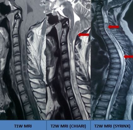
Chiari Malformation With Holocord Syrinx
Contributed by S Munakomi, MD
(Click Image to Enlarge)
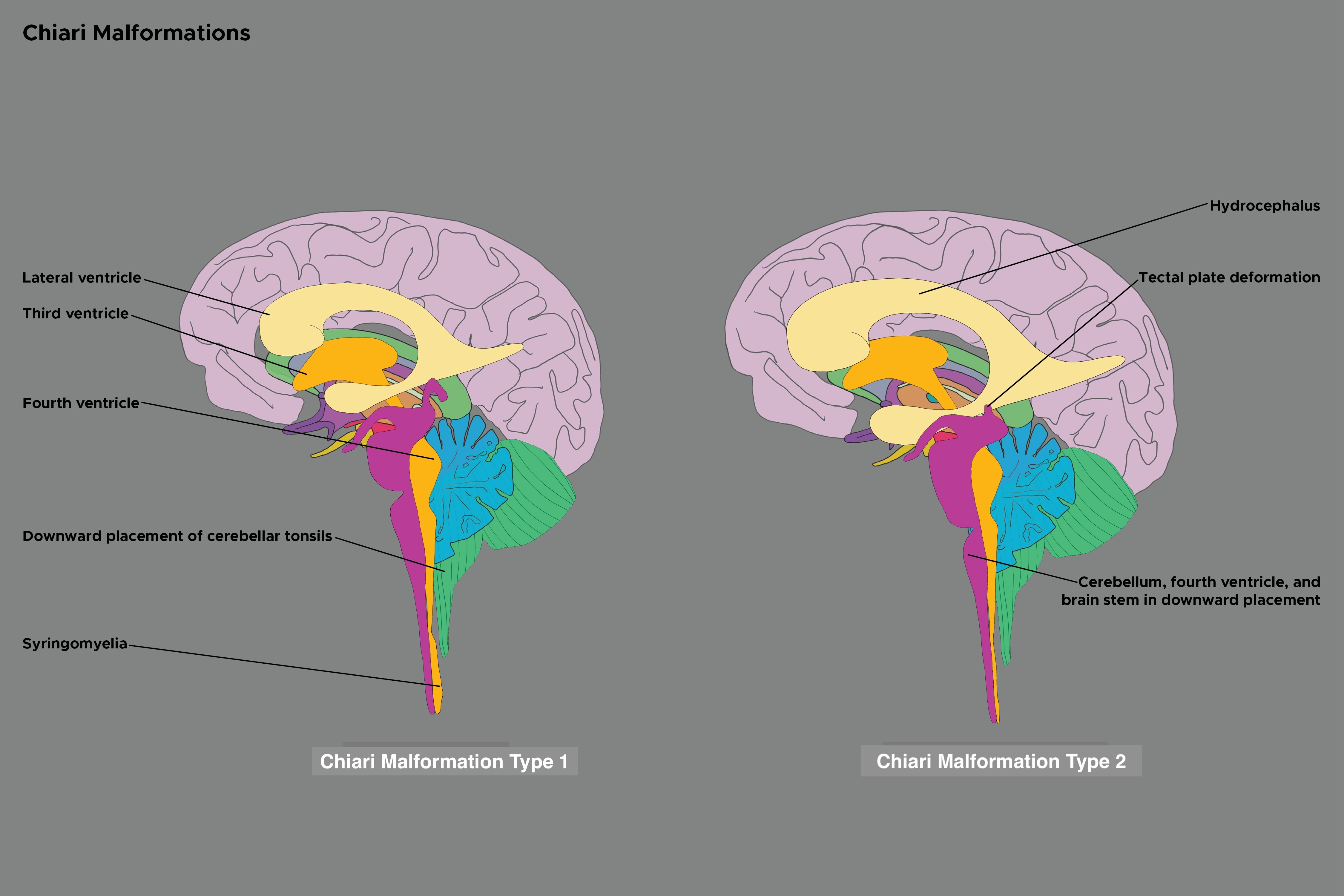
Chiari Malformation Types 1 and 2
Contributed by C Rowe
References
Cools MJ, Wellons JC 3rd, Iskandar BJ. The Nomenclature of Chiari Malformations. Neurosurgery clinics of North America. 2023 Jan:34(1):1-7. doi: 10.1016/j.nec.2022.08.003. Epub 2022 Nov 2 [PubMed PMID: 36424049]
Azahraa Haddad F, Qaisi I, Joudeh N, Dajani H, Jumah F, Elmashala A, Adeeb N, Chern JJ, Tubbs RS. The newer classifications of the chiari malformations with clarifications: An anatomical review. Clinical anatomy (New York, N.Y.). 2018 Apr:31(3):314-322. doi: 10.1002/ca.23051. Epub 2018 Feb 20 [PubMed PMID: 29344999]
Abd-El-Barr MM, Strong CI, Groff MW. Chiari malformations: diagnosis, treatments and failures. Journal of neurosurgical sciences. 2014 Dec:58(4):215-21 [PubMed PMID: 25418275]
Sarnat HB. Disorders of segmentation of the neural tube: Chiari malformations. Handbook of clinical neurology. 2008:87():89-103. doi: 10.1016/S0072-9752(07)87006-0. Epub [PubMed PMID: 18809020]
Sarnat HB. Regional ependymal upregulation of vimentin in Chiari II malformation, aqueductal stenosis, and hydromyelia. Pediatric and developmental pathology : the official journal of the Society for Pediatric Pathology and the Paediatric Pathology Society. 2004 Jan-Feb:7(1):48-60 [PubMed PMID: 15255035]
Marín-Padilla M. Cephalic axial skeletal-neural dysraphic disorders: embryology and pathology. The Canadian journal of neurological sciences. Le journal canadien des sciences neurologiques. 1991 May:18(2):153-69 [PubMed PMID: 2070298]
Marin-Padilla M, Marin-Padilla TM. Morphogenesis of experimentally induced Arnold--Chiari malformation. Journal of the neurological sciences. 1981 Apr:50(1):29-55 [PubMed PMID: 7229658]
Yan H, Han X, Jin M, Liu Z, Xie D, Sha S, Qiu Y, Zhu Z. Morphometric features of posterior cranial fossa are different between Chiari I malformation with and without syringomyelia. European spine journal : official publication of the European Spine Society, the European Spinal Deformity Society, and the European Section of the Cervical Spine Research Society. 2016 Jul:25(7):2202-9. doi: 10.1007/s00586-016-4410-y. Epub 2016 Jan 28 [PubMed PMID: 26821142]
Level 2 (mid-level) evidenceYamada S, Knerium DS, Mandybur GM, Schultz RL, Yamada BS. Pathophysiology of tethered cord syndrome and other complex factors. Neurological research. 2004 Oct:26(7):722-6 [PubMed PMID: 15494111]
Yamada S. Tethered cord syndrome in adults and children. Neurological research. 2004 Oct:26(7):717-8 [PubMed PMID: 15494109]
Truelson SD Jr. What the Index Medicus indexes, and why. Bulletin of the Medical Library Association. 1966 Oct:54(4):329-36 [PubMed PMID: 5922258]
Arnautovic A, Pojskić M, Arnautović KI. Adult Chiari Malformation Type I: Surgical Anatomy, Microsurgical Technique, and Patient Outcomes. Neurosurgery clinics of North America. 2023 Jan:34(1):91-104. doi: 10.1016/j.nec.2022.09.004. Epub [PubMed PMID: 36424069]
Hentati A, Badri M, Bahri K, Zammel I. Acquired Chiari I malformation due to lumboperitoneal shunt: A case report and review of literature. Surgical neurology international. 2019:10():78. doi: 10.25259/SNI-234-2019. Epub 2019 May 10 [PubMed PMID: 31528416]
Level 3 (low-level) evidenceSpeer MC, George TM, Enterline DS, Franklin A, Wolpert CM, Milhorat TH. A genetic hypothesis for Chiari I malformation with or without syringomyelia. Neurosurgical focus. 2000 Mar 15:8(3):E12 [PubMed PMID: 16676924]
Abbott D, Brockmeyer D, Neklason DW, Teerlink C, Cannon-Albright LA. Population-based description of familial clustering of Chiari malformation Type I. Journal of neurosurgery. 2018 Feb:128(2):460-465. doi: 10.3171/2016.9.JNS161274. Epub 2017 Feb 3 [PubMed PMID: 28156254]
Boyles AL, Enterline DS, Hammock PH, Siegel DG, Slifer SH, Mehltretter L, Gilbert JR, Hu-Lince D, Stephan D, Batzdorf U, Benzel E, Ellenbogen R, Green BA, Kula R, Menezes A, Mueller D, Oro' JJ, Iskandar BJ, George TM, Milhorat TH, Speer MC. Phenotypic definition of Chiari type I malformation coupled with high-density SNP genome screen shows significant evidence for linkage to regions on chromosomes 9 and 15. American journal of medical genetics. Part A. 2006 Dec 15:140(24):2776-85 [PubMed PMID: 17103432]
Gonçalves D, Lourenço L, Guardiano M, Castro-Correia C, Sampaio M, Leão M. Chiari Malformation Type I in a Patient with a Novel NKX2-1 Mutation. Journal of pediatric neurosciences. 2019 Jul-Sep:14(3):169-172. doi: 10.4103/jpn.JPN_108_18. Epub 2019 Sep 27 [PubMed PMID: 31649781]
Rosenblum JS, Maggio D, Pang Y, Nazari MA, Gonzales MK, Lechan RM, Smirniotopoulos JG, Zhuang Z, Pacak K, Heiss JD. Chiari Malformation Type 1 in EPAS1-Associated Syndrome. International journal of molecular sciences. 2019 Jun 10:20(11):. doi: 10.3390/ijms20112819. Epub 2019 Jun 10 [PubMed PMID: 31185588]
Milhorat TH, Bolognese PA, Nishikawa M, McDonnell NB, Francomano CA. Syndrome of occipitoatlantoaxial hypermobility, cranial settling, and chiari malformation type I in patients with hereditary disorders of connective tissue. Journal of neurosurgery. Spine. 2007 Dec:7(6):601-9 [PubMed PMID: 18074684]
Level 2 (mid-level) evidenceSaletti V, Viganò I, Melloni G, Pantaleoni C, Vetrano IG, Valentini LG. Chiari I malformation in defined genetic syndromes in children: are there common pathways? Child's nervous system : ChNS : official journal of the International Society for Pediatric Neurosurgery. 2019 Oct:35(10):1727-1739. doi: 10.1007/s00381-019-04319-5. Epub 2019 Jul 30 [PubMed PMID: 31363831]
Capra V, Iacomino M, Accogli A, Pavanello M, Zara F, Cama A, De Marco P. Chiari malformation type I: what information from the genetics? Child's nervous system : ChNS : official journal of the International Society for Pediatric Neurosurgery. 2019 Oct:35(10):1665-1671. doi: 10.1007/s00381-019-04322-w. Epub 2019 Aug 5 [PubMed PMID: 31385087]
Sadler B, Wilborn J, Antunes L, Kuensting T, Hale AT, Gannon SR, McCall K, Cruchaga C, Harms M, Voisin N, Reymond A, Cappuccio G, Brunetti-Pierri N, Tartaglia M, Niceta M, Leoni C, Zampino G, Ashley-Koch A, Urbizu A, Garrett ME, Soldano K, Macaya A, Conrad D, Strahle J, Dobbs MB, Turner TN, Shannon CN, Brockmeyer D, Limbrick DD, Gurnett CA, Haller G. Rare and de novo coding variants in chromodomain genes in Chiari I malformation. American journal of human genetics. 2021 Jan 7:108(1):100-114. doi: 10.1016/j.ajhg.2020.12.001. Epub 2020 Dec 21 [PubMed PMID: 33352116]
Speer MC, Enterline DS, Mehltretter L, Hammock P, Joseph J, Dickerson M, Ellenbogen RG, Milhorat TH, Hauser MA, George TM. Review Article: Chiari Type I Malformation with or Without Syringomyelia: Prevalence and Genetics. Journal of genetic counseling. 2003 Aug:12(4):297-311. doi: 10.1023/A:1023948921381. Epub [PubMed PMID: 26141174]
Langridge B, Phillips E, Choi D. Chiari Malformation Type 1: A Systematic Review of Natural History and Conservative Management. World neurosurgery. 2017 Aug:104():213-219. doi: 10.1016/j.wneu.2017.04.082. Epub 2017 Apr 21 [PubMed PMID: 28435116]
Level 1 (high-level) evidenceHolste KG, Muraszko KM, Maher CO. Epidemiology of Chiari I Malformation and Syringomyelia. Neurosurgery clinics of North America. 2023 Jan:34(1):9-15. doi: 10.1016/j.nec.2022.08.001. Epub 2022 Nov 3 [PubMed PMID: 36424068]
Li Y, Thompson WK, Reuter C, Nillo R, Jernigan T, Dale A, Sugrue LP, ABCD Consortium, Brown J, Dougherty RF, Rauschecker A, Rudie J, Barch DM, Calhoun V, Hagler D, Hatton S, Tanabe J, Marshall A, Sher KJ, Heeringa S, Hermosillo R, Banich MT, Squeglia L, Bjork J, Zucker R, Neale M, Herting M, Sheth C, Huber R, Reeves G, Hettema JM, Howlett KD, Cloak C, Baskin-Sommers A, Rapuano K, Gonzalez R, Karcher N, Laird A, Baker F, James R, Sowell E, Dick A, Hawes S, Sutherland M, Bagot K, Bodurka J, Breslin F, Morris A, Paulus M, Gray K, Hoffman E, Weiss S, Rajapakse N, Glantz M, Nagel B, Ewing SF, Goldstone A, Pfefferbaum A, Prouty D, Rosenberg M, Bookheimer S, Tapert S, Infante M, Jacobus J, Giedd J, Shilling P, Wade N, Uban K, Haist F, Heyser C, Palmer C, Kuperman J, Hewitt J, Cottler L, Isaiah A, Chang L, Edwards S, Ernst T, Heitzeg M, Puttler L, Sripada C, Iacono W, Luciana M, Clark D, Luna B, Schirda C, Foxe J, Freedman E, Mason M, McGlade E, Renshaw P, Yurgelun-Todd D, Albaugh M, Allgaier N, Chaarani B, Potter A, Ivanova M, Lisdahl K, Do E, Maes H, Bogdan R, Anokhin A, Dosenbach N, Glaser P, Heath A, Casey BJ, Gee D, Garavan HP, Dowling G, Brown S. Rates of Incidental Findings in Brain Magnetic Resonance Imaging in Children. JAMA neurology. 2021 May 1:78(5):578-587. doi: 10.1001/jamaneurol.2021.0306. Epub [PubMed PMID: 33749724]
Level 3 (low-level) evidenceDangouloff-Ros V, Roux CJ, Boulouis G, Levy R, Nicolas N, Lozach C, Grevent D, Brunelle F, Boddaert N, Naggara O. Incidental Brain MRI Findings in Children: A Systematic Review and Meta-Analysis. AJNR. American journal of neuroradiology. 2019 Nov:40(11):1818-1823. doi: 10.3174/ajnr.A6281. Epub 2019 Oct 17 [PubMed PMID: 31624116]
Level 1 (high-level) evidenceVernooij MW, Ikram MA, Tanghe HL, Vincent AJ, Hofman A, Krestin GP, Niessen WJ, Breteler MM, van der Lugt A. Incidental findings on brain MRI in the general population. The New England journal of medicine. 2007 Nov 1:357(18):1821-8 [PubMed PMID: 17978290]
Pertl B, Eder S, Stern C, Verheyen S. The Fetal Posterior Fossa on Prenatal Ultrasound Imaging: Normal Longitudinal Development and Posterior Fossa Anomalies. Ultraschall in der Medizin (Stuttgart, Germany : 1980). 2019 Dec:40(6):692-721. doi: 10.1055/a-1015-0157. Epub 2019 Dec 3 [PubMed PMID: 31794996]
Aitken LA, Lindan CE, Sidney S, Gupta N, Barkovich AJ, Sorel M, Wu YW. Chiari type I malformation in a pediatric population. Pediatric neurology. 2009 Jun:40(6):449-54. doi: 10.1016/j.pediatrneurol.2009.01.003. Epub [PubMed PMID: 19433279]
Level 2 (mid-level) evidenceStrahle J, Muraszko KM, Kapurch J, Bapuraj JR, Garton HJ, Maher CO. Chiari malformation Type I and syrinx in children undergoing magnetic resonance imaging. Journal of neurosurgery. Pediatrics. 2011 Aug:8(2):205-13. doi: 10.3171/2011.5.PEDS1121. Epub [PubMed PMID: 21806364]
Ciaramitaro P, Garbossa D, Peretta P, Piatelli G, Massimi L, Valentini L, Migliaretti G, Baldovino S, Roccatello D, Kodra Y, Taruscio D, Interregional Chiari and Syringomyelia Consortium, on behalf of the Interregional Chiari and Syringomyelia Consortium. Syringomyelia and Chiari Syndrome Registry: advances in epidemiology, clinical phenotypes and natural history based on a North Western Italy cohort. Annali dell'Istituto superiore di sanita. 2020 Jan-Mar:56(1):48-58. doi: 10.4415/ANN_20_01_08. Epub [PubMed PMID: 32242535]
Level 3 (low-level) evidenceHeiss JD. Cerebrospinal Fluid Hydrodynamics in Chiari I Malformation and Syringomyelia: Modeling Pathophysiology. Neurosurgery clinics of North America. 2023 Jan:34(1):81-90. doi: 10.1016/j.nec.2022.08.007. Epub 2022 Nov 3 [PubMed PMID: 36424067]
Koyanagi I, Houkin K. Pathogenesis of syringomyelia associated with Chiari type 1 malformation: review of evidences and proposal of a new hypothesis. Neurosurgical review. 2010 Jul:33(3):271-84; discussion 284-5. doi: 10.1007/s10143-010-0266-5. Epub 2010 Jun 8 [PubMed PMID: 20532585]
Ellington M, Francomano CA. Chiari I Malformations and the Heritable Disorders of Connective Tissue. Neurosurgery clinics of North America. 2023 Jan:34(1):61-65. doi: 10.1016/j.nec.2022.09.001. Epub [PubMed PMID: 36424065]
Elster AD, Chen MY. Chiari I malformations: clinical and radiologic reappraisal. Radiology. 1992 May:183(2):347-53 [PubMed PMID: 1561334]
Makoshi Z, Leonard JR. Clinical Manifestations of Chiari I Malformation. Neurosurgery clinics of North America. 2023 Jan:34(1):25-34. doi: 10.1016/j.nec.2022.09.003. Epub [PubMed PMID: 36424061]
Nieswand V, Richter M, Gossrau G. Epidemiology of Headache in Children and Adolescents-Another Type of Pandemia. Current pain and headache reports. 2020 Aug 25:24(10):62. doi: 10.1007/s11916-020-00892-6. Epub 2020 Aug 25 [PubMed PMID: 32840694]
Tubbs RS, Beckman J, Naftel RP, Chern JJ, Wellons JC 3rd, Rozzelle CJ, Blount JP, Oakes WJ. Institutional experience with 500 cases of surgically treated pediatric Chiari malformation Type I. Journal of neurosurgery. Pediatrics. 2011 Mar:7(3):248-56. doi: 10.3171/2010.12.PEDS10379. Epub [PubMed PMID: 21361762]
Level 2 (mid-level) evidenceTubbs RS, Lyerly MJ, Loukas M, Shoja MM, Oakes WJ. The pediatric Chiari I malformation: a review. Child's nervous system : ChNS : official journal of the International Society for Pediatric Neurosurgery. 2007 Nov:23(11):1239-50 [PubMed PMID: 17639419]
Listernick R, Tomita T. Persistent crying in infancy as a presentation of Chiari type I malformation. The Journal of pediatrics. 1991 Apr:118(4 Pt 1):567-9 [PubMed PMID: 2007932]
Level 3 (low-level) evidenceMcGirt MJ, Nimjee SM, Floyd J, Bulsara KR, George TM. Correlation of cerebrospinal fluid flow dynamics and headache in Chiari I malformation. Neurosurgery. 2005 Apr:56(4):716-21; discussion 716-21 [PubMed PMID: 15792510]
Chiapparini L, Saletti V, Solero CL, Bruzzone MG, Valentini LG. Neuroradiological diagnosis of Chiari malformations. Neurological sciences : official journal of the Italian Neurological Society and of the Italian Society of Clinical Neurophysiology. 2011 Dec:32 Suppl 3():S283-6. doi: 10.1007/s10072-011-0695-0. Epub [PubMed PMID: 21800079]
Jokonya L, Makarawo S, Mduluza-Jokonya TL, Ngwende G. Fatal status migrainosus in Chiari 1 malformation. Surgical neurology international. 2019:10():243. doi: 10.25259/SNI_491_2019. Epub 2019 Dec 13 [PubMed PMID: 31893144]
Weig SG, Buckthal PE, Choi SK, Zellem RT. Recurrent syncope as the presenting symptom of Arnold-Chiari malformation. Neurology. 1991 Oct:41(10):1673-4 [PubMed PMID: 1922816]
Selmi F, Davies KG, Weeks RD. Type I Chiari deformity presenting with profound sinus bradycardia: case report and literature review. British journal of neurosurgery. 1995:9(4):543-5 [PubMed PMID: 7576283]
Level 3 (low-level) evidencePetracek LS, Rowe PC. Orthostatic Intolerance and Chiari I Malformation. Neurosurgery clinics of North America. 2023 Jan:34(1):43-54. doi: 10.1016/j.nec.2022.09.002. Epub [PubMed PMID: 36424063]
Albert GW, Menezes AH, Hansen DR, Greenlee JD, Weinstein SL. Chiari malformation Type I in children younger than age 6 years: presentation and surgical outcome. Journal of neurosurgery. Pediatrics. 2010 Jun:5(6):554-61. doi: 10.3171/2010.3.PEDS09489. Epub [PubMed PMID: 20515326]
Okai BK, Jaikumar V, Francois HB, Recker MJ, Reynolds RM. Posterior fossa decompression in patients with Chiari malformation type 1: effect on sleep apnea and follow-up outcomes. Child's nervous system : ChNS : official journal of the International Society for Pediatric Neurosurgery. 2024 Dec:40(12):4075-4082. doi: 10.1007/s00381-024-06623-1. Epub 2024 Oct 4 [PubMed PMID: 39365303]
Jarrell M, Caudill C, Haji F, Leon T, Rozzelle CJ, Maddox MH, Rocque BG. Sleep-disordered breathing in children with Chiari type I malformation. Journal of neurosurgery. Pediatrics. 2024 Oct 1:34(4):393-401. doi: 10.3171/2024.5.PEDS24105. Epub 2024 Jul 12 [PubMed PMID: 38996390]
Marino AC, Farzad F, Jane JA Jr. Chiari I Malformation and Sleep-Disordered Breathing. Neurosurgery clinics of North America. 2023 Jan:34(1):35-41. doi: 10.1016/j.nec.2022.08.005. Epub [PubMed PMID: 36424062]
Abel F, Tahir MZ. Role of sleep study in children with Chiari malformation and sleep disordered breathing. Child's nervous system : ChNS : official journal of the International Society for Pediatric Neurosurgery. 2019 Oct:35(10):1763-1768. doi: 10.1007/s00381-019-04302-0. Epub 2019 Aug 1 [PubMed PMID: 31372737]
Schneider B, Birthi P, Salles S. Arnold-Chiari 1 malformation type 1 with syringohydromyelia presenting as acute tetraparesis: a case report. The journal of spinal cord medicine. 2013 Mar:36(2):161-5. doi: 10.1179/2045772312Y.0000000047. Epub [PubMed PMID: 23809533]
Level 3 (low-level) evidenceTubbs RS, Doyle S, Conklin M, Oakes WJ. Scoliosis in a child with Chiari I malformation and the absence of syringomyelia: case report and a review of the literature. Child's nervous system : ChNS : official journal of the International Society for Pediatric Neurosurgery. 2006 Oct:22(10):1351-4 [PubMed PMID: 16532361]
Level 3 (low-level) evidenceStrahle JM, Taiwo R, Averill C, Torner J, Shannon CN, Bonfield CM, Tuite GF, Bethel-Anderson T, Rutlin J, Brockmeyer DL, Wellons JC, Leonard JR, Mangano FT, Johnston JM, Shah MN, Iskandar BJ, Tyler-Kabara EC, Daniels DJ, Jackson EM, Grant GA, Couture DE, Adelson PD, Alden TD, Aldana PR, Anderson RCE, Selden NR, Baird LC, Bierbrauer K, Chern JJ, Whitehead WE, Ellenbogen RG, Fuchs HE, Guillaume DJ, Hankinson TC, Iantosca MR, Oakes WJ, Keating RF, Khan NR, Muhlbauer MS, McComb JG, Menezes AH, Ragheb J, Smith JL, Maher CO, Greene S, Kelly M, O'Neill BR, Krieger MD, Tamber M, Durham SR, Olavarria G, Stone SSD, Kaufman BA, Heuer GG, Bauer DF, Albert G, Greenfield JP, Wait SD, Van Poppel MD, Eskandari R, Mapstone T, Shimony JS, Dacey RG, Smyth MD, Park TS, Limbrick DD. Radiological and clinical predictors of scoliosis in patients with Chiari malformation type I and spinal cord syrinx from the Park-Reeves Syringomyelia Research Consortium. Journal of neurosurgery. Pediatrics. 2019 Nov 1:24(5):520-527. doi: 10.3171/2019.5.PEDS18527. Epub 2019 Aug 16 [PubMed PMID: 31419800]
Dobkin BH. The adult Chiari malformation. Bulletin of the Los Angeles neurological societies. 1977 Mar:42(1):23-7 [PubMed PMID: 610783]
Level 3 (low-level) evidenceGreenlee JD, Donovan KA, Hasan DM, Menezes AH. Chiari I malformation in the very young child: the spectrum of presentations and experience in 31 children under age 6 years. Pediatrics. 2002 Dec:110(6):1212-9 [PubMed PMID: 12456921]
Level 3 (low-level) evidenceDyste GN, Menezes AH, VanGilder JC. Symptomatic Chiari malformations. An analysis of presentation, management, and long-term outcome. Journal of neurosurgery. 1989 Aug:71(2):159-68 [PubMed PMID: 2746341]
Spina A, Mortini P. The Dark Side of Chiari Malformation. World neurosurgery. 2023 Apr:172():43-45. doi: 10.1016/j.wneu.2023.01.112. Epub 2023 Feb 4 [PubMed PMID: 36746237]
McVige JW, Leonardo J. Neuroimaging and the clinical manifestations of Chiari Malformation Type I (CMI). Current pain and headache reports. 2015 Jun:19(6):18. doi: 10.1007/s11916-015-0491-2. Epub [PubMed PMID: 26017710]
Aboulezz AO, Sartor K, Geyer CA, Gado MH. Position of cerebellar tonsils in the normal population and in patients with Chiari malformation: a quantitative approach with MR imaging. Journal of computer assisted tomography. 1985 Nov-Dec:9(6):1033-6 [PubMed PMID: 4056132]
Spinos E, Laster DW, Moody DM, Ball MR, Witcofski RL, Kelly DL Jr. MR evaluation of Chiari I malformations at 0.15 T. AJR. American journal of roentgenology. 1985 Jun:144(6):1143-8 [PubMed PMID: 3873794]
Pindrik J, McAllister AS, Jones JY. Imaging in Chiari I Malformation. Neurosurgery clinics of North America. 2023 Jan:34(1):67-79. doi: 10.1016/j.nec.2022.08.006. Epub [PubMed PMID: 36424066]
Milhorat TH, Chou MW, Trinidad EM, Kula RW, Mandell M, Wolpert C, Speer MC. Chiari I malformation redefined: clinical and radiographic findings for 364 symptomatic patients. Neurosurgery. 1999 May:44(5):1005-17 [PubMed PMID: 10232534]
Level 2 (mid-level) evidenceBollo RJ, Riva-Cambrin J, Brockmeyer MM, Brockmeyer DL. Complex Chiari malformations in children: an analysis of preoperative risk factors for occipitocervical fusion. Journal of neurosurgery. Pediatrics. 2012 Aug:10(2):134-41. doi: 10.3171/2012.3.PEDS11340. Epub 2012 Jun 22 [PubMed PMID: 22725652]
Bauer DF, Niazi T, Qaiser R, Infinger LK, Vachhrajani S, Ackerman LL, Jackson EM, Jernigan S, Maher CO, Pattisapu JV, Quinsey C, Raskin JS, Rocque BG, Silberstein H. Congress of Neurological Surgeons Systematic Review and Evidence-Based Guidelines for Patients With Chiari Malformation: Diagnosis. Neurosurgery. 2023 Oct 1:93(4):723-726. doi: 10.1227/neu.0000000000002633. Epub 2023 Aug 30 [PubMed PMID: 37646512]
Level 1 (high-level) evidenceHofkes SK, Iskandar BJ, Turski PA, Gentry LR, McCue JB, Haughton VM. Differentiation between symptomatic Chiari I malformation and asymptomatic tonsilar ectopia by using cerebrospinal fluid flow imaging: initial estimate of imaging accuracy. Radiology. 2007 Nov:245(2):532-40 [PubMed PMID: 17890352]
Level 3 (low-level) evidenceIruretagoyena JI, Trampe B, Shah D. Prenatal diagnosis of Chiari malformation with syringomyelia in the second trimester. The journal of maternal-fetal & neonatal medicine : the official journal of the European Association of Perinatal Medicine, the Federation of Asia and Oceania Perinatal Societies, the International Society of Perinatal Obstetricians. 2010 Feb:23(2):184-6. doi: 10.3109/14767050903061769. Epub [PubMed PMID: 19572237]
Level 3 (low-level) evidenceMassimi L, Peretta P, Erbetta A, Solari A, Farinotti M, Ciaramitaro P, Saletti V, Caldarelli M, Canheu AC, Celada C, Chiapparini L, Chieffo D, Cinalli G, Di Rocco F, Furlanetto M, Giordano F, Jallo G, James S, Lanteri P, Lemarchand C, Messing-Jünger M, Parazzini C, Paternoster G, Piatelli G, Poca MA, Prabahkar P, Ricci F, Righini A, Sala F, Sahuquillo J, Stoodley M, Talamonti G, Thompson D, Triulzi F, Zucchelli M, Valentini L, International Experts - Jury of the Chiari & Syringomyelia Consensus Conference, “Milan, November 11–13, 2019”. Diagnosis and treatment of Chiari malformation type 1 in children: the International Consensus Document. Neurological sciences : official journal of the Italian Neurological Society and of the Italian Society of Clinical Neurophysiology. 2022 Feb:43(2):1311-1326. doi: 10.1007/s10072-021-05317-9. Epub 2021 Jun 7 [PubMed PMID: 34097175]
Level 3 (low-level) evidenceEsquivel MFD, Gupta N, O'Brien CA, Bijelić V, Barrowman N, Wilson N, Tu A. Natural history of Chiari I malformation with syrinx and dilatation of the central canal in the pediatric population: the CHEO experience. Child's nervous system : ChNS : official journal of the International Society for Pediatric Neurosurgery. 2024 May:40(5):1443-1448. doi: 10.1007/s00381-023-06273-9. Epub 2024 Jan 12 [PubMed PMID: 38214745]
Yuan C, Du Y, Yao Q, Zhang C, Zhang L, Liu Z, Wang K, Duan W, Wang Z, Wang X, Zeng G, Wu H, Chen Z, Heiss JD, Guan J, Jian FZ. Natural history of Chiari I malformation-syringomyelia: longitudinal cohort study. Journal of neurology, neurosurgery, and psychiatry. 2025 Nov 13:96(12):1204-1214. doi: 10.1136/jnnp-2025-336023. Epub 2025 Nov 13 [PubMed PMID: 40675800]
Ciaramitaro P, Massimi L, Bertuccio A, Solari A, Farinotti M, Peretta P, Saletti V, Chiapparini L, Barbanera A, Garbossa D, Bolognese P, Brodbelt A, Celada C, Cocito D, Curone M, Devigili G, Erbetta A, Ferraris M, Furlanetto M, Gilanton M, Jallo G, Karadjova M, Klekamp J, Massaro F, Morar S, Parker F, Perrini P, Poca MA, Sahuquillo J, Stoodley M, Talamonti G, Triulzi F, Valentini MC, Visocchi M, Valentini L, International Experts Jury of the Chiari Syringomyelia Consensus Conference, Milan, November 11-13, 2019. Diagnosis and treatment of Chiari malformation and syringomyelia in adults: international consensus document. Neurological sciences : official journal of the Italian Neurological Society and of the Italian Society of Clinical Neurophysiology. 2022 Feb:43(2):1327-1342. doi: 10.1007/s10072-021-05347-3. Epub 2021 Jun 15 [PubMed PMID: 34129128]
Level 3 (low-level) evidenceJackson EM, Jernigan S, Raskin JS, Ackerman LL, Infinger LK, Maher CO, Niazi T, Pattisapu JV, Qaiser R, Quinsey C, Rocque BG, Silberstein H, Vachhrajani S, Bauer DF. Congress of Neurological Surgeons Systematic Review and Evidence-Based Guidelines for Patients With Chiari Malformation: Symptoms. Neurosurgery. 2023 Oct 1:93(4):727-730. doi: 10.1227/neu.0000000000002634. Epub 2023 Aug 30 [PubMed PMID: 37646519]
Level 1 (high-level) evidenceSiasios J, Kapsalaki EZ, Fountas KN. Surgical management of patients with Chiari I malformation. International journal of pediatrics. 2012:2012():640127. doi: 10.1155/2012/640127. Epub 2012 Jun 28 [PubMed PMID: 22811732]
Arnautovic A, Splavski B, Boop FA, Arnautovic KI. Pediatric and adult Chiari malformation Type I surgical series 1965-2013: a review of demographics, operative treatment, and outcomes. Journal of neurosurgery. Pediatrics. 2015 Feb:15(2):161-77. doi: 10.3171/2014.10.PEDS14295. Epub 2014 Dec 5 [PubMed PMID: 25479580]
Dherijha MSA, Waqar M, Palin MS, Bukhari S. Foramen magnum decompression in adults with Chiari type 1 malformation: use of intraoperative ultrasound to guide extent of surgery. British journal of neurosurgery. 2024 Aug:38(4):853-856. doi: 10.1080/02688697.2021.1981238. Epub 2021 Oct 4 [PubMed PMID: 34605339]
Hu Y, Zhang M, Duan C, Song D, Wei M, Guo F. A long-term follow-up study of adults with Chiari malformation type I combined with syringomyelia. Frontiers in neurology. 2023:14():1274971. doi: 10.3389/fneur.2023.1274971. Epub 2023 Dec 1 [PubMed PMID: 38107634]
Braga BP, Montgomery EY, Weprin BE, Price AV, Whittemore BA, Pernik MN, Sklar F, De Oliveira Sillero R, Swift DM. Cerebellar tonsil reduction for surgical treatment of Chiari malformation type I in children. Journal of neurosurgery. Pediatrics. 2023 Mar 10:():1-10. doi: 10.3171/2023.1.PEDS22222. Epub 2023 Mar 10 [PubMed PMID: 36905667]
Cai Y, Wang C, Chai S, Li G, Zhang T, Liu Z, Yi D, Chen J, Hu J, Liu K, Xiong N. Prognostic analysis of posterior fossa decompression with or without cerebellar tonsillectomy for Chiari malformation type I: a multicenter retrospective study. Neurosurgical focus. 2023 Mar:54(3):E4. doi: 10.3171/2022.12.FOCUS22626. Epub [PubMed PMID: 36857790]
Level 2 (mid-level) evidenceLou Y, Yang J, Wang L, Chen X, Xin X, Liu Y. The clinical efficacy study of treatment to Chiari malformation type I with syringomyelia under the minimally invasive surgery of resection of Submeningeal cerebellar Tonsillar Herniation and reconstruction of Cisterna magna. Saudi journal of biological sciences. 2019 Dec:26(8):1927-1931. doi: 10.1016/j.sjbs.2019.07.012. Epub 2019 Jul 25 [PubMed PMID: 31885484]
Pan DS, Yang KQ, Li JJ, Wang Z, Zhang JF, Zheng N, Yuan XY, Yu SB, Sui HJ. A new concept and surgical approach for Chiari malformation type I based on the protection and strengthening of the myodural Bridge. Scientific reports. 2025 Mar 19:15(1):9445. doi: 10.1038/s41598-025-92506-7. Epub 2025 Mar 19 [PubMed PMID: 40108288]
Delavari N, Wang AC, Bapuraj JR, Londy F, Muraszko KM, Garton HJL, Maher CO. Intraoperative Phase Contrast MRI Analysis of Cerebrospinal Fluid Velocities During Posterior Fossa Decompression for Chiari I Malformation. Journal of magnetic resonance imaging : JMRI. 2020 May:51(5):1463-1470. doi: 10.1002/jmri.26953. Epub 2019 Oct 30 [PubMed PMID: 31667928]
Amarouche M, Minichini V, Davis H, Giamouriadis A, Bassi S. Syringosubarachnoid shunt: insertion technique. British journal of neurosurgery. 2023 Jun:37(3):476-479. doi: 10.1080/02688697.2019.1700407. Epub 2019 Dec 18 [PubMed PMID: 31852253]
Akbari SHA, Yahanda AT, Ackerman LL, Adelson PD, Ahmed R, Albert GW, Aldana PR, Alden TD, Anderson RCE, Bauer DF, Bethel-Anderson T, Bierbrauer K, Brockmeyer DL, Chern JJ, Couture DE, Daniels DJ, Dlouhy BJ, Durham SR, Ellenbogen RG, Eskandari R, Fuchs HE, Grant GA, Graupman PC, Greene S, Greenfield JP, Gross NL, Guillaume DJ, Hankinson TC, Heuer GG, Iantosca M, Iskandar BJ, Jackson EM, Jallo GI, Johnston JM, Kaufman BA, Keating RF, Khan NR, Krieger MD, Leonard JR, Maher CO, Mangano FT, McComb JG, McEvoy SD, Meehan T, Menezes AH, Muhlbauer MS, O'Neill BR, Olavarria G, Ragheb J, Selden NR, Shah MN, Shannon CN, Shimony JS, Smyth MD, Stone SSD, Strahle JM, Tamber MS, Torner JC, Tuite GF, Tyler-Kabara EC, Wait SD, Wellons JC, Whitehead WE, Park TS, Limbrick DD. Complications and outcomes of posterior fossa decompression with duraplasty versus without duraplasty for pediatric patients with Chiari malformation type I and syringomyelia: a study from the Park-Reeves Syringomyelia Research Consortium. Journal of neurosurgery. Pediatrics. 2022 Jul 1:30(1):39-51. doi: 10.3171/2022.2.PEDS21446. Epub 2022 Apr 15 [PubMed PMID: 35426814]
Sadler B, Skidmore A, Gewirtz J, Anderson RCE, Haller G, Ackerman LL, Adelson PD, Ahmed R, Albert GW, Aldana PR, Alden TD, Averill C, Baird LC, Bauer DF, Bethel-Anderson T, Bierbrauer KS, Bonfield CM, Brockmeyer DL, Chern JJ, Couture DE, Daniels DJ, Dlouhy BJ, Durham SR, Ellenbogen RG, Eskandari R, Fuchs HE, George TM, Grant GA, Graupman PC, Greene S, Greenfield JP, Gross NL, Guillaume DJ, Hankinson TC, Heuer GG, Iantosca M, Iskandar BJ, Jackson EM, Jea AH, Johnston JM, Keating RF, Khan N, Krieger MD, Leonard JR, Maher CO, Mangano FT, Mapstone TB, McComb JG, McEvoy SD, Meehan T, Menezes AH, Muhlbauer M, Oakes WJ, Olavarria G, O'Neill BR, Ragheb J, Selden NR, Shah MN, Shannon CN, Smith J, Smyth MD, Stone SSD, Tuite GF, Wait SD, Wellons JC, Whitehead WE, Park TS, Limbrick DD, Strahle JM. Extradural decompression versus duraplasty in Chiari malformation type I with syrinx: outcomes on scoliosis from the Park-Reeves Syringomyelia Research Consortium. Journal of neurosurgery. Pediatrics. 2021 Aug 1:28(2):167-175. doi: 10.3171/2020.12.PEDS20552. Epub 2021 Jun 18 [PubMed PMID: 34144521]
Hale AT, Adelson PD, Albert GW, Aldana PR, Alden TD, Anderson RCE, Bauer DF, Bonfield CM, Brockmeyer DL, Chern JJ, Couture DE, Daniels DJ, Durham SR, Ellenbogen RG, Eskandari R, George TM, Grant GA, Graupman PC, Greene S, Greenfield JP, Gross NL, Guillaume DJ, Heuer GG, Iantosca M, Iskandar BJ, Jackson EM, Johnston JM, Keating RF, Leonard JR, Maher CO, Mangano FT, McComb JG, Meehan T, Menezes AH, O'Neill B, Olavarria G, Park TS, Ragheb J, Selden NR, Shah MN, Smyth MD, Stone SSD, Strahle JM, Wait SD, Wellons JC, Whitehead WE, Shannon CN, Limbrick DD, Park-Reeves Syringomyelia Research Consortium Investigators. Factors associated with syrinx size in pediatric patients treated for Chiari malformation type I and syringomyelia: a study from the Park-Reeves Syringomyelia Research Consortium. Journal of neurosurgery. Pediatrics. 2020 Jun 1:25(6):629-639. doi: 10.3171/2020.1.PEDS19493. Epub 2020 Mar 6 [PubMed PMID: 32114543]
Durham SR, Fjeld-Olenec K. Comparison of posterior fossa decompression with and without duraplasty for the surgical treatment of Chiari malformation Type I in pediatric patients: a meta-analysis. Journal of neurosurgery. Pediatrics. 2008 Jul:2(1):42-9. doi: 10.3171/PED/2008/2/7/042. Epub [PubMed PMID: 18590394]
Level 1 (high-level) evidenceGiannakaki V, Wildman J, Thejasvin K, Pexas G, Nissen J, Ross N, Mitchell P. Foramen Magnum Decompression for Chiari Malformation Type 1: Is There a Superior Surgical Technique? World neurosurgery. 2023 Feb:170():e784-e790. doi: 10.1016/j.wneu.2022.11.119. Epub 2022 Nov 29 [PubMed PMID: 36455845]
Taylor C, Hall S, Mathad N, Wahab S, Waters R, Chakraborty A. Fourth ventricular stenting in foramen magnum decompression for type 1 Chiari malformations with syringomyelia. Surgical neurology international. 2025:16():271. doi: 10.25259/SNI_40_2025. Epub 2025 Jul 4 [PubMed PMID: 40837313]
Han RK, Medina MP, Giantini-Larsen AM, Chae JK, Cruz A, Garton ALA, Greenfield JP. Fourth ventricular subarachnoid stent for Chiari malformation type I-associated persistent syringomyelia. Neurosurgical focus. 2023 Mar:54(3):E10. doi: 10.3171/2022.12.FOCUS22633. Epub [PubMed PMID: 36857783]
Sun P, Zhou M, Liu Y, Du J, Zeng G. Fourth ventricle stent placement for treatment of type I Chiari malformation in children. Child's nervous system : ChNS : official journal of the International Society for Pediatric Neurosurgery. 2023 Mar:39(3):671-676. doi: 10.1007/s00381-022-05793-0. Epub 2022 Dec 26 [PubMed PMID: 36572815]
Pattisapu JV, Ackerman LL, Infinger LK, Maher CO, Quinsey C, Rocque BG, Silberstein H, Jackson EM, Jernigan S, Niazi T, Qaiser R, Raskin JS, Vachhrajani S, Bauer DF. Congress of Neurological Surgeons Systematic Review and Evidence-Based Guidelines for Patients With Chiari Malformation: Surgical Interventions. Neurosurgery. 2023 Oct 1:93(4):731-735. doi: 10.1227/neu.0000000000002635. Epub 2023 Aug 30 [PubMed PMID: 37646504]
Level 1 (high-level) evidencePatel MS, Botterbush KS, Lackland TN, Prim M, Al-Hammadi N, Shorey M, Mattei TA, Mercier PA. Intraoperative neuromonitoring is not a useful adjunct for Chiari malformation decompressive surgery: a cost-benefit and legal analysis. Child's nervous system : ChNS : official journal of the International Society for Pediatric Neurosurgery. 2025 Jan 31:41(1):96. doi: 10.1007/s00381-024-06738-5. Epub 2025 Jan 31 [PubMed PMID: 39890655]
Sen RD, Martinez V, Eaton J, Holdefer RN, Ellenbogen RG. Intraoperative neuromonitoring for pediatric Chiari decompression: when is it useful? Neurosurgical focus. 2023 Mar:54(3):E9. doi: 10.3171/2022.12.FOCUS22632. Epub [PubMed PMID: 36857781]
Houk JL, Amrhein TJ, Gray L, Malinzak MD, Kranz PG. Differentiation of Chiari malformation type 1 and spontaneous intracranial hypotension using objective measurements of midbrain sagging. Journal of neurosurgery. 2022 Jun 1:136(6):1796-1803. doi: 10.3171/2021.6.JNS211010. Epub 2021 Oct 29 [PubMed PMID: 34715671]
Mea E, Chiapparini L, Leone M, Franzini A, Messina G, Bussone G. Chronic daily headache in the adults: differential diagnosis between symptomatic Chiari I malformation and spontaneous intracranial hypotension. Neurological sciences : official journal of the Italian Neurological Society and of the Italian Society of Clinical Neurophysiology. 2011 Dec:32 Suppl 3():S291-4. doi: 10.1007/s10072-011-0698-x. Epub [PubMed PMID: 21822702]
D'Amico A, Giammalva GR, Furlanis GM, Emanuelli E, Maugeri R, Baro V, Denaro L. Acquired Chiari type I malformation: a late and misunderstood supratentorial over-drainage complication. Child's nervous system : ChNS : official journal of the International Society for Pediatric Neurosurgery. 2023 Feb:39(2):343-351. doi: 10.1007/s00381-022-05775-2. Epub 2022 Dec 1 [PubMed PMID: 36454310]
Kumar R, Cutsforth-Gregory JK, Brinjikji W. Cerebrospinal Fluid Leaks, Spontaneous Intracranial Hypotension, and Chiari I Malformation. Neurosurgery clinics of North America. 2023 Jan:34(1):185-192. doi: 10.1016/j.nec.2022.08.012. Epub 2022 Nov 3 [PubMed PMID: 36424060]
Greenberg JK, Yarbrough CK, Radmanesh A, Godzik J, Yu M, Jeffe DB, Smyth MD, Park TS, Piccirillo JF, Limbrick DD. The Chiari Severity Index: a preoperative grading system for Chiari malformation type 1. Neurosurgery. 2015 Mar:76(3):279-85; discussion 285. doi: 10.1227/NEU.0000000000000608. Epub [PubMed PMID: 25584956]
Yu QS, Li T, Wan M, Zhang L, Qiao GY, Yu XG, Yin YH. Clinical outcome of different surgical approaches for symptomatic Chiari malformation without syringomyelia: a 13-year retrospective study. Neurosurgical review. 2025 Jun 5:48(1):485. doi: 10.1007/s10143-025-03640-9. Epub 2025 Jun 5 [PubMed PMID: 40471450]
Level 2 (mid-level) evidenceDe Vlieger J, Dejaegher J, Van Calenbergh F. Multidimensional, patient-reported outcome after posterior fossa decompression in 79 patients with Chiari malformation type I. Surgical neurology international. 2019:10():242. doi: 10.25259/SNI_377_2019. Epub 2019 Dec 13 [PubMed PMID: 31893143]
Mortazavi A, Almeida ND, Hofmann K, Davidson L, Rotter J, Phan TN, Tsering D, Maxwell C, Karunakaran J, Veznedaroglu E, Caputy AJ, Heiss JD, Sandhu FA, Myseros JS, Oluigbo C, Magge SN, Shields DC, Rosner MK, Chatain GP, Keating RF. Multicenter comparison of Chiari malformation type I presentation in children versus adults. Journal of neurosurgery. Pediatrics. 2024 Jul 1:34(1):49-56. doi: 10.3171/2023.12.PEDS22366. Epub 2024 Feb 23 [PubMed PMID: 38394661]
Nishikawa M, Yamagata T, Naito K, Kunihiro N, Sakamoto H, Hara M, Ohata K, Goto T. Surgical Management of Chiari Malformation Type I Associated with Syringomyelia: Outcome of Surgeries Based on the New Classification and Study of Cerebrospinal Fluid Dynamics. Journal of clinical medicine. 2022 Aug 4:11(15):. doi: 10.3390/jcm11154556. Epub 2022 Aug 4 [PubMed PMID: 35956170]
El-Hajj VG, Öhlén E, Sandvik U, Pettersson-Segerlind J, Atallah E, Jabbour P, Bydon M, Daniels DJ, Elmi-Terander A, Edström E. Long-term outcomes following posterior fossa decompression in pediatric patients with Chiari malformation type 1, a population-based cohort study. Acta neurochirurgica. 2024 Nov 18:166(1):460. doi: 10.1007/s00701-024-06332-3. Epub 2024 Nov 18 [PubMed PMID: 39551853]
Szuflita NS, Phan TN, Boulter JH, Keating RF, Myseros JS. Nonoperative management of enlarging syringomyelia in clinically stable patients after decompression of Chiari malformation type I. Journal of neurosurgery. Pediatrics. 2021 Jul 1:28(1):28-33. doi: 10.3171/2020.12.PEDS20621. Epub 2021 May 21 [PubMed PMID: 34020421]
Aydin L, Dereli D, Kartum TA, Sirinoglu D, Sahin B, Eksi MS, Musluman AM, Yilmaz A. Management of Persistent Syringomyelia in Patients Operated for Chiari Malformation Type 1. World neurosurgery. 2024 Feb:182():e360-e368. doi: 10.1016/j.wneu.2023.11.109. Epub 2023 Nov 25 [PubMed PMID: 38013110]
Atchley TJ, Alam Y, Gross E, Laskay NMB, Estevez-Ordonez D, Schmalz PGR, Fisher WS. Incidence and management of postoperative pseudomeningocele and cerebrospinal fluid leak after Chiari malformation type I decompression. Neurosurgical focus. 2023 Mar:54(3):E8. doi: 10.3171/2022.12.FOCUS22631. Epub [PubMed PMID: 36857794]
Rocque BG, Oakes WJ. Surgical Treatment of Chiari I Malformation. Neurosurgery clinics of North America. 2015 Oct:26(4):527-31. doi: 10.1016/j.nec.2015.06.010. Epub 2015 Aug 4 [PubMed PMID: 26408062]
Lei ZW, Wu SQ, Zhang Z, Han Y, Wang JW, Li F, Shu K. Clinical Characteristics, Imaging Findings and Surgical Outcomes of Chiari Malformation Type I in Pediatric and Adult Patients. Current medical science. 2018 Apr:38(2):289-295. doi: 10.1007/s11596-018-1877-2. Epub 2018 Apr 30 [PubMed PMID: 30074187]
Poca MA, Lopez-Bermeo D, Moncho D, Ferre A, Sanchez-Montañez A, Mestres O, Galve S, Sahuquillo J. Surgical Outcomes in Chiari 1 and Chiari 1.5 Malformation Treated by Posterior Fossa Reconstruction: A Comprehensive Analysis of 110 Pediatric Cases and Literature Review. Journal of clinical medicine. 2024 Jun 30:13(13):. doi: 10.3390/jcm13133852. Epub 2024 Jun 30 [PubMed PMID: 38999418]
Level 3 (low-level) evidenceTubbs RS, Smyth MD, Wellons JC 3rd, Oakes WJ. Distances from the atlantal segment of the vertebral artery to the midline in children. Pediatric neurosurgery. 2003 Dec:39(6):330-4 [PubMed PMID: 14734868]
Farber H, McDowell MM, Alhourani A, Agarwal N, Friedlander RM. Duraplasty Type as a Predictor of Meningitis and Shunting After Chiari I Decompression. World neurosurgery. 2018 Oct:118():e778-e783. doi: 10.1016/j.wneu.2018.07.050. Epub 2018 Jul 17 [PubMed PMID: 30026145]