 Tools
Tools
 Cheyne-Stokes Respirations
Introduction
Cheyne-Stokes respiration (CSR) is a form of periodic breathing characterized by cyclical episodes of central apnea or central hypopnea alternating with a crescendo–decrescendo pattern of hyperventilation.[1] John Cheyne and William Stokes first described this pattern in the early 19th century.[1] CSR has received renewed clinical attention because of its strong association with heart failure and stroke, which are 2 of the leading causes of morbidity and mortality worldwide.[2][3]
Unlike obstructive sleep apnea, which may contribute causally to cardiovascular disease development or progression, CSR is most often a consequence of underlying cardiac or neurologic disease rather than an independent driver of cardiovascular disease.[3] In patients with heart failure, the presence of CSR is associated with worse clinical outcomes, including a higher risk of arrhythmias, hospitalization, and sudden cardiac death.[2][4][5] Dynamic measures of desaturation severity during CSR cycles have been proposed as a more sensitive marker of mortality risk in heart failure than static indices such as the apnea-hypopnea index alone.[5]
In intensive care settings, electrocardiogram-derived CSR and periodic breathing have been associated with cardiorespiratory arrest, underscoring the prognostic significance of this breathing pattern beyond the outpatient population with heart failure. Please see StatPearls' companion reference, "Central Sleep Apnea," for further information. Beyond heart failure and stroke, CSR is increasingly recognized in patients with high cervical spinal cord injury, in whom impairment of chemoreflex and central respiratory control produces a comparable phenotype of breathing instability, as detailed in the Etiology section.[6] Despite improved mechanistic understanding of CSR, treatment options remain limited and primarily target the underlying condition rather than the breathing pattern itself.[1][4] The article reviews the taxonomy, etiology, epidemiology, pathophysiology, clinical evaluation, and treatment of CSR, with an emphasis on subspecialty-level detail relevant to pulmonology, critical care, sleep medicine, and cardiology practice.
Etiology
Register For Free And Read The Full Article
 Search engine and full access to all medical articles
- 10 free questions in your specialty
- Free CME/CE Activities
- Free daily question in your email
- Save favorite articles to your dashboard
- Emails offering discounts
Learn more about a Subscription to StatPearls Point-of-Care
Etiology
CSR is a specific form of periodic breathing defined by waxing and waning airflow or tidal volume in a crescendo–decrescendo pattern occurring between central apneas or central hypopneas.[5] Precise terminology matters because several timing parameters, not just apnea and hyperpnea, carry diagnostic and prognostic meaning.[5] Accordingly, accurate characterization of each component is essential for identifying the CSR waveform.
Central apnea is defined as the cessation of airflow for 10 seconds or longer in the absence of respiratory effort, as indicated by the absence of thoracoabdominal excursion. The article uses this operative definition, which is consistent with the American Academy of Sleep Medicine scoring manual and the companion StatPearls article on central sleep apnea. Please see StatPearls' companion reference, "Central Sleep Apnea," for further information. Central hypopnea is defined as a reduction in airflow of 30% or greater for 10 seconds or longer without associated respiratory effort and accompanied by either oxygen desaturation of 3% or greater or an arousal, according to current American Academy of Sleep Medicine scoring criteria.[7]
In addition to apnea and hypopnea, 4 additional timing parameters define the CSR waveform:
- Apnea length is the duration of central apnea or hypopnea.[5]
- Ventilation length is the hyperventilation phase is the duration of the compensatory hyperpnea that follows. In CSR, the ventilation phase is typically longer than the apnea phase, which helps distinguish CSR from other central sleep apnea phenotypes in which the apnea phase predominates.[5]
- Circulation time is the interval between the end of the apneic or hypopneic event and the nadir of the corresponding oxygen desaturation. This interval approximates the lung-to-chemoreceptor transit time and is prolonged in heart failure due to reduced cardiac output and increased blood volume in the circulatory reservoir.[5]
- Cycle length, also called ventilatory cycle time, is the total duration from the onset of 1 apnea to the onset of the next and equals the apnea length plus the ventilation length. In CSR, the cycle length is typically 40 to 90 seconds.[5]
Diagnostically, Cheyne-Stokes breathing is scored when both of the following criteria are met:
- Three or more consecutive central apneas or hypopneas separated by a crescendo–decrescendo change in breathing amplitude, with a cycle length of 40 seconds or longer
- Five or more such events per hour recorded over 2 hours or longer of monitoring.[7]
Cheyne-Stokes breathing should be distinguished from other central sleep apnea phenotypes, including:
- Idiopathic central sleep apnea
- High-altitude periodic breathing
- Treatment-emergent central sleep apnea
- Drug-induced central sleep apnea (eg, opioid-related central sleep apnea)
In these phenotypes, the crescendo–decrescendo pattern is absent or attenuated, the cycle length is typically shorter, and the apnea phase tends to predominate over the ventilatory phase rather than the reverse. Please see StatPearls' companion reference, "Central Sleep Apnea," for further information.
Heart Failure
CSR is most prevalent in patients with heart failure and reduced ejection fraction, although the breathing pattern also occurs in patients with heart failure and preserved ejection fraction, typically less frequently and with lower severity.[8] Risk factors for CSR in patients with heart failure include male sex, older age, atrial fibrillation, elevated left ventricular filling pressures, prolonged circulation time, and advanced cardiac remodeling.[3] The mechanistic relationship between these risk factors and CSR, particularly the role of circulation time defined above, is discussed further under Pathophysiology.
Stroke
CSR occurs after acute cerebrovascular events in an estimated 20% to 40% of patients, most often during the acute or subacute phase.[2][4] The breathing pattern typically improves over subsequent weeks because central respiratory control mechanisms recover, although CSR may persist in patients with extensive brainstem or bilateral hemispheric injury.[2][4]
Cervical Spinal Cord Injury
Although heart failure and stroke dominate traditional teaching, high-level cervical spinal cord injury is now recognized as a distinct and highly prevalent cause of CSR and central sleep-disordered breathing.[6] Results from a study by Sankari et al found that sleep-disordered breathing is common across the chronic spinal cord injury population, with central-predominant events concentrated in patients with cervical-level injury and obstructive-predominant events more common in patients with thoracic-level injury.[6] Results from a related study by the same group demonstrated that tetraplegia is an independent risk factor for central sleep apnea, implicating loss of segmental sympathetic and diaphragmatic and accessory muscle control, along with heightened peripheral chemoreflex sensitivity, as part of the mechanism.[9] Consequently, clinicians should include CSR in the differential diagnosis for patients with high cervical spinal cord injury who have excessive daytime sleepiness or witnessed apneas, even without heart failure or stroke.[6][9]
Other and Less Common Contributors
Renal failure
Sleep apnea, including a central component, is common in both chronic kidney disease and end-stage kidney disease, independent of comorbid heart failure. Results from a systematic review and meta-analysis of available prevalence studies confirmed that sleep apnea syndrome, including central events, is substantially more common in populations with chronic kidney disease or end-stage kidney disease than in the general population, with prevalence rising further among patients dependent on dialysis.[10] The proposed mechanism converges on the same apneic-threshold framework discussed under Pathophysiology.
Fluid overload and altered chemoreflex sensitivity in uremia narrow the carbon dioxide reserve, and intermittent hemodialysis can acutely destabilize ventilatory control through rapid shifts in fluid status and carbon dioxide buffering capacity.[10] Renal failure is also specifically recognized as a cause of the short-cycle-length variant of central sleep apnea, defined by a cycle length of less than 45 seconds. Recognition of this shorter cycle length should prompt reconsideration of a diagnosis of classic CSR, which typically has a cycle length of 45 to 75 seconds, in patients with concurrent advanced chronic kidney disease.[11]
Atrial fibrillation, independent of heart failure
Atrial fibrillation is associated with central sleep apnea even in patients without structural heart disease. Results from a case-control study comparing patients with idiopathic central sleep apnea, obstructive sleep apnea, and no sleep apnea, all without congestive heart failure, found that the prevalence of atrial fibrillation was markedly higher in the idiopathic central sleep apnea group.[12] The increased prevalence was not explained by hypertension or nocturnal desaturation, both of which were more closely associated with the obstructive sleep apnea group.[12]
These findings suggested that atrial fibrillation and central sleep apnea share a bidirectional relationship independent of ventricular dysfunction, plausibly through loss of normal respiratory modulation of atrioventricular nodal conduction and heart rate variability. Sinus rhythm preserves this modulation, whereas atrial fibrillation alters it.[12] Similar to central sleep apnea associated with renal failure, central sleep apnea associated with atrial fibrillation often manifests with a shorter cycle length than classic CSR associated with heart failure and reduced ejection fraction, providing a useful bedside discriminator when interpreting a polysomnography tracing.[11]
Epidemiology
The true population prevalence of CSR is not well established because most epidemiologic data derive from clinic-referred cohorts with heart failure, stroke, cervical spinal cord injury, chronic kidney disease, or atrial fibrillation rather than from unselected community samples.[13] CSR is considered uncommon in the general population but is common in several specific clinical populations.
Heart Failure
Reported CSR prevalence in patients with heart failure ranges from roughly 25% to 50%, with substantial variation driven by differences in diagnostic technology, including in-laboratory polysomnography compared with home testing, scoring criteria, and heart failure severity and etiology across studies.[3] Prevalence is consistently higher in patients with heart failure and reduced ejection fraction than in patients with heart failure and preserved ejection fraction and rises further with worsening ejection fraction and New York Heart Association functional class.[3][8] Heart failure affects more than 6 million adults in the United States; therefore, even the low end of these prevalence estimates represents a large absolute burden of undiagnosed CSR.[14] CSR is more common in older adults and men, mirroring the sex and age distributions of heart failure and reduced ejection fraction, as well as the chemoreflex hypersensitivity discussed under Pathophysiology.[3][15]
Stroke
Prevalence estimates of 20% to 40% during the acute poststroke period reflect the transient, injury-related nature of CSR after stroke.[2][4] The breathing pattern frequently resolves during recovery, in contrast to the more persistent phenotype observed in patients with chronic heart failure.[2][4]
Cervical Spinal Cord Injury
Results from dedicated cohort studies found sleep-disordered breathing with a central-predominant pattern in a substantial proportion of patients with chronic cervical spinal cord injury, and tetraplegia independently increases risk.[6][9] However, Cheyne-Stokes breathing specifically, rather than central sleep apnea more broadly, has not been quantified as systematically in this population as it has been in patients with heart failure. The limited epidemiologic characterization represents a recognized evidence gap rather than evidence of low prevalence, reflecting the lag between clinical recognition and systematic study.[6]
Renal Failure and Atrial Fibrillation
Sleep apnea, including a central component, is substantially more prevalent in patients with chronic kidney disease or end-stage kidney disease than in the general population, independent of comorbid heart failure.[10][12] Central sleep apnea prevalence is likewise elevated in patients with atrial fibrillation without structural heart disease.[10][12] However, neither association has been characterized as extensively for the Cheyne-Stokes breathing phenotype specifically, rather than central sleep apnea broadly, as has the association with heart failure. This limitation represents another important evidence gap that should be acknowledged.
Pathophysiology
CSR is initiated and sustained by instability in ventilatory control, centered on oscillations in arterial partial pressure of carbon dioxide (PaCO2) around the apneic threshold.[3] Under normal waking conditions, ventilation is governed jointly by cortical (behavioral) control and metabolic (chemoreceptor-mediated) control. Central and peripheral chemoreceptors respond to PaCO2 through negative feedback to match ventilation to metabolic demand, and loss of stability in this homeostatic loop during sleep is the central organizing concept for all forms of central sleep apnea, not CSR alone.[16] The foundational framework for quantifying ventilatory instability was developed by Dempsey and colleagues, who characterized ventilatory responsiveness to carbon dioxide below eupnea as the key determinant of breathing stability during sleep.[17]
Apneic Threshold and Carbon Dioxide Reserve
Resting PaCO2 typically ranges from 40 to 45 mm Hg.[3] The apneic threshold is the PaCO2 level below which the brainstem respiratory pattern generator transiently ceases firing, resulting in central apnea. In most individuals, the apneic threshold is approximately 4 to 6 mm Hg below resting PaCO2.[3][18]
Dempsey termed the gap between eupneic PaCO2 and the apneic threshold the carbon dioxide reserve and demonstrated that this reserve, together with plant gain, defined as the change in PaCO2 produced by a given change in ventilation, and controller gain, defined as chemoreceptor responsiveness above eupnea, determines whether the ventilatory control system remains stable or becomes prone to oscillation.[17] In patients with heart failure, chronically elevated ventilation, driven by pulmonary congestion, augmented chemoreflex sensitivity, and vagal afferent stimulation, lowers resting PaCO2 and narrows the carbon dioxide reserve. The narrowed reserve leaves little margin before a transient hyperventilatory overshoot decreases PaCO2 below the apneic threshold.[17][18]
The Oscillatory Cycle
At the transition from wakefulness to nonrapid eye movement sleep, loss of the wakefulness drive to breathe unmasks the hypocapnia-sensitive apneic threshold.[17][18] Arousals or sleep-stage transitions during stage N1 or N2 sleep produce a transient hyperventilatory overshoot, and the resulting decrease in PaCO2 below the threshold triggers central apnea.[3] PaCO2 then rises during the apnea until it exceeds the recruitment threshold, restimulating the chemoreceptors and producing a compensatory hyperpnea that completes one cycle of the crescendo–decrescendo pattern.[17][18]
In patients with heart failure, prolonged circulation time, reflecting delayed transit of blood and chemoreceptor-sensed blood gas information from the lungs to the carotid bodies and central chemoreceptors, lengthens the overall cycle and amplifies the oscillation because ventilatory corrections remain delayed relative to the true blood gas state.[3][5] This mechanism explains the definitions of circulation time and cycle length introduced earlier. Reduced cardiac output and an expanded circulatory reservoir delay transmission of the blood gas signal from the lungs to the chemoreceptors, thereby extending the cycle length toward the 45- to 90-second range characteristic of CSR rather than the shorter cycles observed in other central sleep apnea phenotypes.[5]
Cervical Spinal Cord Injury
The mechanistic pattern in patients with high cervical spinal cord injury differs from that in patients with heart failure in the process that narrows the carbon dioxide reserve, although both conditions converge on the same final common pathway.[6][9] Loss of supraspinal modulation of sympathetic outflow and altered peripheral afferent traffic appear to increase peripheral chemoreflex sensitivity in patients with cervical spinal cord injury. This mechanism narrows the carbon dioxide reserve via a pathway distinct from the volume overload and augmented chemosensitivity observed in heart failure, but the endpoint is identical: once PaCO2 crosses the apneic threshold during sleep, central apnea occurs regardless of the upstream mechanism that narrows the reserve.[9][18] The shared pathway provides the rationale for studying acetazolamide and buspirone, agents that widen the carbon dioxide reserve or reduce chemoreflex gain, in both central sleep apnea associated with heart failure and that associated with spinal cord injury. The pharmacologic evidence is discussed in the Treatment section.[6][9]
Toxicokinetics
Certain medications and substances can alter central respiratory control and may contribute to central sleep apnea or periodic breathing patterns resembling Cheyne-Stokes respiration. Drugs that depress the central nervous system, particularly opioids such as methadone and morphine, can suppress brainstem respiratory centers and reduce respiratory responsiveness to changes in carbon dioxide and oxygen levels. Reduced chemosensitivity may destabilize ventilatory control and promote episodes of central apnea.[14]
Other substances that depress central nervous system activity can produce similar effects. Sedative-hypnotic medications may suppress respiratory drive during sleep, whereas alcohol can further impair central respiratory regulation through generalized central nervous system depression. Anesthetic agents may also alter ventilatory control and respiratory rhythm generation by modulating neuronal activity within brainstem respiratory networks and reducing chemoreceptor responsiveness to carbon dioxide and hypoxia. In susceptible individuals, particularly patients with underlying heart failure or neurologic disease, these pharmacologic effects may exacerbate ventilatory instability and contribute to periodic breathing or central sleep apnea patterns.[16][19]
History and Physical
Patients with heart failure who develop CSR may present with dyspnea, fatigue, reduced exercise tolerance, and nocturnal awakenings, reflecting the underlying cardiac dysfunction rather than the breathing pattern itself.[2] A history of ischemic heart disease, prior myocardial infarction, longstanding hypertension, atrial fibrillation, or cardiomyopathy raises the pretest likelihood of CSR.[2] Additionally, patients may report symptoms of sleep disruption, including excessive daytime sleepiness or nonrestorative sleep. Bed partners may observe the characteristic cyclical breathing pattern, with alternating hyperventilation and apnea, which is often a more reliable historical clue than the patient's own symptom report because subjective sleepiness in CSR associated with heart failure does not reliably correlate with objective severity on polysomnography.[2][4] CSR may also occur in patients with acute stroke or other neurologic disorders affecting central respiratory control, and the breathing pattern may be witnessed during sleep or periods of decreased consciousness.[2][4]
In patients presenting with sleep-disordered breathing, obstructive sleep apnea should remain in the differential diagnosis. A history of obesity, loud snoring, or witnessed apneas favors obstructive sleep apnea over CSR, and a formal sleep evaluation is required to distinguish the 2 definitively.[2] When the typical risk factors for heart failure or stroke are absent, a careful medication history is essential because central nervous system depressants, particularly opioids and sedative-hypnotic medications, can independently produce central sleep apnea or periodic breathing.[14][19] Clinicians should also actively consider CSR or central sleep apnea in patients with high cervical spinal cord injury who have any of these symptoms, even without heart failure or stroke, because of the elevated baseline prevalence in this population.[6][9]
On examination, findings typically reflect the underlying disease rather than CSR itself. Findings associated with heart failure may include jugular venous distention, pulmonary crackles, peripheral edema, or an S3 gallop, whereas patients with neurologic disease may have focal neurologic deficits or altered mental status.[2][6] Patients with cervical spinal cord injury may have findings consistent with the level and completeness of the injury, including a diaphragmatic-predominant breathing pattern or orthostatic hypotension.[2][6]
Targeted History and Examination to Distinguish CSR From Mimics
Because several other abnormal breathing patterns can be confused with CSR, targeted questions and examination maneuvers can help narrow the differential diagnosis before polysomnography is obtained:
- Witnessed breathing pattern: Ask the bed partner specifically whether the crescendo–decrescendo pattern of waxing-and-waning breathing is present, rather than an irregular cluster-and-pause pattern, which suggests Biot or ataxic breathing, or continuous deep breathing without apneic pauses, which suggests Kussmaul respiration.[20] A history of sudden breathing-pattern change in a hospitalized patient should raise concern for Biot breathing caused by an acute medullary process rather than CSR, which typically evolves more gradually with progression of heart failure or stroke.[20]
- Respiratory effort during the pause: Respiratory effort is best assessed by direct observation or a bed partner's description of chest and abdominal wall movement during the apneic phase. Persistent effort against an obstructed airway favors obstructive sleep apnea, whereas absent effort favors a central process, including CSR or another central sleep apnea phenotype. Please see StatPearls' companion reference, "Central Sleep Apnea," for further information.[21]
- Signs and symptoms of elevated intracranial pressure or brainstem injury: Headache, vomiting, altered consciousness, or focal cranial nerve findings in a patient with an irregular breathing pattern should prompt urgent neuroimaging to exclude a structural brainstem or medullary lesion presenting with Biot or apneustic breathing. Clinicians should not reflexively attribute these findings to CSR.[20]
- Symptoms of metabolic acidosis: Polyuria, polydipsia, abdominal pain, or a history of diabetes mellitus or advanced renal failure in a patient with deep, rapid, continuous breathing should prompt evaluation for Kussmaul respiration caused by diabetic ketoacidosis or uremia rather than CSR. The absence of apneic pauses is the key discriminator and should be assessed explicitly rather than assumed.[20]
- Level of consciousness and toxidrome screening: In a patient with complete, abrupt cessation of breathing without preceding oscillation, clinicians should directly assess for a history of severe neurologic injury, overdose, or another cause of primary apnea. Relevant findings may include empty medication containers, a known ingestion, or changes in the Glasgow Coma Scale score because primary apnea lacks the cyclic hyperventilation that precedes true CSR apneas.[20]
- Level and completeness of spinal cord injury: For patients with known cervical spinal cord injury, ask specifically about the injury level and completeness because these characteristics directly influence the pretest probability of a central-predominant rather than obstructive-predominant sleep-disordered breathing pattern. High cervical injuries involving C1 through C4 carry the greatest risk of central apnea.[6][9]
Evaluation
Cheyne-Stokes respiration, or Cheyne-Stokes breathing, is characterized by alternating central apnea or hypopnea and hyperventilation in a crescendo–decrescendo pattern, most often occurring during nonrapid eye movement sleep, particularly stages N1 and N2, but occasionally observed during wakefulness in patients with more severe disease. The pattern is confirmed on polysomnography (PSG), which demonstrates cyclic variation in airflow and respiratory effort accompanied by oscillating oxygen saturation (see Image. Polysomnography of Cheyne-Stokes Respiration With Labeled Apnea Length, Circulation Time). In mechanically ventilated individuals, the same cyclical pattern may be visible as oscillations in tidal volume and minute ventilation on ventilator waveforms. Please see StatPearls' companion reference, "Central Sleep Apnea," for further information.
The apnea-hyperpnea cycle length is typically 45 to 90 seconds, longer than that observed in other central sleep apnea phenotypes. Additionally, the pattern may become more pronounced in the supine position and during sleep-stage transitions.[5] Diagnostic scoring follows the American Academy of Sleep Medicine and International Classification of Sleep Disorders, Third Edition, criteria detailed in the Etiology and Taxonomy sections: 3 or more consecutive central apneas or hypopneas separated by a crescendo–decrescendo pattern with a cycle length of 40 seconds or longer and 5 or more such events per hour over 2 or more hours of recording. Please see StatPearls' companion reference, "Central Sleep Apnea," for further information.[AASM. The AASM Manual for the Scoring of Sleep and Associated Events]
In-laboratory PSG remains the diagnostic gold standard because it provides simultaneous data on airflow, respiratory effort, oxygen saturation, and sleep staging, which are needed to confidently distinguish central from obstructive events. Historically, home sleep apnea testing with conventional portable monitors had limited ability to distinguish central from obstructive events and could underestimate the severity of central sleep apnea in patients with chronic heart failure, making in-laboratory PSG essentially mandatory for suspected Cheyne-Stokes respiration.[22] However, peripheral arterial tonometry–based ambulatory devices have changed this approach.
Results from a multicenter validation study of the WatchPAT system across 11 sleep centers found that an enhanced peripheral arterial tonometry algorithm, incorporating systolic upstroke pulse-wave analysis together with dedicated snoring and body-position sensors, correlated strongly with the apnea-hypopnea index derived from PSG and meaningfully differentiated central from obstructive events, a capability lacking in earlier-generation peripheral arterial tonometry devices.[23] Consequently, ambulatory peripheral arterial tonometry–based testing may be considered a reasonable option for detecting central sleep apnea or Cheyne-Stokes respiration in appropriately selected patients, particularly when access to in-laboratory polysomnography is limited. However, in-laboratory PSG remains preferred when the diagnosis is uncertain, concurrent sleep-staging detail is clinically necessary, or treatment titration, such as positive airway pressure titration, is planned concurrently.
Please see StatPearls' companion reference, "Central Sleep Apnea," for further information.[23] Because CSR frequently signals underlying cardiovascular or neurologic disease, evaluation should extend beyond the sleep study to include assessment of the suspected underlying condition. Additional evaluation may include echocardiography and assessment for heart failure, neuroimaging, or autonomic assessment specific to spinal cord injury, depending on the clinical presentation.[2][3][6]
Treatment / Management
The cornerstone of CSR treatment is optimization of the underlying disorder. Guideline-directed medical therapy for heart failure can reduce CSR severity and, more importantly, improve patient-centered outcomes.[24] The 2025 American Academy of Sleep Medicine clinical practice guideline on the treatment of central sleep apnea in adults reinforces this principle while updating recommendations for the devices and medications discussed below.[25](A1)
Positive Airway Pressure Therapy
Continuous positive airway pressure (CPAP) and adaptive servoventilation remain the 2 principal noninvasive positive airway pressure modalities for CSR.[26] CPAP can stabilize breathing by improving oxygenation and reducing pulmonary congestion. Results from studies showed improvements in nocturnal oxygenation, left ventricular function, and functional capacity among patients with heart failure and CSR, although CPAP has not consistently improved long-term survival.[24](B3)
Adaptive Servoventilation: Updated Evidence
Results from the 2015 Treatment of Sleep-Disordered Breathing With Predominant Central Sleep Apnea by Adaptive Servo Ventilation in Patients With Heart Failure trial found that adaptive servoventilation increased cardiovascular mortality in patients with heart failure and reduced ejection fraction, a left ventricular ejection fraction of 45% or less, and predominant central sleep apnea, leading to a contraindication in that specific population that remains standard teaching.[27] However, the contraindication does not establish that adaptive servoventilation is unsafe in all patients with central sleep apnea associated with heart failure. Two recent developments have meaningfully updated the evidence, which follow:
- Adaptive Servo Ventilation for Sleep Apnea in Heart Failure trial, ADVENT-HF: Results from this 2024 multicenter, multinational phase 3 randomized controlled trial, which enrolled patients with heart failure and reduced ejection fraction and either obstructive or central sleep apnea, found that adaptive servoventilation had no effect on the primary composite cardiovascular outcome or mortality while effectively and safely eliminating sleep-disordered breathing. The trial used a different adaptive servoventilation algorithm and device generation than the Treatment of Sleep-Disordered Breathing With Predominant Central Sleep Apnea by Adaptive Servo Ventilation in Patients With Heart Failure (SERVE-HF) trial and did not reproduce the earlier excess-mortality signal.[28]
- 2025 European Respiratory Society and European Sleep Research Society statement: A joint statement reviewing cardiovascular end points, echocardiographic parameters, exercise capacity, and patient-reported outcomes across newer trials and registries concluded that current-generation adaptive servoventilation has no negative cardiovascular impact and may improve patient-reported outcomes in patients receiving optimized treatment for heart failure across a range of ejection fractions, including an ejection fraction of 30% to 45%.[29] (A1)
Collectively, the current evidence supports a more nuanced approach than a categorical contraindication to adaptive servoventilation. Adaptive servoventilation should still be avoided in patients who resemble the population enrolled in the SERVE-HF trial, including those with heart failure and reduced ejection fraction, a left ventricular ejection fraction of 45% or less, and central sleep apnea as the predominant sleep-disordered breathing pattern treated with earlier-generation algorithms, according to the original American Academy of Sleep Medicine safety signal. However, results from the newer trial and statement suggested that clinicians should not reflexively extrapolate this restriction to all patients with central sleep apnea associated with heart failure or to current-generation devices.[25][28][30](A1)
Supplemental Oxygen
Nocturnal supplemental oxygen reduces central apneas and improves oxygen saturation in some patients with central sleep apnea associated with heart failure.[26](B3)
Transvenous Phrenic Nerve Stimulation
Transvenous phrenic nerve stimulation is a United States Food and Drug Administration–approved option for central sleep apnea, including CSR. An implanted device stimulates the phrenic nerve during sleep to stabilize respiratory rhythm, and trial results showed improvements in the apnea-hypopnea index, oxygen saturation, and sleep quality.[31] The 2025 American Academy of Sleep Medicine guideline now explicitly recommends transvenous phrenic nerve stimulation over no stimulation in adults with primary central sleep apnea or central sleep apnea due to heart failure. The guideline characterizes this recommendation as conditional due to the treatment's invasiveness, cost, and limited accessibility.[25](A1)
Pharmacologic Therapy
Pharmacologic options for CSR remain limited by a sparse evidence base, and no agent currently has United States Food and Drug Administration approval specifically for this indication.[32] Results from a 2023 Cochrane systematic review that pooled 4 crossover randomized controlled trials and 1 parallel randomized controlled trial involving 68 participants evaluated acetazolamide, buspirone, theophylline, and triazolam for central sleep apnea and found insufficient evidence to support routine use of any pharmacologic agent. Accordingly, the medications discussed below are physiologically plausible and mechanistically relevant but have not changed routine clinical practice.[32](A1)
- Acetazolamide, a carbonic anhydrase inhibitor, induces mild metabolic acidosis that lowers the apneic threshold, thereby widening the carbon dioxide reserve described under Pathophysiology and reducing the frequency of central apneas.[33] Results from a double-blind, prospective crossover study in patients with stable systolic heart failure showed that a single bedtime dose improved central sleep apnea and related daytime symptoms.[33] Results from studies in patients with chronic cervical spinal cord injury also showed that acetazolamide reduced susceptibility to hypocapnic central sleep apnea, consistent with the shared apneic-threshold mechanism discussed in Pathophysiology.[29]
- Buspirone is an anxiolytic with respiratory-stimulant properties mediated through effects on chemoreflex sensitivity. Results from a randomized crossover trial in patients with heart failure found that buspirone reduced chemoreflex gain and central apneas. Results from a separate study in patients with chronic spinal cord injury found reduced susceptibility to hypocapnic central sleep apnea.[34][35]
- Theophylline is a nonselective adenosine receptor antagonist; theophylline reduced central apneas during sleep, as shown in findings from older case reports and small studies. However, a narrow therapeutic index and the risk of cardiac arrhythmias limit its use, and theophylline is not considered standard treatment. The evidence supports considering theophylline only as 1 of several agents evaluated in small studies, rather than as the sole pharmacologic option.[36][37] (A1)
None of these agents is recommended as first-line treatment. Mechanistic support and limited clinical evidence restrict their potential role to second-line or adjunctive treatment pending results from larger trials. This cautious framing is consistent with both the Cochrane review and the 2025 American Academy of Sleep Medicine (AASM) guideline.[25][32](A1)
Management Algorithm
The following sequence summarizes the suggested approach:
- Confirm diagnosis and phenotype: Use in-laboratory PSG or validated peripheral arterial tonometry–based home sleep apnea testing in appropriately selected individuals to confirm that the AASM criteria for CSR or Cheyne-Stokes breathing are met. Identify the underlying cause, including heart failure, stroke, cervical spinal cord injury, renal failure, atrial fibrillation, or opioid or central nervous system depressant use.
- Treat the underlying condition first: Optimize guideline-directed medical therapy for heart failure, treat stroke and support neurologic recovery, and discontinue or reduce causative central nervous system depressants when clinically feasible.
- Treat persistent CSR: When CSR persists despite optimized treatment of the underlying disorder, initiate a trial of continuous positive airway pressure as first-line therapy. When continuous positive airway pressure is ineffective or not tolerated, and the patient does not resemble the population enrolled in the SERVE-HF trial, consider adaptive servoventilation according to current AASM guidance. When adaptive servoventilation is contraindicated or unsuitable, consider supplemental oxygen, transvenous phrenic nerve stimulation, or bilevel positive airway pressure in spontaneous-timed mode. Acetazolamide or buspirone may be considered as adjunctive treatment in refractory disease after an explicit discussion of the limited evidence.
- Reassess periodically: Reevaluate CSR as the underlying disease changes because severity may vary with heart failure status and recovery after stroke. Treatment efficacy, tolerance, and ongoing indication should also be reviewed at follow-up evaluations.
Differential Diagnosis
CSR represents a specific form of periodic breathing. Periodic breathing is a broader term describing oscillations in ventilation that may occur in several conditions, including high-altitude periodic breathing, heart failure, and central sleep apnea syndromes (see Table. Differential Diagnosis of Cheyne-Stokes Respiration). Not all forms of periodic breathing meet the diagnostic criteria for CSR, which requires a characteristic cyclic crescendo–decrescendo pattern of tidal volume followed by central apnea, with a typical cycle length of approximately 45 to 90 seconds.
Please see StatPearls' companion reference, "Abnormal Respirations," for further information. Definitive differentiation between these breathing patterns often requires polysomnography combined with clinical correlation, particularly in patients with underlying cardiovascular or neurologic disease. Recognition of these respiratory patterns is important because the underlying causes, prognosis, and treatment strategies differ significantly from those of CSR.[20]
Other Central Sleep Apnea Syndromes
Other forms of central sleep apnea may present with recurrent apneas during sleep but lack the characteristic crescendo–decrescendo ventilation pattern observed in CSR. Examples include idiopathic central sleep apnea, high-altitude periodic breathing, and central sleep apnea associated with medications, particularly chronic opioid use. PSG may demonstrate central apneas in these disorders, which typically have shorter cycle lengths and more irregular ventilatory patterns.
Obstructive sleep apnea
Obstructive sleep apnea must also be distinguished from CSR. In obstructive sleep apnea, airflow ceases because of upper airway obstruction despite persistent respiratory effort, whereas CSR involves central apneas characterized by the absence of respiratory effort. PSG differentiates these conditions because respiratory effort belts can identify continued thoracoabdominal effort during obstructive events.
Biot breathing
Biot breathing, also known as ataxic breathing, is characterized by irregular clusters of breaths of similar depth followed by unpredictable periods of apnea. Unlike CSR, Biot breathing does not demonstrate the gradual crescendo–decrescendo pattern of ventilation characteristic of CSR. Biot breathing is typically associated with medullary injury, increased intracranial pressure, or brainstem lesions.
Kussmaul respiration
Kussmaul respiration is a pattern of deep, rapid, and labored breathing. The breathing pattern develops as a compensatory response to metabolic acidosis, most commonly from diabetic ketoacidosis or severe renal failure. Unlike CSR, Kussmaul breathing is continuous and does not alternate with periods of apnea.
Apneustic breathing
Apneustic breathing is characterized by prolonged inspiratory pauses followed by inadequate expiration. This breathing pattern is most commonly associated with damage to the pontine respiratory centers, particularly in patients with pontine stroke or traumatic brain injury. Apneustic breathing lacks the cyclical waxing and waning ventilation characteristic of CSR.
Primary apnea
Primary apnea refers to complete cessation of breathing without preceding ventilatory oscillations. The pattern may occur in severe neurologic injury, drug intoxication, or central sleep apnea syndromes. Unlike CSR, primary apnea does not demonstrate cyclic hyperventilation preceding the apneic event.
Table. Differential Diagnosis of Cheyne-Stokes Respiration
| Breathing Pattern | Ventilatory Pattern | Apnea Present | Typical Cycle Length | Typical Clinical Context |
Key Features Differentiating From Cheyne-Stokes |
|
Cheyne-Stokes respiration |
Crescendo–decrescendo tidal volume followed by central apnea |
Yes | ~45–90 seconds | Heart failure, stroke, neurologic disease | Cyclic waxing and waning ventilation |
|
Biot (ataxic) breathing |
Irregular clusters of breaths of similar depth followed by unpredictable apnea |
Yes | Irregular |
Brainstem injury, increased intracranial pressure, medullary lesions |
Irregular pattern without crescendo–decrescendo |
| Kussmaul respiration | Deep, rapid, labored breathing | No | Continuous | Metabolic acidosis (diabetic ketoacidosis, renal failure) | Continuous hyperventilation without apnea |
| Apneustic breathing |
Prolonged inspiratory pause followed by inadequate expiration |
Usually not rhythmic apnea |
Irregular | Pontine stroke or pontine injury | Inspiratory hold pattern |
| Primary apnea | Complete cessation of breathing | Yes | Variable |
Severe central nervous system depression, drug overdose, severe neurologic injury |
No preceding ventilatory oscillation |
|
Obstructive sleep apnea |
Airflow cessation despite respiratory effort | Yes | Variable | Obesity, upper airway obstruction | Respiratory effort present during apnea |
|
Other central apnea |
Recurrent central apneas without waxing–waning pattern |
Yes | Usually shorter | High altitude, opioid use | No crescendo–decrescendo pattern |
Pertinent Studies and Ongoing Trials
The treatment recommendations above rest on a specific, evolving chain of trial evidence, summarized here in roughly chronological order to clarify the rationale for current practice.
Continuous Positive Airway Pressure: The Foundational Negative Trial
The Canadian Continuous Positive Airway Pressure for Patients With Central Sleep Apnea and Heart Failure (CANPAP) trial randomized 258 patients with heart failure and central sleep apnea to continuous positive airway pressure or no continuous positive airway pressure and found that, although continuous positive airway pressure reduced central sleep apnea, improved nocturnal oxygenation, increased ejection fraction, lowered norepinephrine levels, and increased 6-minute walk distance, it did not improve transplant-free survival.[38] A post hoc analysis of CANPAP found that patients in whom continuous positive airway pressure reduced central sleep apnea to an apnea-hypopnea index of less than 15 within 3 months had improved ejection fraction and transplant-free survival compared with patients in whom central sleep apnea remained above this threshold.[39] These hypothesis-generating findings suggested that early and effective suppression of central sleep apnea, rather than continuous positive airway pressure itself, may be the operative variable and influenced the design of subsequent adaptive servoventilation trials.
Adaptive Servoventilation: The Trial Sequence Discussed in Treatment
Results from the SERVE-HF trial in 2015 found cardiovascular mortality with adaptive servoventilation in patients with heart failure and reduced ejection fraction and predominant central sleep apnea, establishing the current contraindication in that specific population.[27] Results from the 2024 ADVENT-HF trial subsequently found no positive or negative mortality signal with a different adaptive servoventilation algorithm in a broader population of patients with heart failure and reduced ejection fraction and either obstructive or central sleep apnea.[28] The 2025 European Respiratory Society and European Sleep Research Society statement synthesized these and other datasets and concluded that current-generation adaptive servoventilation had no negative cardiovascular impact and improved patient-reported outcomes in patients receiving optimized heart failure treatment.[30] The 2025 AASM guideline reflects this updated evidence base in its current recommendations.[25]
Pharmacotherapy: The 2023 Cochrane Review
As detailed in Treatment, results from the Cochrane review, which pooled trials of acetazolamide, buspirone, theophylline, and triazolam, involving 68 participants across 5 small studies, found insufficient evidence to support any of these agents.[32] This limited evidence should guide interpretation when acetazolamide or buspirone is prescribed off-label for refractory disease. Accordingly, pharmacotherapy remains adjunctive rather than established treatment.
Device Therapy
Results from trials of transvenous phrenic nerve stimulation showed improvements in the apnea-hypopnea index, oxygenation, and sleep quality. The 2025 AASM guideline now includes an explicit recommendation for this therapy.[25][31]
Emerging Evidence
Optimization of guideline-directed medical therapy is the first-line intervention for CSR, and the drug classes included in modern heart failure treatment continue to generate new data relevant to CSR. Results from a 2025 single-center prospective cohort study evaluating sodium-glucose cotransporter 2 inhibitor initiation in 60 patients with acute decompensated heart failure and reduced ejection fraction showed significant reductions in CSR burden, overall apnea-hypopnea index, and oxygen desaturation index at 3 months.[40] These findings suggested that the respiratory benefit may be partly independent of recovery in ejection fraction. Because the study was a single-center cohort rather than a randomized controlled trial, the findings remain hypothesis-generating rather than practice-changing. Nevertheless, the expanding role of sodium-glucose cotransporter 2 inhibitors in guideline-directed medical therapy for heart failure makes the potential effect of these agents on CSR an important area for further investigation.[40]
Prognosis
The prognosis of CSR largely depends on the underlying condition, most commonly heart failure or neurologic disease.[3] In patients with heart failure, CSR is associated with increased mortality, higher rates of arrhythmias, and a greater risk of sudden cardiac death.[41] Recurrent hypoxemia, sleep fragmentation, and increased sympathetic nervous system activation may contribute to adverse cardiovascular remodeling and heart failure progression, thereby worsening outcomes.[3][41]
CSR is considered an important marker of disease severity in patients with heart failure, particularly those with reduced ejection fraction, and recent phenotyping work has made this association more quantifiable.[41] Results from a SERVE-HF substudy analysis of 280 patients with systolic heart failure and central sleep apnea found that CSR cycle length, lung-to-periphery circulation time, and time to peak flow were significantly longer in patients who subsequently experienced a serious adverse event, including death, a life-saving cardiovascular intervention, or unplanned hospitalization for heart failure, than in those who did not, independent of adaptive servoventilation treatment allocation.[42] These findings align directly with the taxonomy and pathophysiology discussed earlier in the article: circulation time and cycle length are not merely descriptive waveform parameters but may carry independent prognostic significance, as a more prolonged circulatory delay likely reflects more severe impairment of cardiac output.[5][42]
However, the authors noted that further validation is needed before these measures can be adopted as independent risk-stratification tools.[42] In addition to cardiovascular outcomes, affected patients frequently experience impaired sleep quality, excessive daytime sleepiness, and reduced exercise tolerance, which can further worsen overall functional status.[3] Results from rare reports described CSR occurring in the upright position, which may reflect severe cardiovascular dysregulation or advanced heart failure and represent a potentially ominous clinical sign.[43]
Prognosis Outside the Classic Heart Failure and Stroke Context
In patients with cervical spinal cord injury, the prognostic significance of CSR or central sleep apnea specifically has not been characterized as well as in patients with heart failure. However, results from a case report provided a clinically instructive example of the potential importance of sleep-disordered breathing in this population. A patient with complete C4 spinal cord injury had daily early-morning episodes of autonomic dysreflexia that were ultimately attributed to previously undiagnosed obstructive sleep apnea and resolved after initiation of continuous positive airway pressure.
The report raised the question of whether routine sleep testing should be considered in patients with spinal cord injury because sleep-related triggers of autonomic dysreflexia may otherwise be overlooked.[44] Although this single report does not provide population-level outcome data, the findings illustrate a mechanistically important distinction. In patients with heart failure, CSR is associated directly with mortality and hospitalization, whereas in patients with spinal cord injury, sleep-disordered breathing may serve as a modifiable trigger for autonomic dysreflexia, a distinct cardiovascular syndrome associated with spinal cord injury, rather than as an established marker of mortality risk. Longitudinal outcome data specifically tied to the CSR phenotype, rather than to central sleep apnea associated with spinal cord injury more broadly, remain limited, and this relationship requires further study.[6][9]
Similarly, in patients with renal failure or central sleep apnea associated with atrial fibrillation, prognosis is more directly related to the severity of the underlying disease, including dialysis dependence, atrial fibrillation burden, and stroke risk, than to independently published CSR-specific outcome data. The absence of dedicated prognostic studies represents an evidence gap rather than evidence of no association. As in patients with spinal cord injury, the clinical association is established, but CSR-specific prognostic data remain less developed than the evidence available for heart failure.[10][12]
Complications
Persistent CSR can lead to several physiologic and clinical complications, largely related to recurrent cycles of apnea, hypoxemia, and sleep fragmentation.[3] These disturbances may result in sympathetic nervous system activation, blood pressure variability, and increased cardiovascular stress.[3] In patients with heart failure, CSR has been associated with worsening cardiac function, arrhythmias, and an increased risk of sudden cardiac death.[41]
Recurrent nocturnal hypoxemia and arousals may also contribute to daytime fatigue, impaired cognitive function, and reduced quality of life.[3] In severe disease, prolonged ventilatory instability may lead to fluctuations in arterial carbon dioxide levels and acid–base disturbances, particularly in patients with advanced cardiopulmonary disease.[3] However, CSR itself is generally considered a marker of severe underlying disease rather than a direct cause of mortality.[41]
Poststroke Complications
In patients recovering from acute stroke, CSR has a distinct complication profile associated with functional recovery rather than cardiac events. Results from a prospective study of 68 patients with a first lacunar stroke found that those with CSR had significantly higher apnea-hypopnea and central apnea indices than those without CSR, along with higher stroke severity scores on the Canadian Neurological Scale, greater impairment on the Barthel Index, and longer hospital stays.[45] These findings suggested that CSR after stroke is not merely incidental but is associated with more severe neurologic injury and a more complicated inpatient recovery course. This complication pathway differs from the risks of cardiac arrhythmia and sudden death associated with CSR in patients with heart failure.[45]
Cervical Spinal Cord Injury Complications
As discussed under Prognosis, sleep-disordered breathing in patients with cervical spinal cord injury carries a complication risk that is mechanistically distinct from those associated with heart failure and stroke. Results from a case report demonstrated that previously undiagnosed sleep apnea triggered daily episodes of autonomic dysreflexia in a patient with complete cervical spinal cord injury.[44] Autonomic dysreflexia is a potentially dangerous and underrecognized cardiovascular complication, and the episodes resolved after treatment with continuous positive airway pressure. Because episodes of autonomic dysreflexia can occur silently during sleep, clinicians may overlook this complication unless they specifically consider sleep-disordered breathing as a trigger in patients with high cervical spinal cord injury and unexplained or poorly controlled autonomic dysreflexia.[6][44]
Postoperative and Rehabilitation Care
General Postoperative Care
Patients with CSR or suspected central sleep apnea require careful postoperative respiratory monitoring, particularly after exposure to opioids, sedatives, and anesthetic agents, which can worsen ventilatory instability and increase the risk of respiratory depression.[46] Continuous monitoring with pulse oximetry and, when available, capnography may be appropriate in patients at higher-risk, especially those with heart failure, neurologic disease, or coexisting sleep-disordered breathing.[46] In patients already treated with positive airway pressure therapy, postoperative continuation or early resumption of their usual positive airway pressure device should be considered once clinically feasible.[41] Because CSR represents a form of central sleep apnea, diagnostic evaluation and treatment planning often require in-laboratory polysomnography, particularly when new or worsening central respiratory events are suspected after hospitalization.[41]
After intensive care unit admission or mechanical ventilation, persistent periodic breathing or recurrent central apneas should prompt reassessment for contributing factors such as heart failure exacerbation, recent cerebrovascular injury, opioid exposure, residual sedatives, or hypocapnia.[41] During ventilator weaning and after extubation, clinicians should minimize excessive sedation, use opioids cautiously, and monitor for recurrent apnea or unstable breathing patterns.[41] Supplemental oxygen may improve hypoxemia but does not correct the underlying ventilatory instability.[41] Rehabilitation and follow-up care should focus on treating the underlying disorder, particularly optimizing heart failure therapy or supporting neurologic recovery, reviewing medications, and referring the patient for a sleep medicine evaluation if abnormal breathing patterns persist after discharge.[41]
Postoperative Care Following Transvenous Phrenic Nerve Stimulator Implantation
Patients undergoing implantation of a transvenous phrenic nerve stimulator require care that differs from general postoperative CSR monitoring, as the stimulator is an implanted device positioned near cardiac structures rather than a noninvasive treatment.[31] Standard surgical site precautions apply in the immediate postoperative period, including monitoring for implantation site infection, hematoma, and lead-related complications.[31]
- Delayed activation: Unlike continuous positive airway pressure or adaptive servoventilation, which provide immediate treatment, transvenous phrenic nerve stimulation is not activated immediately. Activation is deliberately delayed for approximately 1 month after implantation to allow the stimulation lead to heal in place. Clinicians should clearly communicate this delay to the patient to prevent expectations of immediate symptomatic improvement.[47]
- Device-specific precautions: Patients with a newly implanted transvenous phrenic nerve stimulator should not undergo magnetic resonance imaging or diathermy. Clinicians should clearly document these restrictions in the medical record and communicate them to the patient, as they affect future imaging and procedural decisions.[47] In patients with an additional implanted cardiac device (pacemaker or defibrillator), specialized interrogation testing is required after tranvenous phrenic nerve stimulator implantation to confirm that the devices do not interact or interfere with each other.[47]
- Follow-up cadence: Structured device follow-up, typically every 3 months initially and at longer intervals once therapy is established and stable, allows clinicians to monitor stimulation efficacy, lead integrity, and battery status. Results from long-term follow-up data showed a median device longevity of more than 3 years before battery depletion.[47] Patients should also be counseled that mild discomfort from phrenic nerve stimulation itself is a recognized, generally tolerable effect once therapy begins, distinct from a surgical complication.[47]
Consultations
Evaluation and treatment of CSR often require an interdisciplinary approach.[3] Referral to a sleep medicine specialist is recommended for patients with suspected central sleep apnea or periodic breathing who require diagnostic evaluation with PSG and a sleep-disordered breathing assessment. In most cases, sleep studies are performed in the outpatient setting after stabilization of the underlying condition. Please see StatPearls' companion reference, "Central Sleep Apnea," for further information.
Consultation with cardiology, particularly a specialist in heart failure, is important for patients with CSR associated with heart failure because optimizing guideline-directed medical therapy may reduce ventilatory instability and improve overall outcomes.[24] Neurology consultation may be appropriate in patients with CSR occurring after stroke, traumatic brain injury, or other neurologic disorders, because central respiratory instability may result from injury to brainstem respiratory control mechanisms.[2][45] In hospitalized individuals or those with significant respiratory compromise, consultation with pulmonology or critical care specialists may support ventilatory treatment and monitoring and help identify cardiopulmonary or neurologic contributors to abnormal breathing patterns.[3]
Additional consultations depend on the suspected underlying cause. In patients with cervical spinal cord injury, consultation with physical medicine and rehabilitation specialists is valuable for coordinating sleep evaluation with the broader rehabilitation course and treating complications specific to spinal cord injury, such as autonomic dysreflexia, that may be triggered or worsened by unrecognized sleep-disordered breathing.[6][44] In patients with renal failure or atrial fibrillation as a suspected cause of central sleep apnea, consultation with nephrology or cardiac electrophysiology, respectively, may help address the underlying disease process directly because CSR-specific outcome data in these populations remain more limited than data in patients with heart failure; consequently, treatment is guided largely by addressing the underlying condition.[10][12]
Deterrence and Patient Education
Patients and caregivers should be educated to recognize abnormal breathing patterns, including cyclical breathing with periods of apnea followed by progressively deeper respirations. Early recognition may facilitate timely medical evaluation, particularly in patients with heart failure, stroke, or other neurologic disease, who are at increased risk of developing CSR. Education should emphasize the importance of optimizing treatment of underlying conditions, especially heart failure and cerebrovascular disease. Patients and caregivers should seek medical attention if symptoms of worsening heart failure develop, including increasing shortness of breath, orthopnea, rapid weight gain, lower extremity edema, worsening fatigue, or reduced exercise tolerance. Similarly, new neurologic symptoms, including weakness, difficulty speaking, confusion, or changes in consciousness, should prompt urgent medical evaluation.
Patients prescribed positive airway pressure therapy, such as CPAP or adaptive servoventilation, should receive instructions regarding proper device use, mask fit, and consistent use. Caregivers should be aware of signs that therapy may be ineffective, including persistent loud snoring while the patient is wearing the mask, frequent mask leaks, air blowing into the eyes, persistent daytime sleepiness, or continued witnessed apneic episodes during sleep. Persistent symptoms may indicate poor mask fit, inadequate pressure settings, or the need for treatment reassessment and should prompt follow-up with a sleep medicine specialist. Caregivers should also be advised to discuss new medications with the treating clinician, particularly medications with sedative or respiratory depressant effects, including opioids, benzodiazepines, and sedative-hypnotic agents, because these agents may worsen ventilatory instability and increase the risk of central sleep apnea.
Lifestyle measures that improve cardiovascular health may also help reduce the severity of CSR in some patients. Regular physical activity and participation in supervised cardiac rehabilitation programs may improve functional capacity and optimize heart failure treatment, thereby indirectly reducing ventilatory instability during sleep. Although weight reduction is a key intervention for obstructive sleep apnea, it plays a less direct role in CSR; nevertheless, maintaining a healthy weight and engaging in regular physical activity may improve cardiovascular and overall health.
Pearls and Other Issues
Pearls regarding CSR include:
- CSR is a form of periodic breathing characterized by cyclic crescendo–decrescendo ventilation followed by central apnea, with a cycle length of typically 45 to 90 seconds. This cycle length is longer than that of other central sleep apnea phenotypes and may carry prognostic significance rather than merely serve as a descriptive feature.[5][42]
- CSR most commonly occurs in patients with heart failure or neurologic disease, particularly after stroke, but is also increasingly recognized in patients with cervical spinal cord injury, chronic kidney disease, and atrial fibrillation independent of heart failure.[3][6][10][12]
- The presence of CSR in patients with heart failure is associated with worse clinical outcomes, including increased sympathetic activation, arrhythmias, and a higher risk of hospitalization and mortality.[41]
- Recurrent cycles of apnea, hypoxemia, and sleep arousal contribute to autonomic activation, fluctuations in blood pressure, and increased cardiovascular stress.[3][41]
- Patients with heart failure and CSR often have a lower left ventricular ejection fraction, higher natriuretic peptide levels, and increased circulating catecholamine levels, reflecting more advanced disease severity.[41]
- CSR should be recognized as a marker of underlying cardiopulmonary or neurologic disease, and treatment should focus primarily on optimizing the underlying condition, particularly guideline-directed medical therapy for heart failure.[24][25]
- In patients with cervical spinal cord injury, sleep-disordered breathing can trigger autonomic dysreflexia, which is a distinct, potentially dangerous, and easily missed complication. Clinicians should consider this association in patients with high cervical injuries and unexplained or poorly controlled episodes of autonomic dysreflexia.[6][44]
- Positive airway pressure therapies remain central to treatment, although the evidence has evolved. CPAP improves physiologic parameters without a proven survival benefit. Adaptive servoventilation, once broadly avoided in SERVE-HF, has since been reassessed by the ADVENT-HF trial and the 2025 European Respiratory Society and European Sleep Research Society statement. Results from these sources found no adverse safety signal with current-generation devices, although adaptive servoventilation remains contraindicated in patients resembling the original trial population, including those with heart failure and reduced ejection fraction, a left ventricular ejection fraction of 45% or less, and central sleep apnea–predominant disease.[27][28][30]
- Transvenous phrenic nerve stimulation and pharmacologic agents (acetazolamide, buspirone) are guideline-endorsed but explicitly second-line-or-later options given limited evidence certainty and, for phrenic nerve stimulation, the invasiveness and cost of an implanted device.[25][32]
- Recognition of CSR is clinically important because the breathing pattern may signal deterioration of cardiovascular or neurologic status and prompt reassessment of the patient’s underlying disease and treatment strategy.[41]
Enhancing Healthcare Team Outcomes
Treating patients with CSR requires a coordinated interprofessional approach, particularly because the condition most commonly occurs in patients with advanced heart failure or neurologic disease. Clinicians, advanced practice clinicians, nurses, respiratory therapists, sleep medicine specialists, and pharmacists each play an important role in recognizing abnormal breathing patterns and addressing the underlying condition contributing to ventilatory instability. Cardiologists and heart failure specialists are central to optimizing guideline-directed medical therapy for heart failure, which may reduce the severity of periodic breathing and improve overall cardiovascular outcomes. Pulmonologists and sleep medicine specialists assist with diagnostic evaluation using polysomnography and guide treatment with positive airway pressure therapy or other interventions for sleep-disordered breathing.
Critical care clinicians and respiratory therapists may be involved in the care of hospitalized patients, particularly those requiring ventilatory support or monitoring for unstable breathing patterns. Nurses and advanced practice clinicians play an essential role in identifying abnormal respiratory patterns, monitoring consistent use of prescribed therapy, and educating patients and caregivers about symptoms that may indicate worsening disease. Pharmacists contribute by reviewing medications that may worsen respiratory instability, including opioids, sedative-hypnotic medications, and other respiratory depressants. Effective communication among team members, along with patient education and coordinated follow-up with cardiology and sleep medicine services, can improve patient safety, consistent treatment use, and overall clinical outcomes.
Media
(Click Image to Enlarge)
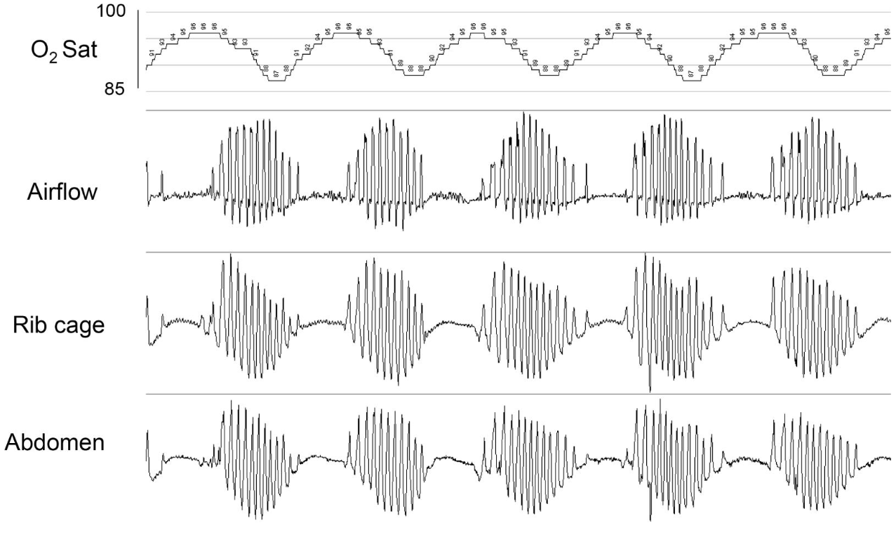
Polysomnography of Cheyne-Stokes Respiration. The periodic crescendo-decrescendo ventilation is characteristic of Cheyne-Stokes respiration, featuring cyclical fluctuations in airflow and thoracic-abdominal effort followed by central apneas and corresponding systemic oxygen desaturation.
StatPearls
References
Vargas-Ramirez L, Gonzalez-Garcia M, Franco-Reyes C, Bazurto-Zapata MA. Severe sleep apnea, Cheyne-Stokes respiration and desaturation in patients with decompensated heart failure at high altitude. Sleep science (Sao Paulo, Brazil). 2018 May-Jun:11(3):146-151. doi: 10.5935/1984-0063.20180028. Epub [PubMed PMID: 30455846]
Kim Y, Kim S, Ryu DR, Lee SY, Im KB. Factors Associated with Cheyne-Stokes Respiration in Acute Ischemic Stroke. Journal of clinical neurology (Seoul, Korea). 2018 Oct:14(4):542-548. doi: 10.3988/jcn.2018.14.4.542. Epub 2018 Sep 6 [PubMed PMID: 30198229]
Matthes S, Randerath W. Assessment of sleep disordered breathing in patients with heart failure. Breathe (Sheffield, England). 2022 Sep:18(3):220153. doi: 10.1183/20734735.0153-2022. Epub 2022 Nov 15 [PubMed PMID: 36865658]
Pinna GD, Robbi E, Terzaghi M, Corbellini D, La Rovere MT, Maestri R. Temporal relationship between arousals and Cheyne-Stokes respiration with central sleep apnea in heart failure patients. Clinical neurophysiology : official journal of the International Federation of Clinical Neurophysiology. 2018 Sep:129(9):1955-1963. doi: 10.1016/j.clinph.2018.05.029. Epub 2018 Jun 30 [PubMed PMID: 30015085]
Jamil SM, Owens RL, Lipford MC, Shafazand S, Marron RM, Vega Sanchez ME, Lam MT, Sunwoo BY, Schmickl C, Orr JE, Sharma S, Sankari A, Javaheri S, Bertisch S, Dupuy-McCauley K, Kolla B, Imayama I, Prasad B, Guzman E, Hayes MM, Wang T. ATS Core Curriculum 2020. Adult Sleep Medicine. ATS scholar. 2020 Dec 30:1(4):476-494. doi: 10.34197/ats-scholar.2020-0017RE. Epub 2020 Dec 30 [PubMed PMID: 33870314]
Sankari A, Bascom A, Oomman S, Badr MS. Sleep disordered breathing in chronic spinal cord injury. Journal of clinical sleep medicine : JCSM : official publication of the American Academy of Sleep Medicine. 2014 Jan 15:10(1):65-72. doi: 10.5664/jcsm.3362. Epub 2014 Jan 15 [PubMed PMID: 24426822]
Berry RB, Brooks R, Gamaldo C, Harding SM, Lloyd RM, Quan SF, Troester MT, Vaughn BV. AASM Scoring Manual Updates for 2017 (Version 2.4). Journal of clinical sleep medicine : JCSM : official publication of the American Academy of Sleep Medicine. 2017 May 15:13(5):665-666. doi: 10.5664/jcsm.6576. Epub 2017 May 15 [PubMed PMID: 28416048]
Grimm C, Viniol C, Degerli M, Hildebrandt O, Kesper K, Koehler U, Grimm W, Parahuleva M. Sleep-disordered breathing in patients with heart failure with preserved left ventricular ejection fraction. Sleep & breathing = Schlaf & Atmung. 2026 Mar 13:30(2):. doi: 10.1007/s11325-026-03635-w. Epub 2026 Mar 13 [PubMed PMID: 41824116]
Sankari A, Bascom AT, Chowdhuri S, Badr MS. Tetraplegia is a risk factor for central sleep apnea. Journal of applied physiology (Bethesda, Md. : 1985). 2014 Feb 1:116(3):345-53. doi: 10.1152/japplphysiol.00731.2013. Epub 2013 Oct 10 [PubMed PMID: 24114704]
Pisano A, Zoccali C, Bolignano D, D'Arrigo G, Mallamaci F. Sleep apnoea syndrome prevalence in chronic kidney disease and end-stage kidney disease patients: a systematic review and meta-analysis. Clinical kidney journal. 2024 Jan:17(1):sfad179. doi: 10.1093/ckj/sfad179. Epub 2023 Nov 14 [PubMed PMID: 38186876]
Level 1 (high-level) evidenceNaughton MT. PRO: Persistent Central Sleep Apnea/Hunter-Cheyne-Stokes Breathing, Despite Best Guideline-Based Therapy of Heart Failure With Reduced Ejection Fraction, Is a Compensatory Mechanism and Should Not Be Suppressed. Journal of clinical sleep medicine : JCSM : official publication of the American Academy of Sleep Medicine. 2018 Jun 15:14(6):909-914. doi: 10.5664/jcsm.7146. Epub 2018 Jun 15 [PubMed PMID: 29860966]
Leung RS, Huber MA, Rogge T, Maimon N, Chiu KL, Bradley TD. Association between atrial fibrillation and central sleep apnea. Sleep. 2005 Dec:28(12):1543-6 [PubMed PMID: 16408413]
Ishikawa O, Oks M. Central Sleep Apnea. Clinics in geriatric medicine. 2021 Aug:37(3):469-481. doi: 10.1016/j.cger.2021.04.009. Epub 2021 Jun 4 [PubMed PMID: 34210451]
Farney RJ, Walker JM, Cloward TV, Rhondeau S. Sleep-disordered breathing associated with long-term opioid therapy. Chest. 2003 Feb:123(2):632-9 [PubMed PMID: 12576394]
Martin SS, Aday AW, Almarzooq ZI, Anderson CAM, Arora P, Avery CL, Baker-Smith CM, Barone Gibbs B, Beaton AZ, Boehme AK, Commodore-Mensah Y, Currie ME, Elkind MSV, Evenson KR, Generoso G, Heard DG, Hiremath S, Johansen MC, Kalani R, Kazi DS, Ko D, Liu J, Magnani JW, Michos ED, Mussolino ME, Navaneethan SD, Parikh NI, Perman SM, Poudel R, Rezk-Hanna M, Roth GA, Shah NS, St-Onge MP, Thacker EL, Tsao CW, Urbut SM, Van Spall HGC, Voeks JH, Wang NY, Wong ND, Wong SS, Yaffe K, Palaniappan LP, American Heart Association Council on Epidemiology and Prevention Statistics Committee and Stroke Statistics Subcommittee. 2024 Heart Disease and Stroke Statistics: A Report of US and Global Data From the American Heart Association. Circulation. 2024 Feb 20:149(8):e347-e913. doi: 10.1161/CIR.0000000000001209. Epub 2024 Jan 24 [PubMed PMID: 38264914]
Hao X, Yang Y, Liu J, Zhang D, Ou M, Ke B, Zhu T, Zhou C. The Modulation by Anesthetics and Analgesics of Respiratory Rhythm in the Nervous System. Current neuropharmacology. 2024:22(2):217-240. doi: 10.2174/1570159X21666230810110901. Epub [PubMed PMID: 37563812]
Dempsey JA, Smith CA, Przybylowski T, Chenuel B, Xie A, Nakayama H, Skatrud JB. The ventilatory responsiveness to CO(2) below eupnoea as a determinant of ventilatory stability in sleep. The Journal of physiology. 2004 Oct 1:560(Pt 1):1-11 [PubMed PMID: 15284345]
Level 3 (low-level) evidenceJavaheri S, Dempsey JA. Central sleep apnea. Comprehensive Physiology. 2013 Jan:3(1):141-63. doi: 10.1002/cphy.c110057. Epub [PubMed PMID: 23720283]
Gavidia R, Emenike A, Meng A, Jansen EC, Hershner S, Goldstein C, Fetterolf J, Dunietz GL. The influence of opioids and nonopioid central nervous system active medications on central sleep apnea: a case-control study. Journal of clinical sleep medicine : JCSM : official publication of the American Academy of Sleep Medicine. 2021 Jan 1:17(1):55-60. doi: 10.5664/jcsm.8826. Epub [PubMed PMID: 32964833]
Level 2 (mid-level) evidenceCherniack NS, Fishman AP. Abnormal breathing patterns. Disease-a-month : DM. 1975 Jul:():1-45 [PubMed PMID: 238083]
Rana AM, Sankari A. Central Sleep Apnea. StatPearls. 2026 Jan:(): [PubMed PMID: 35201727]
Li S, Xu L, Dong X, Zhang X, Keenan BT, Han F, Bi T, Chang Y, Yu Y, Zhou B, Pack AI, Kuna ST. Home sleep apnea testing of adults with chronic heart failure. Journal of clinical sleep medicine : JCSM : official publication of the American Academy of Sleep Medicine. 2021 Jul 1:17(7):1453-1463. doi: 10.5664/jcsm.9224. Epub [PubMed PMID: 33688828]
Pillar G, Berall M, Berry R, Etzioni T, Shrater N, Hwang D, Ibrahim M, Litman E, Manthena P, Koren-Morag N, Rama A, Schnall RP, Sheffy K, Spiegel R, Tauman R, Penzel T. Detecting central sleep apnea in adult patients using WatchPAT-a multicenter validation study. Sleep & breathing = Schlaf & Atmung. 2020 Mar:24(1):387-398. doi: 10.1007/s11325-019-01904-5. Epub 2019 Aug 11 [PubMed PMID: 31402439]
Level 1 (high-level) evidenceDrager LF, McEvoy RD, Barbe F, Lorenzi-Filho G, Redline S, INCOSACT Initiative (International Collaboration of Sleep Apnea Cardiovascular Trialists). Sleep Apnea and Cardiovascular Disease: Lessons From Recent Trials and Need for Team Science. Circulation. 2017 Nov 7:136(19):1840-1850. doi: 10.1161/CIRCULATIONAHA.117.029400. Epub [PubMed PMID: 29109195]
Badr MS, Khayat RN, Allam JS, Hyer S, Mustafa RA, Naughton MT, Patil S, Pien GW, Randerath W, Won C. Treatment of central sleep apnea in adults: an American Academy of Sleep Medicine clinical practice guideline. Journal of clinical sleep medicine : JCSM : official publication of the American Academy of Sleep Medicine. 2025 Dec 1:21(12):2181-2191. doi: 10.5664/jcsm.11858. Epub [PubMed PMID: 40820608]
Level 1 (high-level) evidenceWolf J, Narkiewicz K. Managing comorbid cardiovascular disease and sleep apnea with pharmacotherapy. Expert opinion on pharmacotherapy. 2018 Jun:19(9):961-969. doi: 10.1080/14656566.2018.1476489. Epub 2018 May 24 [PubMed PMID: 29792524]
Level 3 (low-level) evidenceCowie MR, Woehrle H, Wegscheider K, Angermann C, d'Ortho MP, Erdmann E, Levy P, Simonds AK, Somers VK, Zannad F, Teschler H. Adaptive Servo-Ventilation for Central Sleep Apnea in Systolic Heart Failure. The New England journal of medicine. 2015 Sep 17:373(12):1095-105. doi: 10.1056/NEJMoa1506459. Epub 2015 Sep 1 [PubMed PMID: 26323938]
Bradley TD, Logan AG, Lorenzi Filho G, Kimoff RJ, Durán Cantolla J, Arzt M, Redolfi S, Parati G, Kasai T, Dunlap ME, Delgado D, Yatsu S, Bertolami A, Pedrosa R, Tomlinson G, Marin Trigo JM, Tantucci C, Floras JS, ADVENT-HF Investigators. Adaptive servo-ventilation for sleep-disordered breathing in patients with heart failure with reduced ejection fraction (ADVENT-HF): a multicentre, multinational, parallel-group, open-label, phase 3 randomised controlled trial. The Lancet. Respiratory medicine. 2024 Feb:12(2):153-166. doi: 10.1016/S2213-2600(23)00374-0. Epub 2023 Dec 21 [PubMed PMID: 38142697]
Level 1 (high-level) evidenceGinter G, Sankari A, Eshraghi M, Obiakor H, Yarandi H, Chowdhuri S, Salloum A, Badr MS. Effect of acetazolamide on susceptibility to central sleep apnea in chronic spinal cord injury. Journal of applied physiology (Bethesda, Md. : 1985). 2020 Apr 1:128(4):960-966. doi: 10.1152/japplphysiol.00532.2019. Epub 2020 Feb 20 [PubMed PMID: 32078469]
Randerath WJ, Schiza SE, Arzt M, Bonsignore MR, Heinzer R, Lombardi C, Lyssens R, Parati G, Penzel T, Pepin JL, Pevernagie D, Ryan S, Vignatelli L, Schwarz EI. European Respiratory Society and European Sleep Research Society statement on the treatment of central sleep apnoea with adaptive servo-ventilation. The European respiratory journal. 2025 Aug:66(2):. pii: 2500263. doi: 10.1183/13993003.00263-2025. Epub 2025 Aug 22 [PubMed PMID: 40571320]
Baumert M, Immanuel S, McKane S, Linz D. Transvenous phrenic nerve stimulation for the treatment of central sleep apnea reduces episodic hypoxemic burden. International journal of cardiology. 2023 May 1:378():89-95. doi: 10.1016/j.ijcard.2023.02.041. Epub 2023 Feb 24 [PubMed PMID: 36841294]
Rocha A, Pinto ACPN, Pachito DV, Drager LF, Lorenzi-Filho G, Atallah ÁN. Pharmacological treatment for central sleep apnoea in adults. The Cochrane database of systematic reviews. 2023 Feb 27:2(2):CD012922. doi: 10.1002/14651858.CD012922.pub2. Epub 2023 Feb 27 [PubMed PMID: 36861808]
Level 1 (high-level) evidenceJavaheri S. Acetazolamide improves central sleep apnea in heart failure: a double-blind, prospective study. American journal of respiratory and critical care medicine. 2006 Jan 15:173(2):234-7 [PubMed PMID: 16239622]
Level 1 (high-level) evidenceGiannoni A, Borrelli C, Mirizzi G, Richerson GB, Emdin M, Passino C. Benefit of buspirone on chemoreflex and central apnoeas in heart failure: a randomized controlled crossover trial. European journal of heart failure. 2021 Feb:23(2):312-320. doi: 10.1002/ejhf.1854. Epub 2020 May 22 [PubMed PMID: 32441857]
Level 1 (high-level) evidenceMaresh S, Prowting J, Vaughan S, Kruppe E, Alsabri B, Yarandi H, Badr MS, Sankari A. Buspirone decreases susceptibility to hypocapnic central sleep apnea in chronic SCI patients. Journal of applied physiology (Bethesda, Md. : 1985). 2020 Oct 1:129(4):675-682. doi: 10.1152/japplphysiol.00435.2020. Epub 2020 Aug 20 [PubMed PMID: 32816639]
Wolf J, Świerblewska E, Jasiel-Wojculewicz H, Gockowski K, Wyrzykowski B, Somers VK, Narkiewicz K. Theophylline therapy for Cheyne-Stokes respiration during sleep in a 41-year-old man with refractory arterial hypertension. Chest. 2014 Jul:146(1):e8-e10. doi: 10.1378/chest.13-2897. Epub [PubMed PMID: 25010981]
Level 3 (low-level) evidencePesek CA, Cooley R, Narkiewicz K, Dyken M, Weintraub NL, Somers VK. Theophylline therapy for near-fatal Cheyne-Stokes respiration. A case report. Annals of internal medicine. 1999 Mar 2:130(5):427-30 [PubMed PMID: 10068417]
Level 3 (low-level) evidenceBradley TD, Logan AG, Kimoff RJ, Sériès F, Morrison D, Ferguson K, Belenkie I, Pfeifer M, Fleetham J, Hanly P, Smilovitch M, Tomlinson G, Floras JS, CANPAP Investigators. Continuous positive airway pressure for central sleep apnea and heart failure. The New England journal of medicine. 2005 Nov 10:353(19):2025-33 [PubMed PMID: 16282177]
Level 1 (high-level) evidenceArzt M, Floras JS, Logan AG, Kimoff RJ, Series F, Morrison D, Ferguson K, Belenkie I, Pfeifer M, Fleetham J, Hanly P, Smilovitch M, Ryan C, Tomlinson G, Bradley TD, CANPAP Investigators. Suppression of central sleep apnea by continuous positive airway pressure and transplant-free survival in heart failure: a post hoc analysis of the Canadian Continuous Positive Airway Pressure for Patients with Central Sleep Apnea and Heart Failure Trial (CANPAP). Circulation. 2007 Jun 26:115(25):3173-80 [PubMed PMID: 17562959]
Level 1 (high-level) evidenceKalaydzhiev P, Velikova T, Davidkova Y, Voynova G, Borizanova A, Spasova N, Georgieva N, Ilieva R, Kinova E, Goudev A. Effects of SGLT2 Inhibitors on Sleep Apnea Parameters and Cheyne-Stokes Respiration in Patients with Acute Decompensated Heart Failure: A Prospective Cohort Study. Biomedicines. 2025 Jun 14:13(6):. doi: 10.3390/biomedicines13061474. Epub 2025 Jun 14 [PubMed PMID: 40564192]
Randerath W, Baillieul S, Tamisier R. Central sleep apnoea: not just one phenotype. European respiratory review : an official journal of the European Respiratory Society. 2024 Jan 31:33(171):. doi: 10.1183/16000617.0141-2023. Epub 2024 Mar 27 [PubMed PMID: 38537948]
Javed F, Tamisier R, Pepin JL, Cowie MR, Wegscheider K, Angermann C, d'Ortho MP, Erdmann E, Simonds AK, Somers VK, Teschler H, Levy P, Armitstead J, Woehrle H. Association of serious adverse events with Cheyne-Stokes respiration characteristics in patients with systolic heart failure and central sleep apnoea: A SERVE-Heart Failure substudy analysis. Respirology (Carlton, Vic.). 2020 Mar:25(3):305-311. doi: 10.1111/resp.13613. Epub 2019 Jun 20 [PubMed PMID: 31218793]
Fudim M, Soloveva A. Upright Cheyne-Stokes Respiration in Heart Failure: An Ominous Sign of Cardiovascular Dysregulation. Journal of the American College of Cardiology. 2020 Oct 27:76(17):2038-2039. doi: 10.1016/j.jacc.2020.06.089. Epub [PubMed PMID: 33092740]
Alsaqobi A. Obstructive sleep apnoea triggering autonomic dysreflexia: Isn't it time for sleep studies to be routine in SCI? Brain & spine. 2024:4():102725. doi: 10.1016/j.bas.2023.102725. Epub 2023 Dec 12 [PubMed PMID: 38510615]
Bonnin-Vilaplana M, Arboix A, Parra O, García-Eroles L, Montserrat JM, Massons J. Cheyne-stokes respiration in patients with first-ever lacunar stroke. Sleep disorders. 2012:2012():257890. doi: 10.1155/2012/257890. Epub 2012 Apr 23 [PubMed PMID: 23471518]
Lam T, Nagappa M, Wong J, Singh M, Wong D, Chung F. Continuous Pulse Oximetry and Capnography Monitoring for Postoperative Respiratory Depression and Adverse Events: A Systematic Review and Meta-analysis. Anesthesia and analgesia. 2017 Dec:125(6):2019-2029. doi: 10.1213/ANE.0000000000002557. Epub [PubMed PMID: 29064874]
Level 1 (high-level) evidenceFox H, Oldenburg O, Javaheri S, Ponikowski P, Augostini R, Goldberg LR, Stellbrink C, Mckane S, Meyer TE, Abraham WT, Costanzo MR. Long-term efficacy and safety of phrenic nerve stimulation for the treatment of central sleep apnea. Sleep. 2019 Oct 21:42(11):. doi: 10.1093/sleep/zsz158. Epub [PubMed PMID: 31634407]