 Tools
Tools
 Embryology, Central Nervous System
Introduction
Central nervous system (CNS) embryology is a broad subject. This topic summarizes CNS organogenesis and reviews the framework of embryology, the embryogenesis of the brain and spinal cord, various in-utero tests for CNS anomalies, and problems that may be encountered during embryogenesis, with particular attention to the CNS. The CNS system involves 3 germinal layers: ectoderm, mesoderm, and endoderm.
The ectoderm is the primary driver of CNS development during embryogenesis. It differentiates into surface ectoderm, which forms the epidermis, hair, and nails, and neural ectoderm, which gives rise to the neural tube and neural crest. These structures subsequently develop into the brain, spinal cord, and peripheral nerves. The endoderm gives rise to the lining of the gastrointestinal and respiratory systems. It also gives rise to abdominal organs such as the liver, pancreas, and bladder. The mesoderm differentiates into 3 primary regions, each with distinct developmental contributions. The paraxial mesoderm consists primarily of somites, which develop into the axial skeleton, dermis, and skeletal muscles. The intermediate mesoderm gives rise to the gonads, kidneys, and other urogenital structures. The lateral plate mesoderm is further divided into parietal and visceral layers, with the parietal mesoderm forming the limb skeleton and the visceral mesoderm contributing to the muscular wall of the gut tube.
Embryological Transformations
Because these changes do not occur at once, embryology is a complicated subject. The following embryonic development timeline, with particular attention to the CNS, provides a clearer understanding of the process.
- Weeks 1 to 3: Zygote formation, blastocyst, and gastrulation occur.
- Mid-fourth week: Embryo is linear and uniform; notochord formation occurs.
- Late-fourth week: Many forms of differential growth occur; upper limb buds always develop before the lower limb buds.
- Fifth week: Limb buds are more pronounced.
- Sixth week: Can begin to see eyes and auricular hillocks, which develops into the external ears.
- Seventh week: Formation of eyes, ears, and fingers.
- Late eighth week: Formation of all organ systems.
- Nine to 12 weeks (11 to 14 gestational age): Embryo has a large head and a small body, and the body grows to catch up with the limbs. The genitalia can be recognized during this period, giving a chance for parents to find out the gender of the embryo.
- Thirteen to 16 weeks (15 to 18 gestational age): Coordinated limb developments and ossification of skull occur; Ovaries differentiate and contain primordial ovarian follicles that contain oogonia; the eyes face anteriorly, and ears are in place.
- Seventeen to 20 weeks (18 to 22 gestational ages): Eyebrows and head hair are visible at 20 weeks.
- Twenty-one to 25 weeks (23 to 27 gestational age): Type II pneumocytes secrete surfactant. It is after this stage that babies are considered viable.
- Twenty-six to 29 weeks (28 to 31 gestational age): Eyelids open; the quantity of white fat increases. The CNS has matured and can control breathing and temperature regulation. Additionally, the bone marrow takes over (from the yolk sac) as the major site of erythropoiesis.
- Thirty to 34 weeks (32 to 36 gestational age): Maturation and growth of organs occur.
- Thirty-five to 38 weeks (37 to 40 gestational age): Baby now has a firm grasp with hands. Testes may have descended in males.[1][2]
Development
Register For Free And Read The Full Article
 Search engine and full access to all medical articles
- 10 free questions in your specialty
- Free CME/CE Activities
- Free daily question in your email
- Save favorite articles to your dashboard
- Emails offering discounts
Learn more about a Subscription to StatPearls Point-of-Care
Development
Embryogenesis: Weeks 2 to 8
Beginning with the trilaminar germ disc, which refers to the epiblast and hypoblast, the epiblast cells undergo an epithelial-mesenchymal transition that replaces the hypoblast. They also proliferate in the middle layer to form the mesoderm, where they remain mesenchymal and differentiate into connective tissue. The primitive streak then appears superiorly from the thickened region of the ectoderm. It grows caudal to the cranial and induces notochord formation. The ectoderm then invaginates as cells migrate to form the primitive node and primitive pit, where the notochordal process is formed.
- The primitive pit is a depression at the center of the primitive node, which is an opening in the notochordal canal.
- Neurulation refers to the folding of the neural plate. The neural plate folds, via induction from the notochord, into the neural tube, which then becomes the neuroectoderm, which finally forms the CNS, namely the brain and spinal cord; the brain from the cranial two-thirds of the segment and spinal cord from the caudal one-third of the segment.
- Neural Crest cells form dorsal root ganglia and connective tissue in the head and neck.
- The notochord defines the longitudinal axis of the embryo and contributes to parts of the intervertebral discs, but it does not form the spinal cord or vertebrae. The notochordal process develops above the primitive node and elongates caudally, extending toward the cranial end of the embryo.
The CNS is derived from the neuroectoderm: the notochord induces the formation of the neural plate (thickening of the ectodermal layer), which further differentiates into neural folds with a neural groove between them, leading to the formation of the neural tube (via neurulation).
Spinal Cord
The spinal cord is formed from the neural plate and now contains 3 layers:
- The ventricular layer that lines the central canal.
- The mantle layer contains neuronal bodies, which eventually form the gray matter.
- Marginal layer that contains axons and eventually forms the white matter.[3][4]
While this topic summarizes the embryological changes that occur within the CNS, the peripheral nervous system (PNS) is formed from neuroepithelial cells. These cells travel from the pia mater to the ventricular layer of the spinal cord, where they differentiate and migrate to form glioblasts (support cells, Schwann cells), neurons, and ependymal cells. As this information is often tested on boards, the myelin sheath, composed of support cells, wraps around axons and insulates neurons, increasing the speed of neuronal conduction.
- Myelination of peripheral axons occurs via the neurolemma, which arises from Schwann cells (derived from neural crest cells).
- Myelination of CNS axons occurs via oligodendrocytes, which are neuroepithelium derivatives.
Three membranous layers cover the whole CNS:
- Dura mater: derived from surrounding mesenchyme and is tough and durable.
- Arachnoid mater: derived from neural crest; forms as a single layer with Pia mater.
- Pia mater: derived from neural crest; intimately covers the CNS.
Brain
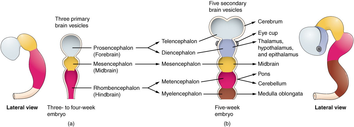
During brain formation, there are 3 primary brain vesicles that differentiate into 5 secondary brain vesicles (see Image. Brain Vesicles).
- Prosencephalon, which becomes the forebrain: This later develops into the cerebral hemispheres, which contain structures underneath, such as the epithalamus, thalamus, and hypothalamus. This section of the brain is responsible for consciousness, sensorimotor transformation, and sensory integration.
- Mesencephalon, which becomes the midbrain: This part of the brain undergoes little structure reorganization compared to the spinal cord and other brain vesicles.
- Rhombencephalon, which becomes the hindbrain: This part can be further divided into 3 segments:
- Metencephalon: The dorsal growth of the cerebellum (integrates sensory information to fine-tune output)
- Caudal myelencephalon: Similar to the structure of the spinal cord with a “closed” central canal of Medulla
- Rostral myelencephalon: “Open part” of medulla; cerebrospinal fluid is produced via choroid plexus and leaks into the subarachnoid space.
Finally, the hypophysis gives rise to the pituitary gland, which has 2 origins. The posterior pituitary is an outgrowth of the hypothalamus and, therefore, has a direct connection. On the other hand, the anterior pituitary is an ectodermal growth from the mouth. It depends on a dense capillary network and communicates with the brain via the vascular system.[5][6]
Testing
Non-invasive prenatal testing can be performed as early as 10 weeks of gestation and analyzes cell-free DNA in maternal plasma to help identify certain fetal anomalies or genetic conditions. Amniocentesis, typically performed between 14 and 20 weeks, involves sampling amniotic fluid and serves as a diagnostic test to screen for fetal anomalies. First-trimester testing assesses the risk of trisomy 21, trisomy 18, trisomy 13, and other neural tube defects. Alpha-fetoprotein levels in amniotic fluid are elevated in central nervous system and ventral abdominal wall anomalies and decreased in fetuses with trisomy 21, trisomy 18, or other chromosomal defects. Anatomy sonography can be performed throughout pregnancy, with particular emphasis between 16 and 22 weeks, to estimate fetal weight and gestational age using measurements of head circumference, biparietal diameter, femur length (from epiphysis to epiphysis), and abdominal circumference. Although beyond the scope of this topic, a Quad-screen test can also be performed in the second trimester. The components of this screening test include alpha-fetoprotein, hCG, Estriol, and Inhibin-A.[7]
Pathophysiology
Embryogenesis can be complex and may result in mild or severe defects (pathophysiological changes). Teratogenesis is defined as any external factor that can influence the growth of the embryo. Embryos are highly susceptible and critical between weeks 3 and 8, when organ systems develop. Dysraphism is the failure of fusion between the symmetric halves of an anatomical structure. These include, and are not limited to, spina bifida malformations.
- Spina bifida occulta occurs when the vertebral column fails to fuse, but other layers develop normally. It is the least severe form of dysraphism and usually affects the lumbosacral region (S1 to S2 most commonly). It can be associated with moles, angiomas, lipomas, and abnormal hair growth in the affected area.
- Spina bifida aperta occurs when there is an incomplete fusion of the skin with or without a cyst. The spinal cord is still covered by the arachnoid mater, thus preserving the subarachnoid space and preventing leakage of the cerebrospinal fluid.
- Spina bifida cystica is the most severe form of dysraphism. Patients may develop urinary or fecal incontinence. 80% of these lesions occur in the lumbosacral region.
Dysraphism in the cranium causes malformations analogous to spina bifida:
- Encephalocele: Protrusion of the brain into the subarachnoid space. It can be associated with Chiari III malformation, in which part of the cerebellum protrudes, and the spinal cord becomes twisted. This is commonly associated with cleft lip and palate.
- Anencephaly is characterized by the absence of the cerebral cortex and thalamic structures, with the cerebellum, brainstem, and spinal cord present (though they may be deformed). This can occur due to failure of notochord signaling, which is necessary for median hinge point formation or for the induction of neural crest cell maturation.
- Holoprosencephaly: Failure of features to form along the midline of the face. Features include a single central incisor, cyclopia, or an unpaired cerebral hemisphere.
- Craniorachischisis totalis is when the entire neural plate fails to fold, and the CNS is open to the amniotic cavity. These are often associated with stillborn fetuses.[8]
Clinical Significance
Pathophysiological processes that can occur during embryogenesis are rare and do not occur very often; when they do, the newborn is alive rather than stillborn. However, the newborn may require certain surgeries to correct the craniofacial anomalies before significant damage occurs.[9] Furthermore, those babies born with spina bifida require further evaluation with an ultrasound and surgical correction of the spinal cord to prevent herniation or other complications. Clinically, to avoid abnormal embryonic development, the mother should avoid teratogens or any external factors that can influence the baby's growth, especially during weeks 3 through 8 of embryogenesis. Significant teratogens include alcohol, tobacco use, certain prescription drugs, and illicit drugs. Most importantly, women seeking to get pregnant or who are pregnant take multivitamins, especially folic acid supplements to aid with neurodevelopment.[10]
Media
(Click Image to Enlarge)
Brain Vesicles
Access for free at https://openstax.org/books/anatomy-and-physiology-2e/pages/1-introduction
The Embryologic Perspective by Rice University (CC by 4.0 https://creativecommons.org/licenses/by/4.0/)
References
Donovan MF, Arbor TC, Bordoni B. Embryology, Yolk Sac. StatPearls. 2024 Jan:(): [PubMed PMID: 32310425]
Khan YS, Ackerman KM. Embryology, Week 1. StatPearls. 2024 Jan:(): [PubMed PMID: 32119449]
Gouti M, Metzis V, Briscoe J. The route to spinal cord cell types: a tale of signals and switches. Trends in genetics : TIG. 2015 Jun:31(6):282-9. doi: 10.1016/j.tig.2015.03.001. Epub 2015 Mar 27 [PubMed PMID: 25823696]
Kaplan KM, Spivak JM, Bendo JA. Embryology of the spine and associated congenital abnormalities. The spine journal : official journal of the North American Spine Society. 2005 Sep-Oct:5(5):564-76 [PubMed PMID: 16153587]
Stiles J, Jernigan TL. The basics of brain development. Neuropsychology review. 2010 Dec:20(4):327-48. doi: 10.1007/s11065-010-9148-4. Epub 2010 Nov 3 [PubMed PMID: 21042938]
Level 3 (low-level) evidenceGilmore JH, Knickmeyer RC, Gao W. Imaging structural and functional brain development in early childhood. Nature reviews. Neuroscience. 2018 Feb 16:19(3):123-137. doi: 10.1038/nrn.2018.1. Epub [PubMed PMID: 29449712]
Dorney E, Black KI. Preconception care. Australian journal of general practice. 2018 Jul:47(7):424-429. doi: 10.31128/AJGP-02-18-4485. Epub [PubMed PMID: 30114868]
Shamji MF, Ibrahim A. IMAGES IN CLINICAL MEDICINE. Cervical Meningocele. The New England journal of medicine. 2015 Jul 23:373(4):e4. doi: 10.1056/NEJMicm1408098. Epub [PubMed PMID: 26200992]
Zakhary GM, Montes DM, Woerner JE, Notarianni C, Ghali GE. Surgical correction of craniosynostosis. A review of 100 cases. Journal of cranio-maxillo-facial surgery : official publication of the European Association for Cranio-Maxillo-Facial Surgery. 2014 Dec:42(8):1684-91. doi: 10.1016/j.jcms.2014.05.014. Epub 2014 Jun 4 [PubMed PMID: 24969768]
Level 2 (mid-level) evidencevan Gelder MM, de Jong-van den Berg LT, Roeleveld N. Drugs associated with teratogenic mechanisms. Part II: a literature review of the evidence on human risks. Human reproduction (Oxford, England). 2014 Jan:29(1):168-83. doi: 10.1093/humrep/det370. Epub 2013 Oct 9 [PubMed PMID: 24108217]
Level 3 (low-level) evidence