 Tools
Tools
 Cellulitis
Introduction
Cellulitis is a common bacterial skin infection, with over 14 million documented cases in the United States annually. This condition accounts for approximately $3.7 billion in ambulatory care costs and 650,000 hospitalizations annually.[1] Cellulitis typically presents as a poorly demarcated, warm, erythematous area with associated edema and tenderness to palpation (see Image. Cellulitis).
Cellulitis is an acute bacterial infection causing inflammation of the deep dermis and surrounding subcutaneous tissue. The infection is without an abscess or purulent discharge.[2] Beta-hemolytic streptococci most commonly cause cellulitis, typically group A streptococcus (Streptococcus pyogenes), followed by methicillin-sensitive Staphylococcus aureus (MSSA). Patients who are immunocompromised, colonized with methicillin-resistant Staphylococcus aureus (MRSA), bitten by animals, or have comorbidities such as diabetes mellitus may become infected with other bacteria.[3] If the clinician correctly identifies and promptly treats cellulitis, the condition resolves with appropriate antibiotic treatment.
Etiology
Register For Free And Read The Full Article
 Search engine and full access to all medical articles
- 10 free questions in your specialty
- Free CME/CE Activities
- Free daily question in your email
- Save favorite articles to your dashboard
- Emails offering discounts
Learn more about a Subscription to StatPearls Point-of-Care
Etiology
The skin is a protective barrier that prevents normal skin flora and other microbial pathogens from reaching the subcutaneous tissue and lymphatic system. When a break in the skin occurs, normal skin flora and other bacteria enter the dermis and subcutaneous tissue. Introducing these bacteria below the skin surface can lead to an acute superficial infection affecting the deep dermis and subcutaneous tissue, causing cellulitis. Cellulitis most commonly results from infection with group A beta-hemolytic streptococcus (ie, Streptococcus pyogenes).[4]
Risk factors for cellulitis include any cause of skin barrier breakdown, such as skin injuries, surgical incisions, intravenous site punctures, fissures between toes, insect bites, animal bites, and other skin infections.[5] Patients with comorbidities such as diabetes mellitus, venous insufficiency, peripheral arterial disease, and lymphedema are at higher risk of developing cellulitis.[6] See Image. Venous Insufficiency and Lipodermatosclerosis.
Epidemiology
Cellulitis is relatively common and most often occurs in middle-aged and older adults. When comparing men and women, there is no statistically significant difference in the incidence of cellulitis. There are approximately 50 cases per 1000 patient-years.[7]
Pathophysiology
Cellulitis is characterized by erythema, warmth, edema, and tenderness to palpation resulting from cytokine and neutrophil response from bacteria breaching the epidermis.[8] The cytokines and neutrophils are recruited to the affected area after bacteria have penetrated the skin, leading to an epidermal response. This response includes the production of antimicrobial peptides and keratinocyte proliferation and is postulated to produce the characteristic exam findings in cellulitis.[9] Group A Streptococci, the most common bacteria to cause cellulitis, can also produce virulence factors such as pyrogenic exotoxins (A, B, C, and F) and streptococcal superantigen that can lead to a more pronounced and invasive disease.[10]
History and Physical
Patients with cellulitis typically present with a poorly demarcated area of erythema that is warm, swollen, and tender to the touch. Constitutional symptoms, including fever, malaise, and fatigue, may also be present, reflecting systemic involvement in moderate to severe cases.[11] When evaluating patients with cellulitis, clinicians should obtain a comprehensive history of the presenting illness, focusing on the circumstances surrounding the onset of skin changes. Key questions include recent travel, trauma or injuries, history of intravenous drug use, and any insect or animal bites to the affected area. A thorough medical history should also be reviewed to identify chronic conditions that increase the risk of cellulitis, such as diabetes mellitus, venous insufficiency, peripheral vascular disease, chronic tinea pedis, and lymphedema.
The affected area should be thoroughly inspected for skin breakdown and outlined with a marker to monitor progression. The area should be palpated to assess for fluctuance, which may indicate a developing abscess. Clinicians should also note any warmth, tenderness, or purulent drainage during gentle palpation. Cellulitis can occur in any area of the body, but most commonly affects the lower extremities (see Image. Cellulitis of the Extremity). This condition is rarely bilateral; in cases of lower extremity cellulitis, careful examination between the interspaces of the toes is recommended.[12] Clinicians should also assess sensation and verify pulses in the affected extremities to monitor for potential compartment syndrome. Additionally, it is important to document the presence of vesicles, bullae, peau d’orange, or lymphadenopathy.
Evaluation
Cellulitis is diagnosed clinically based on spreading erythematous inflammation of the deep dermis and subcutaneous tissue. This condition presents with worsening erythema, edema, warmth, and tenderness. Two of the 4 criteria (warmth, erythema, edema, or tenderness) are required to make the diagnosis. Cellulitis's most common presentation is on the lower extremities, but it can affect any body area; it is most often unilateral and rarely (if ever) presents bilaterally. The patient's skin should be thoroughly evaluated to find the potential source for the cellulitis by looking for microabrasions of the skin secondary to injuries, insect bites, pressure ulcers, or injection sites. If cellulitis affects the patient's lower extremities, careful evaluation should be made to look between the patient's toes for fissuring or tinea pedis.
Additionally, cellulitis can affect the lymphatic system and cause underlying lymphadenopathy. The associated edema with cellulitis can lead to the formation of vesicles, bullae, and edema surrounding hair follicles, leading to peau d'orange.[13] The Infectious Disease Society of America practice guidelines recommend against imaging the infected area except in patients with febrile neutropenia. Consider blood cultures only in patients who are immunocompromised, have experienced an immersion injury, or have had animal bites.[12] Blood cultures are also necessary when a patient has signs of systemic infection.[10]
Treatment / Management
Patients presenting with mild cellulitis and no systemic signs of infection should receive antibiotics effective against streptococcal species. Although a less common cause, MSSA should also be considered in the choice of therapy. The oral antibiotic therapy duration should be at least 5 days. In nonpurulent cellulitis, patients should receive cephalexin 500 mg every 6 hours. If they have a severe allergic reaction to beta-lactamase inhibitors, they should receive clindamycin 300 mg to 450 mg every 6 hours.[14](B3)
In patients with purulent cellulitis, MRSA colonization, cellulitis associated with an abscess or extensive puncture wounds, or a history of intravenous drug use, patients should receive antibiotics that treat MRSA as well. Cellulitis with MRSA risk factors should be treated with trimethoprim-sulfamethoxazole 800 mg/160 mg twice daily for 5 days, and cephalexin 500 mg every 6 hours. If a patient is allergic to trimethoprim-sulfamethoxazole, treat with clindamycin 300 mg to 450 mg every 6 hours. A longer duration of antibiotic treatment may be a consideration in patients who show minimal improvement with antibiotic therapy within 48 hours.
Hospitalization with the initiation of systemic antibiotics may be necessary for patients who present with systemic signs of infection (defined as 2 or more systemic inflammatory response criteria, such as fever above 38° C, heart rate greater than 90 beats/min, respiratory rate over 20 breaths/min, leukocytosis with white blood cell count above 12,000/mm³, leukopenia with white blood cell count below 4000/mm³, or bandemia ≥10%).[12] Admission should also be considered for those who have failed outpatient treatment, are immunocompromised, exhibit rapidly progressing erythema, are unable to tolerate oral medications, or have cellulitis overlying or near an indwelling medical device.(A1)
Patients requiring intravenous antibiotics should first receive treatment targeting group A strep. If there are no risk factors for MRSA, begin with intravenous cefazolin, then switch to oral cephalexin when possible, for a total of 5 days of therapy. If MRSA risk factors are present, start with intravenous vancomycin and later transition to oral trimethoprim-sulfamethoxazole. For immunocompromised individuals who need hospital-level intravenous therapy, broader treatment may be required, such as combining vancomycin with piperacillin-tazobactam or using a carbapenem. The clinician should obtain blood cultures if a patient is exhibiting signs of systemic toxicity, has persistent cellulitis despite adequate treatment, or has unique exposures such as animal bites or water-associated injuries.[15]
Atypical organisms can cause cellulitis in particular situations. If exposed to a dog or cat bite, patients risk developing cellulitis secondary to Pasteurella multocida. Cellulitis can also be caused by Vibrio vulnificus if secondary to an injury involving exposure to water, such as a cut from an oyster shell. Diabetic individuals and patients with diabetic foot ulcers are at risk for Pseudomonas aeruginosa. Those who are immunocompromised are at risk for Pseudomonas aeruginosa and Cryptococcus. Suppose patients have significant edema with a known cause for the edema. In that case, the underlying condition should receive proper treatment to decrease the edema and prevent future episodes of cellulitis. Patients should be instructed to keep the affected area elevated.[12](A1)
Differential Diagnosis
Cellulitis is a common infection of the deep dermis and subcutaneous tissue, often affecting the lower extremities. Still, it can be difficult to distinguish because many other conditions can appear similar.[16] Erysipelas is sometimes considered a form of cellulitis (see Image. Erysipelas). However, it is a more superficial infection affecting the upper dermis and superficial lymphatic system. Bright red erythema, elevation of the affected skin, and well-demarcated borders can help to diagnose erysipelas and distinguish it from cellulitis, which tends to be more mildly erythematous (pink) and flat with less distinct boundaries. Erysipelas may also have streaking when superficial lymphatics are involved. This condition most commonly results from the exotoxins released from group A strep (Streptococcus pyogenes). The first-line treatment for erysipelas is amoxicillin or cephalexin.[17]
Chronic venous stasis dermatitis is a long-standing, bilateral, inflammatory dermatosis secondary to chronic venous insufficiency and typically involves the medial malleoli. This appears on the lower extremities and manifests as erythema with scaling, peripheral edema, and hyperpigmentation. Treatment focuses on treating the underlying chronic venous insufficiency and its sequelae, such as lower extremity edema.[1]
Necrotizing fasciitis is a rare infection of the fascia that leads to necrosis of the subcutaneous tissue. The characteristic presentation includes fevers, erythema, edema, pain out of proportion to the exam, and crepitus. This qualifies as a surgical emergency and requires surgical debridement immediately. Imaging may be obtained to help confirm the diagnosis of necrotizing fasciitis, but it should not delay surgical intervention. Computed tomography imaging revealing subcutaneous gas in the soft tissue is highly specific for necrotizing fasciitis. Septic arthritis, or an infected joint, can involve any joint but typically occurs at the knee. Patients present with swelling, warmth, pain, and decreased joint mobility. Treatment is joint aspiration and antibiotics directed at the most common pathogens.
Deep vein thrombosis is typically unilateral and presents with tenderness, erythema, warmth, and edema. This often affects the lower extremities. Patients commonly have the presence of risk factors for deep vein thrombosis, such as a history of immobility, active cancer, or a family history of venous thromboembolism. Deep vein thrombosis rarely manifests with fevers or leukocytosis, but they can be present. Ultrasound imaging is used to confirm the diagnosis.[5]
Prognosis
If the clinician promptly identifies cellulitis and initiates treatment with the correct antibiotic, patients can expect to notice an improvement in signs and symptoms within 48 hours. Annual recurrence of cellulitis occurs in about 8% to 20% of patients, with overall recurrence rates reaching as high as 49%.[1][9] Recurrence is preventable with prompt treatment of cuts or abrasions, proper hand hygiene, and effective treatment of any underlying comorbidities. There is approximately an 18% failure rate with initial antibiotic treatment. Overall, cellulitis has a good prognosis.[18]
Complications
Without prompt diagnosis and treatment, cellulitis could lead to several complications. If the bacterial infection reaches the bloodstream, it could lead to bacteremia. Bacteremia is diagnosable by obtaining blood cultures in patients who exhibit systemic symptoms. The clinician should obtain identification and susceptibilities from the blood cultures and tailor antibiotics accordingly.[19] Failure to identify and treat bacteremia from cellulitis can lead to endocarditis, an infection of the heart's inner lining (endocardium).
Patients with cellulitis and 2 or more systemic inflammatory response syndrome criteria (fever over 100.4° F, tachypnea, tachycardia, or abnormal white cell count) are diagnosed with sepsis. If cellulitis moves from the deep dermis and subcutaneous tissue to the bone, it can lead to osteomyelitis. Cellulitis that leads to bacteremia, endocarditis, or osteomyelitis requires longer antibiotics and possibly surgery.[1]
Deterrence and Patient Education
Patients should be informed to take prescribed antibiotics as indicated. Keep the area clean and dry. When possible, they should elevate the area above the level of their heart to reduce edema. Cellulitis should start resolving within 24 to 48 hours after initiating antibiotics. The clinician may mark the area of erythema, and patients should return if they notice the erythema beginning to spread or not responding to antibiotics, develop persistent fevers, begin developing significant bullae, or if pain worsens.[12] Patients should additionally maintain good hand hygiene and adequately clean any future abrasions in their skin.[20]
Enhancing Healthcare Team Outcomes
Treatment and prevention of cellulitis require an interprofessional approach involving the patient, primary healthcare professionals, pharmacists, and wound care nurses. Pharmacists, ideally with infectious disease expertise, play an important role in collaborating with clinicians to select the most appropriate antibiotic regimen. Most patients can be cared for as outpatients; however, those requiring hospitalization for intravenous antibiotics benefit from coordinated input across the healthcare team.
Clinicians are responsible for accurately determining the severity and type of cellulitis (purulent vs nonpurulent). Nurses monitor progression by outlining areas of erythema, tracking symptoms, and reporting changes. Pharmacists assist with dosing and monitoring, particularly for medications with renal toxicity risk, such as vancomycin. Wound care nurses provide education on skin care, limb elevation, and maintaining mobility to reduce complications like deep vein thrombosis. Both nurses and pharmacists should counsel patients on medication adherence to promote treatment success. A collaborative, team-based approach is essential for effective treatment, reducing recurrence, and improving long-term outcomes.[21][22]
Media
(Click Image to Enlarge)
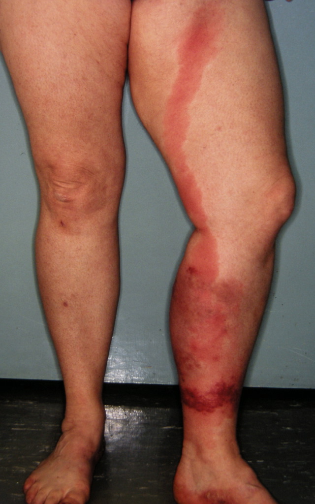
Cellulitis. This image shows the characteristic reddening of the skin seen in cellulitis.
(Click Image to Enlarge)
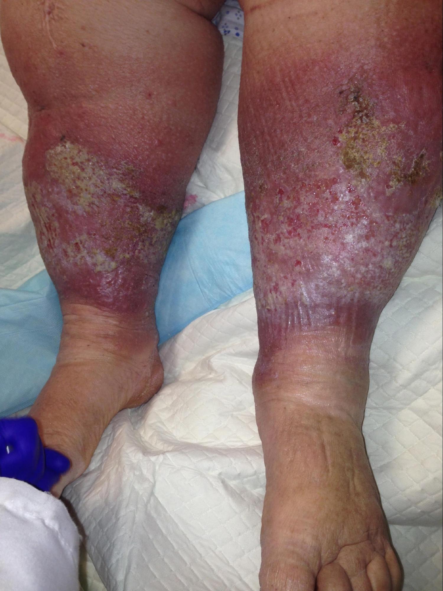
Chronic Venous Insufficiency and Lipodermatosclerosis. Common clinical features of chronic venous insufficiency include rubor, atrophy blanche, and lipodermatosclerosis.
Contributed by MA Dreyer, DPM, FACFAS
(Click Image to Enlarge)
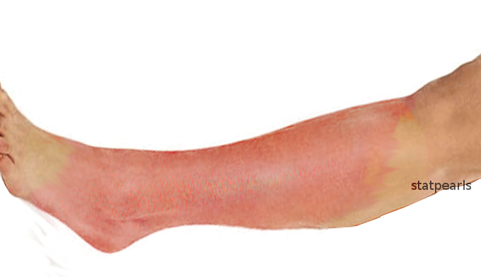
Cellulitis of the Extremity. Cellulitis can occur on any area of the body, but most commonly affects the lower extremities.
Contributed by O Chaigasame, MD
(Click Image to Enlarge)
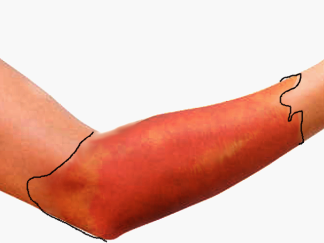
Erysipelas. This condition is a superficial infection affecting the upper dermis and superficial lymphatic system. Bright red erythema, elevation of the affected skin, and well-demarcated borders can help to diagnose erysipelas and distinguish it from cellulitis.
Contributed by O Chaigasame, MD
References
Raff AB, Kroshinsky D. Cellulitis: A Review. JAMA. 2016 Jul 19:316(3):325-37. doi: 10.1001/jama.2016.8825. Epub [PubMed PMID: 27434444]
Bystritsky RJ. Cellulitis. Infectious disease clinics of North America. 2021 Mar:35(1):49-60. doi: 10.1016/j.idc.2020.10.002. Epub [PubMed PMID: 33494874]
Cranendonk DR, Lavrijsen APM, Prins JM, Wiersinga WJ. Cellulitis: current insights into pathophysiology and clinical management. The Netherlands journal of medicine. 2017 Nov:75(9):366-378 [PubMed PMID: 29219814]
Liu C, Bayer A, Cosgrove SE, Daum RS, Fridkin SK, Gorwitz RJ, Kaplan SL, Karchmer AW, Levine DP, Murray BE, J Rybak M, Talan DA, Chambers HF, Infectious Diseases Society of America. Clinical practice guidelines by the infectious diseases society of america for the treatment of methicillin-resistant Staphylococcus aureus infections in adults and children. Clinical infectious diseases : an official publication of the Infectious Diseases Society of America. 2011 Feb 1:52(3):e18-55. doi: 10.1093/cid/ciq146. Epub 2011 Jan 4 [PubMed PMID: 21208910]
Level 1 (high-level) evidenceQuirke M, Ayoub F, McCabe A, Boland F, Smith B, O'Sullivan R, Wakai A. Risk factors for nonpurulent leg cellulitis: a systematic review and meta-analysis. The British journal of dermatology. 2017 Aug:177(2):382-394. doi: 10.1111/bjd.15186. Epub 2017 Jul 25 [PubMed PMID: 27864837]
Level 1 (high-level) evidenceKaye KS, Petty LA, Shorr AF, Zilberberg MD. Current Epidemiology, Etiology, and Burden of Acute Skin Infections in the United States. Clinical infectious diseases : an official publication of the Infectious Diseases Society of America. 2019 Apr 8:68(Suppl 3):S193-S199. doi: 10.1093/cid/ciz002. Epub [PubMed PMID: 30957165]
McNamara DR, Tleyjeh IM, Berbari EF, Lahr BD, Martinez JW, Mirzoyev SA, Baddour LM. Incidence of lower-extremity cellulitis: a population-based study in Olmsted county, Minnesota. Mayo Clinic proceedings. 2007 Jul:82(7):817-21 [PubMed PMID: 17605961]
Level 2 (mid-level) evidenceRrapi R, Chand S, Kroshinsky D. Cellulitis: A Review of Pathogenesis, Diagnosis, and Management. The Medical clinics of North America. 2021 Jul:105(4):723-735. doi: 10.1016/j.mcna.2021.04.009. Epub [PubMed PMID: 34059247]
Richmond JM, Harris JE. Immunology and skin in health and disease. Cold Spring Harbor perspectives in medicine. 2014 Dec 1:4(12):a015339. doi: 10.1101/cshperspect.a015339. Epub 2014 Dec 1 [PubMed PMID: 25452424]
Level 3 (low-level) evidenceCunningham MW. Pathogenesis of group A streptococcal infections. Clinical microbiology reviews. 2000 Jul:13(3):470-511 [PubMed PMID: 10885988]
Han J, Faletsky A, Mostaghimi A. Cellulitis. JAMA dermatology. 2020 Dec 1:156(12):1384. doi: 10.1001/jamadermatol.2020.2083. Epub [PubMed PMID: 32965485]
Stevens DL, Bisno AL, Chambers HF, Dellinger EP, Goldstein EJ, Gorbach SL, Hirschmann JV, Kaplan SL, Montoya JG, Wade JC. Practice guidelines for the diagnosis and management of skin and soft tissue infections: 2014 update by the infectious diseases society of America. Clinical infectious diseases : an official publication of the Infectious Diseases Society of America. 2014 Jul 15:59(2):147-59. doi: 10.1093/cid/ciu296. Epub 2014 Jun 18 [PubMed PMID: 24947530]
Level 1 (high-level) evidenceLong B, Gottlieb M. Diagnosis and Management of Cellulitis and Abscess in the Emergency Department Setting: An Evidence-Based Review. The Journal of emergency medicine. 2022 Jan:62(1):16-27. doi: 10.1016/j.jemermed.2021.09.015. Epub 2021 Oct 14 [PubMed PMID: 34657784]
Peghin M, Graziano E, Rovelli C, Grossi PA. Prevention and treatment of recurrent cellulitis. Current opinion in infectious diseases. 2023 Apr 1:36(2):95-101. doi: 10.1097/QCO.0000000000000903. Epub 2023 Feb 9 [PubMed PMID: 36853755]
Level 3 (low-level) evidenceTorres J, Avalos N, Echols L, Mongelluzzo J, Rodriguez RM. Low yield of blood and wound cultures in patients with skin and soft-tissue infections. The American journal of emergency medicine. 2017 Aug:35(8):1159-1161. doi: 10.1016/j.ajem.2017.05.039. Epub 2017 May 26 [PubMed PMID: 28592371]
Chuat C, Clerc O, Donzé J. [Cellulitis : management recommendations]. Revue medicale suisse. 2024 Nov 13:20(895):2131-2134. doi: 10.53738/REVMED.2024.20.895.2131. Epub [PubMed PMID: 39544061]
Rodrigues MA, Caetano M, Amorim I, Selores M. [Non-Necrotizing Acute Dermo-Hypodermal Infections: Erysipela and Infectious Cellulitis]. Acta medica portuguesa. 2021 Mar 1:34(3):217-228. doi: 10.20344/amp.12642. Epub 2021 Mar 1 [PubMed PMID: 33971117]
Obaitan I, Dwyer R, Lipworth AD, Kupper TS, Camargo CA Jr, Hooper DC, Murphy GF, Pallin DJ. Failure of antibiotics in cellulitis trials: a systematic review and meta-analysis. The American journal of emergency medicine. 2016 Aug:34(8):1645-52. doi: 10.1016/j.ajem.2016.05.064. Epub 2016 May 26 [PubMed PMID: 27344098]
Level 1 (high-level) evidenceGunderson CG, Martinello RA. A systematic review of bacteremias in cellulitis and erysipelas. The Journal of infection. 2012 Feb:64(2):148-55. doi: 10.1016/j.jinf.2011.11.004. Epub 2011 Nov 11 [PubMed PMID: 22101078]
Level 1 (high-level) evidenceSwartz MN. Clinical practice. Cellulitis. The New England journal of medicine. 2004 Feb 26:350(9):904-12 [PubMed PMID: 14985488]
Singh M, Negi A, Zadeng Z, Verma R, Gupta P. Long-Term Ophthalmic Outcomes in Pediatric Orbital Cellulitis: A Prospective, Multidisciplinary Study From a Tertiary-Care Referral Institute. Journal of pediatric ophthalmology and strabismus. 2019 Sep 1:56(5):333-339. doi: 10.3928/01913913-20190807-01. Epub [PubMed PMID: 31545868]
Gibbons JA, Smith HL, Kumar SC, Duggins KJ, Bushman AM, Danielson JM, Yost WJ, Wadle JJ. Antimicrobial stewardship in the treatment of skin and soft tissue infections. American journal of infection control. 2017 Nov 1:45(11):1203-1207. doi: 10.1016/j.ajic.2017.05.013. Epub 2017 Jul 18 [PubMed PMID: 28732743]