 Tools
Tools
 Cavernous Sinus Syndromes
Introduction
Understanding the clinical management of cavernous sinus syndrome (CSS) requires a thorough grasp of cavernous sinus anatomy. The cavernous sinus is small but complex and contains several important structures. The cavernous sinus is not a venous plexus, but rather a true dural venous sinus.[1] he cavernous sinus is bordered by the temporal bone of the skull and the sphenoid bone and lies lateral to the sella turcica. The inferior and lateral walls and the roof of the cavernous sinus are extensions of the dura mater. A thin collagen layer may or may not line the medial wall. The blood-filled space contains the internal carotid artery (carotid siphon) surrounded by sympathetic fibers, as well as cranial nerves (CNs). In particular, CN III (oculomotor nerve), CN IV (trochlear nerve), and 2 branches of CN V (trigeminal nerve), CN V1 (ophthalmic) and CN V2 (maxillary), pass through this blood-filled space (see Image. Anatomy of the Cavernous Sinus).[2] Cavernous sinus syndrome is any disease process involving the cavernous sinus. CCA is characterized by signs and symptoms that may include ophthalmoplegia, chemosis, proptosis, Horner syndrome, or trigeminal sensory loss.
Etiology
Register For Free And Read The Full Article
 Search engine and full access to all medical articles
- 10 free questions in your specialty
- Free CME/CE Activities
- Free daily question in your email
- Save favorite articles to your dashboard
- Emails offering discounts
Learn more about a Subscription to StatPearls Point-of-Care
Etiology
Determining the etiology of cavernous sinus syndrome remains difficult, despite improvements in diagnostic techniques, including imaging and analysis of blood and cerebrospinal fluid. Diagnosing cavernous sinus syndrome from cavernous sinus tissue is also difficult. Neoplastic (metastatic or primary), infectious, inflammatory, vascular, and traumatic processes are the principal causes of cavernous sinus syndrome. Jefferson first classified the disorder in 1938 into 3 types: anterior, middle, and posterior cavernous sinus syndrome. The Jefferson classification is based on the extent of trigeminal nerve involvement. In 1996, Ishikawa emphasized the lack of a clinical-anatomical correlation in the Jefferson classification and proposed a new classification of cavernous sinus syndrome. Ishikawa classified cavernous sinus syndrome into 3 separate parts: anterior, middle, and posterior. The Ishikawa classification uses the optic canal and the maxillary nerve as anatomical landmarks. Results from a study of 162 patients with cavernous sinus lesions showed that the Ishikawa classification was superior for identifying and localizing cavernous sinus lesions.[3] However, results from a study of 73 consecutive patients with cavernous sinus syndrome at a tertiary care center in Northern India showed that the Ishikawa classification was not superior to the Jefferson classification for identifying etiology.[4]
Epidemiology
Estimates of the incidence of CCS are not well documented in the literature. Results from an analysis of 151 cases from Los Angeles in 1996 showed 89 (59%) were men.[5] The average age was 39 years, with a range of 15 to 72 years. Tumors were the most common cause of cavernous sinus syndrome, accounting for 30%, followed by trauma at 24%. Other series excluded trauma as a cause of cavernous sinus syndrome. Results from a series of 126 patients in Barcelona, Spain, in 2007 showed that 42% were men.[6] The average age was 55.5 years, with a range of 40 to 70 years. Tumors were again the most common cause of cavernous sinus syndrome, accounting for 63%. According to this study, the second most common cause was vascular disease, accounting for 20%. Results from a 2017 study of 73 patients at tertiary care centers in Northern India showed that 64% were men. The average age was 44 years, with a range of 11 to 70 years. Tumors remained the most common cause of cavernous sinus syndrome, accounting for 30%; however, unlike previously reported series from the Western world, the prevalence of fungal infections was 24.6%, and the prevalence of Tolosa-Hunt syndrome was also much higher at 23.2%.[6] Overall, cavernous sinus syndrome did not predominate by sex. Patients may present at any age, with reported ages ranging from 20 to 80 years.
Pathophysiology
Cavernous Sinus Tumors
- Tumors were the most frequent cause of CCS, as previously mentioned
- Tumors may be primary or metastatic and have a wide differential, including meningiomas, schwannomas of CN III, IV, V1/V2 (the most common), or VI, haemangiomas, haemangiopericytomas, and metastatic disease (ie, the perineural spread of tumor through neural foramina) from lung, breast, or prostate.
- Tumors also may arise from either a pituitary tumor or by locally spreading, such as nasopharyngeal carcinoma with intracranial extension (more common in Southeast Asia).[7]
Cavernous sinus vascular pathologies:
- Vascular pathologies in the CS include carotid cavernous fistulas (CCFs), carotid cavernous aneurysms (CCAs), and cavernous sinus thrombosis (CST).
- CCAs do not carry a major risk of subarachnoid hemorrhage. CCFs may arise spontaneously or from secondary causes such as trauma, CCAs, and venous thrombosis.
- CCFs can be classified as indirect, low-flow, or dural CCFs (an indirect communication between the cavernous sinus and branches of the internal or external carotid artery within the adjacent dura). They can also be classified as being direct or high-flow CCFs (a direct connection between the intracavernous carotid artery and the cavernous sinus).[8]
- The causes of CST are aseptic or infectious. Infections can spread from a nasal furuncle (50%), sphenoidal or ethmoidal sinus (30%), as well as dental infections (10%). Aseptic causes typically occur after surgery or trauma. The most common causative microorganism is Staphylococcus aureus, followed by streptococci of the milleri group, which include Streptococcus constellatus, Streptococcus intermedius, and Streptococcus anginosus. Septic cavernous sinus thrombosis is a rare but serious complication of cavernous sinus infections.[9]
Infections
- Bacterial infection sources are explained above. One patient with tuberculosis was described.
- Fungal infection is the second most common cause of CCS in Northern India. Out of 18 fungal infections, Aspergillosis accounted for 44.4%, mucormycosis accounted for 22.2%, and other fungal infections accounted for 33.3%.[6]
Inflammation
- An inflammatory process of unknown etiology causes Tolosa-Hunt syndrome. Inflammation causes swelling, pressure, and then dysfunction of the structures, particularly of the cranial nerves.
- Sarcoidosis or granulomatosis with polyangiitis (formerly Wegener granulomatosis) can also predispose patients to cavernous sinus syndrome.
- Herpes zoster can cause inflammation.
History and Physical
Symptoms that occur across different etiologies of CCS share common features, such as headache (up to 90%), diplopia (up to 90%), painless or painful ophthalmoplegia, ptosis, proptosis, chemosis, facial sensory loss, visual loss, fever, facial asymmetry, hearing loss, and seizure. Assessment should include determining whether symptoms are unilateral or bilateral, whether symptom onset is acute, subacute, or chronic, and whether the condition is painful or painless. Physical examination findings depend on the etiology and the extent of cavernous sinus involvement. The following signs may be seen in patients with CCS:
- Involvement of cranial nerves with motor and/or sensory findings: the most common are CN III (85%), CN VI (70%), CN V1 and/or V2, and CN IV
- Reflexes: Decreased or absent corneal, direct and/or indirect light reflexes
- Ophthalmoplegia: painful or painless
- Chemosis: the swelling (or edema) of the conjunctiva
- Proptosis, protrusion or displacement of an eye:
- Ocular and cranial bruits
- Funduscopic findings: Papilledema, pallor and/or retinal hemorrhages
- Visual findings: Visual field cut, blurry vision, diplopia and/or vision loss
Common symptoms and signs on presentation can accompany the specific etiologies below:
- Cavernous sinus tumors may cause isolated or combined ophthalmoplegia, painful ophthalmoplegia, anesthesia in CN III, unitemporal or bitemporal visual field defects, acromegaly, and galactorrhea.
- Carotid-cavernous fistulas may cause ocular bruit auscultated over the globe in high-flow carotid-cavernous fistulas, proptosis, chemosis, conjunctival injection, ocular or orbital pain, headache, diplopia, and blurry vision.
- Cavernous sinus thrombosis may present with signs of an infectious process involving the paranasal sinuses or orbital cellulitis, including conjunctival injection, chemosis, and proptosis.
- Tolosa-Hunt syndrome may cause unilateral symptoms (bilateral symptoms in 4% to 5%) [11], painful ophthalmoplegia, and diplopia from cranial mono- or polyneuropathy (most commonly CN III), as well as Horner syndrome if the periarterial sympathetic fibers are involved.[10]
- Sarcoidosis may cause systemic signs, uveitis, ophthalmoplegia, and facial diplegia.
- Herpes zoster also may present with acute zoster ophthalmicus, typical skin lesions, and keratitis.
Evaluation
The diagnosis of CCS is challenging due to the wide range of possible etiologies. Imaging studies of the orbit, sella, and parasellar region play a major role in diagnosis. Precontrast and postcontrast scans are advisable. Contrast-enhanced CT scans provide better visualization of bone and calcium. MRI provides better detail of all soft tissues contained in the sinuses. CT angiography (CTA), CT venography (CTV), magnetic resonance angiography (MRA), MR venography (MRV), and conventional digital subtraction angiography help visualize all vascular structures in detail. Routine, cytologic, infectious, or inflammatory examinations of serum and cerebrospinal fluid (CSF) may be helpful as well.
Cavernous Sinus Tumors
- Clinicians should identify the source of the mass and determine whether the lesion is primary, metastatic, or due to local spread.
- CT, MRI, and chest/abdomen/pelvic CT
- A lumbar puncture with cytologic examination
- A biopsy of the cavernous sinus tumors is rarely needed for diagnosis of primary tumors if no tumors are spreading from another primary source.
Cavernous Sinus Vascular
- Vascular etiologies can be seen on CTA, MRI, MRA, and angiography.
- Conventional digital subtraction angiography is the gold-standard test for diagnosis of CCF. CCFs can also be seen on orbital or transcranial ultrasonography.
- Intraocular pressure should be checked in patients with ocular sequelae.
- Infectious workup, D-dimer, and MRV or CTV should be performed if there is a suspicion for CST.
- An aseptic thrombosis may be associated with hypercoagulable states as well as lymphoproliferative disorders. The patient may need further hematological workup.
Infections
- Further infectious workup should include rapid plasma reagin, HIV testing, fluorescent treponemal antibody testing, Lyme serology, QuantiFERON-TB Gold testing, and cultures of serum for bacteria, fungi, and mycobacteria.
- Cerebrospinal fluid workup should include protein, glucose, cell counts, cytology, and Lyme and syphilis serology.
Inflammation
- Clinicians should investigate additional systemic inflammatory and granulomatous processes after other common causes, such as tumor or vascular etiologies, have been excluded.
- Workup should include an erythrocyte sedimentation rate, C-reactive protein, angiotensin-converting enzyme, antinuclear antibody, anti-dsDNA antibody, anti-Sm antibody, anti-neutrophil cytoplasmic antibody, serum protein electrophoresis with immunotyping, CSF protein, glucose, cell counts, cytology, and angiotensin-converting enzyme.
- The specific diagnostic criteria for Tolosa-Hunt syndrome, as recommended by the International Headache Society, are summarized. A unilateral headache localized around the ipsilateral brow and eye, oculomotor paresis for less than 2 weeks, granulomatous inflammation of the cavernous sinus, superior orbital fissure, or orbit, established by MRI or biopsy, paresis of one or more of the ipsilateral CN III, IV, and/or VI, and symptoms not accounted for by an alternative diagnosis. [11] Glucocorticoid administration has diagnostic as well as therapeutic utility.
Treatment / Management
Cavernous Sinus Tumors
- Tumors in the cavernous sinus represent a major challenge for surgical resection because proximity to critical neurologic structures makes complete excision difficult and increases the risk of complications. Radiotherapy is important for providing excellent tumor control and avoiding the risk of surgical procedures.[12]
- Results from 3 large series of about 400 patients with cavernous sinus meningiomas treated with stereotactic radiosurgery at doses of 12-14 Gy showed a 5-year tumor control rate ranging from 94% to 98%.[13][14][15]
- Transsphenoidal surgery is the preferred treatment for most kinds of pituitary adenomas. Gamma knife surgery (GKS) is a common treatment for recurrent or residual pituitary adenomas.[16]
- Radiotherapy may offer transient relief, particularly in nasopharyngeal cancer.
- Whole radiotherapy may offer a transient improvement of metastatic lesions. (B2)
Cavernous Sinus Vascular
- Results from a Japanese cohort showed that carotid cavernous aneurysms had the lowest rupture rate among unruptured cerebral aneurysms.[17]
- Treatment of unruptured intracranial aneurysms was evaluated in the 2003 International Study of Unruptured Intracranial Aneurysms, which performed cost-effectiveness analyses showing that treatment was ineffective or not cost-effective for large aneurysms greater than 25 mm located in the cavernous carotid artery in patients aged 40 years.[18]
- The management of CCFs depends on their classification, symptom onset, and risk of long-term neurological impairment. Most (20% to 60%)indirect CCFs will close spontaneously.[19] Direct CCFs should be closed if symptomatic and at risk of progression with attendant morbidity because these CCFs are unlikely to close spontaneously.
- Endovascular treatment with transarterial or transvenous embolization is the preferred approach for closure of CCFs.
- Surgery can be considered when endovascular treatment is not possible or is unsuccessful. Surgery includes placement of packing, sealing with fascia and glue, suturing or clipping the fistula.
- Stereotactic radiosurgery is indicated when an endovascular approach is not feasible, and surgical intervention is difficult or carries a risk of significant morbidity.[20]
- Management of increased intraocular pressure with topical agents, β-blockers, acetazolamide, and intravenous corticosteroids may provide some relief. If vision loss is threatened or intraocular pressure remains elevated, fistula closure is required to achieve favorable pressure control.
- The management of CST should include antimicrobial therapy with/without surgical drainage of the sinuses or mastoid, and antithrombotic therapy.
- Patients with CST should be treated for 3 to 4 weeks with intravenous antibiotic(s), or for 6 to 7 weeks based on the clinical picture. Treatment should be broad-spectrum, with vancomycin vs. nafcillin, plus a third-generation cephalosporin and metronidazole, until a definite pathogen is identified.[21] Antifungal therapy with amphotericin B is required in developing countries.
- Retrospective analysis suggests that treatment with heparin may reduce mortality in carefully selected cases of septic cavernous-sinus thrombosis.[22]
- Corticosteroids in the acute phase of cerebral venous thrombosis (CVT) were not useful and were detrimental in patients without parenchymal cerebral lesions. Findings from the International Study on Cerebral Veins and Dural Sinus Thrombosis (ISCVT) do not support the use of corticosteroids in CVT.[23] The potential benefit of using the corticosteroid on CST would be decreased inflammation and vasogenic edema surrounding cranial nerves and orbital structures. (B2)
Inflammation
- Glucocorticoids have been used to manage Tolosa-Hunt syndrome since the 1960s.[24] Initial high-dose glucocorticoids for 2 to 4weeks, followed by a gradual taper for at least 4 to 6 weeks up to several months. Close clinical follow-up with repeat MRI every 1 to 2 months is necessary to be sure, followed by a gradual taper lasting 4 to 6 weeks, or even several months. Close clinical follow-up with repeat MRI every 1 to 2 months is necessary to ensure the glucocorticoid treatment remains effective and to detect any evidence of another etiology, as seen in a case report describing the progression of Tolosa-Hunt syndrome to CCF.[25][26]
- Radiotherapy and immunosuppressive medications like cyclosporine, azathioprine, methotrexate, mycophenolate mofetil, and infliximab have been tried in many case reports with benefits.[27][28][29]
- Other inflammatory causes of cavernous sinus syndrome may respond to treatment of the underlying systemic inflammation or vasculitis. (B2)
Differential Diagnosis
The differential diagnosis includes:
- Carotid-cavernous aneurysms
- Carotid-cavernous fistulas
- Cavernous sinus thrombosis
- Chondromas
- Herpes zoster
- Lymphomas
- Meningiomas
- Neurofibromas
- Sarcoidosis
- Tuberculosis
Enhancing Healthcare Team Outcomes
CSS is difficult to diagnose and manage. Because there are many causes of CSS, it is best managed by an interprofessional team that includes a specialty-trained neurology nurse, neurologist, ophthalmologist, neurosurgeon, radiologist, internist, ear, nose, and throat surgeon, and infectious disease specialist. The majority of these patients require monitoring by the intensive care unit nurse. The treatment of CSS depends on the cause. Because of the many structures that run through the cavernous sinus, some residual visual and neurological deficits may persist after treatment.
Media
(Click Image to Enlarge)
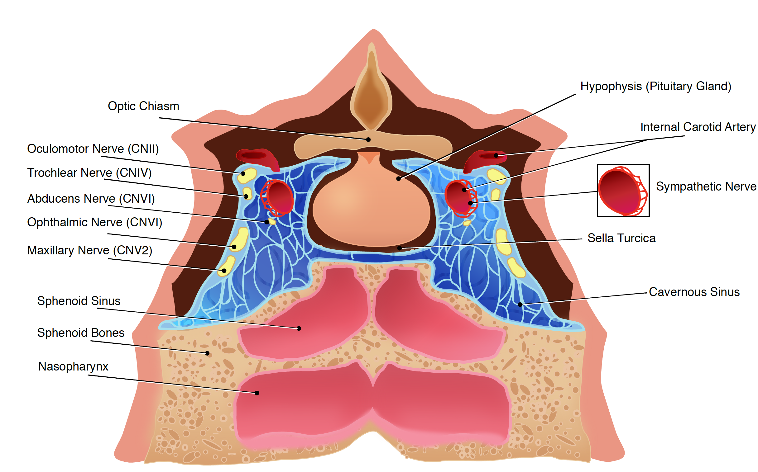
Anatomy of the cavernous sinus Contributed by Okkes Kuybu, MD and Diana
References
Yasuda A, Campero A, Martins C, Rhoton AL Jr, de Oliveira E, Ribas GC. Microsurgical anatomy and approaches to the cavernous sinus. Neurosurgery. 2008 Jun:62(6 Suppl 3):1240-63. doi: 10.1227/01.neu.0000333790.90972.59. Epub [PubMed PMID: 18695545]
Charbonneau F, Williams M, Lafitte F, Héran F. No more fear of the cavernous sinuses! Diagnostic and interventional imaging. 2013 Oct:94(10):1003-16. doi: 10.1016/j.diii.2013.08.012. Epub 2013 Oct 5 [PubMed PMID: 24099909]
Yoshihara M, Saito N, Kashima Y, Ishikawa H. The Ishikawa classification of cavernous sinus lesions by clinico-anatomical findings. Japanese journal of ophthalmology. 2001 Jul-Aug:45(4):420-4 [PubMed PMID: 11485777]
Bhatkar S, Goyal MK, Takkar A, Modi M, Mukherjee KK, Singh P, Radotra BD, Singh R, Lal V. Which Classification of Cavernous Sinus Syndrome is Better - Ishikawa or Jefferson? A Prospective Study of 73 Patients. Journal of neurosciences in rural practice. 2016 Dec:7(Suppl 1):S68-S71. doi: 10.4103/0976-3147.196448. Epub [PubMed PMID: 28163507]
Keane JR. Cavernous sinus syndrome. Analysis of 151 cases. Archives of neurology. 1996 Oct:53(10):967-71 [PubMed PMID: 8859057]
Level 3 (low-level) evidenceBhatkar S, Goyal MK, Takkar A, Mukherjee KK, Singh P, Singh R, Lal V. Cavernous sinus syndrome: A prospective study of 73 cases at a tertiary care centre in Northern India. Clinical neurology and neurosurgery. 2017 Apr:155():63-69. doi: 10.1016/j.clineuro.2017.02.017. Epub 2017 Feb 22 [PubMed PMID: 28260625]
Level 3 (low-level) evidenceAzarpira N, Taghipour M, Pourjebely M. Nasopharyngeal carcinoma with skull base erosion cytologic findings. Iranian Red Crescent medical journal. 2012 Aug:14(8):492-4 [PubMed PMID: 23105987]
Level 3 (low-level) evidenceBarrow DL, Spector RH, Braun IF, Landman JA, Tindall SC, Tindall GT. Classification and treatment of spontaneous carotid-cavernous sinus fistulas. Journal of neurosurgery. 1985 Feb:62(2):248-56 [PubMed PMID: 3968564]
Weerasinghe D, Lueck CJ. Septic Cavernous Sinus Thrombosis: Case Report and Review of the Literature. Neuro-ophthalmology (Aeolus Press). 2016 Dec:40(6):263-276 [PubMed PMID: 27928417]
Level 3 (low-level) evidenceCakirer S. MRI findings in Tolosa-Hunt syndrome before and after systemic corticosteroid therapy. European journal of radiology. 2003 Feb:45(2):83-90 [PubMed PMID: 12536085]
Zhang X, Zhou Z, Steiner TJ, Zhang W, Liu R, Dong Z, Wang X, Wang R, Yu S. Validation of ICHD-3 beta diagnostic criteria for 13.7 Tolosa-Hunt syndrome: Analysis of 77 cases of painful ophthalmoplegia. Cephalalgia : an international journal of headache. 2014 Jul:34(8):624-32. doi: 10.1177/0333102413520082. Epub 2014 Jan 29 [PubMed PMID: 24477599]
Level 2 (mid-level) evidencePollock BE, Stafford SL, Link MJ, Garces YI, Foote RL. Single-fraction radiosurgery for presumed intracranial meningiomas: efficacy and complications from a 22-year experience. International journal of radiation oncology, biology, physics. 2012 Aug 1:83(5):1414-8. doi: 10.1016/j.ijrobp.2011.10.033. Epub 2011 Dec 29 [PubMed PMID: 22209154]
Level 2 (mid-level) evidenceLee JY, Niranjan A, McInerney J, Kondziolka D, Flickinger JC, Lunsford LD. Stereotactic radiosurgery providing long-term tumor control of cavernous sinus meningiomas. Journal of neurosurgery. 2002 Jul:97(1):65-72 [PubMed PMID: 12134934]
Level 2 (mid-level) evidenceNicolato A, Foroni R, Alessandrini F, Bricolo A, Gerosa M. Radiosurgical treatment of cavernous sinus meningiomas: experience with 122 treated patients. Neurosurgery. 2002 Nov:51(5):1153-9; discussion 1159-61 [PubMed PMID: 12383360]
Spiegelmann R, Cohen ZR, Nissim O, Alezra D, Pfeffer R. Cavernous sinus meningiomas: a large LINAC radiosurgery series. Journal of neuro-oncology. 2010 Jun:98(2):195-202. doi: 10.1007/s11060-010-0173-1. Epub 2010 Apr 20 [PubMed PMID: 20405308]
Level 2 (mid-level) evidenceSheehan JP, Pouratian N, Steiner L, Laws ER, Vance ML. Gamma Knife surgery for pituitary adenomas: factors related to radiological and endocrine outcomes. Journal of neurosurgery. 2011 Feb:114(2):303-9. doi: 10.3171/2010.5.JNS091635. Epub 2010 Jun 11 [PubMed PMID: 20540596]
UCAS Japan Investigators, Morita A, Kirino T, Hashi K, Aoki N, Fukuhara S, Hashimoto N, Nakayama T, Sakai M, Teramoto A, Tominari S, Yoshimoto T. The natural course of unruptured cerebral aneurysms in a Japanese cohort. The New England journal of medicine. 2012 Jun 28:366(26):2474-82. doi: 10.1056/NEJMoa1113260. Epub [PubMed PMID: 22738097]
Wiebers DO, Whisnant JP, Huston J 3rd, Meissner I, Brown RD Jr, Piepgras DG, Forbes GS, Thielen K, Nichols D, O'Fallon WM, Peacock J, Jaeger L, Kassell NF, Kongable-Beckman GL, Torner JC, International Study of Unruptured Intracranial Aneurysms Investigators. Unruptured intracranial aneurysms: natural history, clinical outcome, and risks of surgical and endovascular treatment. Lancet (London, England). 2003 Jul 12:362(9378):103-10 [PubMed PMID: 12867109]
Level 2 (mid-level) evidencede Keizer R. Carotid-cavernous and orbital arteriovenous fistulas: ocular features, diagnostic and hemodynamic considerations in relation to visual impairment and morbidity. Orbit (Amsterdam, Netherlands). 2003 Jun:22(2):121-42 [PubMed PMID: 12789591]
O'Leary S, Hodgson TJ, Coley SC, Kemeny AA, Radatz MW. Intracranial dural arteriovenous malformations: results of stereotactic radiosurgery in 17 patients. Clinical oncology (Royal College of Radiologists (Great Britain)). 2002 Apr:14(2):97-102 [PubMed PMID: 12069135]
Desa V, Green R. Cavernous sinus thrombosis: current therapy. Journal of oral and maxillofacial surgery : official journal of the American Association of Oral and Maxillofacial Surgeons. 2012 Sep:70(9):2085-91. doi: 10.1016/j.joms.2011.09.048. Epub 2012 Feb 9 [PubMed PMID: 22326173]
Level 3 (low-level) evidenceSouthwick FS, Richardson EP Jr, Swartz MN. Septic thrombosis of the dural venous sinuses. Medicine. 1986 Mar:65(2):82-106 [PubMed PMID: 3512953]
Level 3 (low-level) evidenceCanhão P, Cortesão A, Cabral M, Ferro JM, Stam J, Bousser MG, Barinagarrementeria F, ISCVT Investigators. Are steroids useful to treat cerebral venous thrombosis? Stroke. 2008 Jan:39(1):105-10 [PubMed PMID: 18063833]
Level 2 (mid-level) evidenceHUNT WE, MEAGHER JN, LEFEVER HE, ZEMAN W. Painful opthalmoplegia. Its relation to indolent inflammation of the carvernous sinus. Neurology. 1961 Jan:11():56-62 [PubMed PMID: 13716871]
Zurawski J, Akhondi H. Tolosa-Hunt syndrome--a rare cause of headache and ophthalmoplegia. Lancet (London, England). 2013 Sep 7:382(9895):912. doi: 10.1016/S0140-6736(13)61442-7. Epub [PubMed PMID: 24012271]
Level 3 (low-level) evidenceSugano H, Iizuka Y, Arai H, Sato K. Progression of Tolosa-Hunt syndrome to a cavernous dural arteriovenous fistula: a case report. Headache. 2003 Feb:43(2):122-6 [PubMed PMID: 12558766]
Level 3 (low-level) evidenceWilmut I, Haley CS, Simons JP, Webb R. The potential role of molecular genetic manipulation in the improvement of reproductive performance. Journal of reproduction and fertility. Supplement. 1992:45():157-73 [PubMed PMID: 1304029]
Level 3 (low-level) evidenceSmith JR, Rosenbaum JT. A role for methotrexate in the management of non-infectious orbital inflammatory disease. The British journal of ophthalmology. 2001 Oct:85(10):1220-4 [PubMed PMID: 11567968]
Level 2 (mid-level) evidenceHalabi T, Sawaya R. Successful Treatment of Tolosa-Hunt Syndrome after a Single Infusion of Infliximab. Journal of clinical neurology (Seoul, Korea). 2018 Jan:14(1):126-127. doi: 10.3988/jcn.2018.14.1.126. Epub [PubMed PMID: 29629550]