 Tools
Tools
 Carotid Artery Stenosis
Introduction
Stroke is the third leading cause of death in developed countries and the leading cause of morbidity. The majority of strokes (85%) are ischemic in nature, and the most common source of occlusion is thought to be intracranial embolic disease from the ipsilateral carotid artery. Recurrent strokes are a significant concern, as these often occur in the same vascular territory as the initial stroke and are associated with 65% mortality.[1]
Determining the difference between hemodynamic and embolic causes in the presence of internal carotid artery stenosis is important for identifying the source of ischemia, which usually presents as a classic stroke or transient ischemic attack (TIA). Symptoms may include contralateral motor or sensory deficits and amaurosis fugax. Hemodynamic compromise may present similarly to a classic stroke or TIA, but may also be less predictable and atypical. Patients have reported symptoms, eg, limb shaking, retinal claudication, headache from large pulsatile external carotid artery collaterals, syncope, and generalized fatigue. One-third of all strokes are related to cervical carotid disease. The mechanism of cervical carotid stroke is usually embolization from a carotid bifurcation plaque, but hemodynamic compromise due to stenosis may also play a role. The risks of embolization and hemodynamic compromise increase with the degree of carotid artery stenosis.[2][3][4]
The benefit of carotid endarterectomy depends on the degree of stenosis, with greater benefit associated with higher levels of stenosis. Carotid endarterectomy is not a benign procedure, with an estimated 4% to 7% risk of stroke and death within the first 30 days of surgery. This is related to many factors, but primarily to the manipulation of intraluminal plaque. Periprocedural morbidity can also be difficult to quantify accurately because the natural history of stroke and TIA also results in a certain percentage of recurrent stroke and death.[1] Therefore, determining the stenosis levels at which the risk of carotid endarterectomy is outweighed by the benefit of medical management alone is essential. Optimal medical management should be applied to all patients and includes lipid-lowering agents, hypertension control, and diabetes management.
Timing of Carotid Endarterectomy
The timing of intervention for carotid stenosis remains controversial, as conflicting evidence exists regarding the risks and benefits across different treatment windows. The landmark North American Symptomatic Carotid Endarterectomy Trial (NASCET) and European Carotid Surgery Trial (ECST), both published in 1991, demonstrated that carotid endarterectomy for symptomatic carotid stenosis performed within 6 months of a symptomatic event reduces the rate of recurrent stroke. The greatest clinical benefit occurred when carotid endarterectomy was performed within 2 weeks of the ischemic event. These findings led to recommendations that carotid endarterectomy be performed within 14 days of an ischemic event, with some guidelines recommending an even shorter window, including carotid endarterectomy within 48 hours of the index event.[5]
A later analysis from the Swedish Vascular Registry (Swedvasc), published in 2012, reported higher perioperative complication rates when carotid endarterectomy was performed within 48 hours of the ischemic event compared with when performed 3 to 7 days after the event (11.5% versus 3.6%). This study also found no increased risk of embolic events when carotid endarterectomy was performed at 8 to 14 days after the event.[5][6] More recent guideline-based approaches recommend first classifying the index event as a TIA or a stroke, then tailoring the timing accordingly. Carotid endarterectomy within 48 hours may be considered after a TIA, whereas carotid endarterectomy performed between 3 and 7 days may be considered after a stroke.[4] An important exception includes patients with recurrent or worsening TIAs, also referred to as crescendo TIAs, who benefit from early intervention to reduce further neurologic compromise.[7] Multiple patient-specific factors continue to influence decision-making regarding optimal timing of carotid endarterectomy.
Asymptomatic Carotid Artery Stenosis
Another major area of controversy involves significant carotid artery stenosis in asymptomatic patients, defined as patients without neurologic symptoms for the preceding 6 months. Three randomized controlled trials evaluated carotid endarterectomy versus optimal medical management in asymptomatic carotid stenosis, typically defined as stenosis greater than 50%: the Veterans Affairs Cooperative Study Group, the Asymptomatic Carotid Atherosclerosis Study (ACAS), and the Asymptomatic Carotid Surgery Trial-1 (ACST-1). These studies generally demonstrated a reduction in stroke risk among patients undergoing carotid endarterectomy; however, important limitations were present, including a low incidence of ischemic stroke in both treatment groups, restriction of participation to surgeons with low complication rates in 2 trials, and substantial advances in medical therapy since the trials were conducted.[8][9]
Asymptomatic carotid stenosis is commonly defined as a narrowing of the proximal internal carotid artery by 50% or more at its cervical origin due to atherosclerosis. This diagnosis applies to patients without a history of recent ischemic stroke or transient ischemic attack involving the ipsilateral carotid territory within the preceding 6 months. Carotid stenosis is generally defined as 50% or greater narrowing, although many studies use 60% or greater, with stenosis exceeding 70% classified as severe carotid stenosis.[10] Recognition and management remain clinically important because asymptomatic carotid stenosis may not appear to be a decisive risk factor for ischemic stroke, yet it serves as a strong marker of systemic atherosclerotic disease and is associated with increased risk of coronary artery disease and mortality.[11]
Carotid artery stenosis represents a major cause of ischemic stroke due to atherosclerotic disease.[12] The presence or absence of recent transient ischemic attack or stroke plays a central role in major treatment decisions. Management of symptomatic carotid stenosis follows a more uniform and straightforward decision pathway for carotid revascularization.[13] In contrast, significant controversy exists regarding the management of asymptomatic carotid stenosis for revascularization, resulting in multiple differing guideline recommendations.[14][15][16][17] Mechanistically, hemodynamic compromise may result in transient ischemic attacks or stroke, while embolization from carotid bifurcation plaque represents the most common cause of cervical carotid stroke. Although risks of embolization and hemodynamic compromise increase with greater stenosis severity, more recent studies demonstrate that plaque composition and structural characteristics are strong predictors of stroke risk and clinical behavior in carotid artery stenosis.[18][19][20][21]
Over the past 3 decades, the best medical therapy for asymptomatic carotid stenosis has changed substantially. During the Asymptomatic Carotid Atherosclerosis Study (ACAS) era in the 1990s, recommended medical therapy primarily consisted of aspirin alone. Contemporary optimal medical therapy now includes multiple antiplatelet strategies, strict control of blood pressure and blood glucose, lipid-lowering agents, and comprehensive lifestyle modification, including smoking cessation, obesity management, regular exercise, and dietary optimization. These advances have significantly reduced stroke risk in medically managed patients. The 5-year ipsilateral stroke risk decreased from 11% in the 1995 ACAS trial to 3.6% in the 2010 Asymptomatic Carotid Surgery Trial-1 (ACST-1). A systematic review and meta-analysis of 11 studies demonstrated a decline in stroke risk from 2.8% to 1.4% between 1985 and 2007. Best medical therapy alone now reduces annual ipsilateral stroke risk to approximately 1%.[22] A 2023 systematic review analyzing 73 studies and 28,265 patients with asymptomatic carotid stenosis managed medically showed that ipsilateral ischemic stroke decreased by 24% every 5 years of recruitment.[23]
Given the markedly reduced stroke risk with modern medical therapy, the benefit of carotid revascularization procedures, including carotid endarterectomy and carotid artery stenting, has become more limited in asymptomatic patients. However, select high-risk patients may still benefit from intervention. These include patients with severe stenosis (80% to 99%), microembolic signals detected on transcranial Doppler, echolucent or ulcerated plaques on duplex ultrasound, reduced cerebrovascular reserve, documented progression of stenosis, and silent embolic infarcts identified on neuroimaging.[22][24]
Carotid Artery Stenosis and Coronary Artery Disease
Because carotid stenosis reflects systemic atherosclerosis, a substantial proportion of patients with severe carotid disease also have concomitant CAD. In patients requiring coronary artery bypass graft surgery (CABG) who also have severe (>70%) but asymptomatic carotid stenosis, the timing of carotid intervention relative to cardiac surgery remains an important clinical question. A randomized controlled trial involving 185 patients demonstrated significant benefit when carotid endarterectomy was performed either before or at the same time as CABG, compared with patients undergoing carotid endarterectomy 1 to 3 months after CABG. The delayed carotid endarterectomy group experienced a significantly higher risk of perioperative or delayed stroke.[25]
Etiology
Register For Free And Read The Full Article
 Search engine and full access to all medical articles
- 10 free questions in your specialty
- Free CME/CE Activities
- Free daily question in your email
- Save favorite articles to your dashboard
- Emails offering discounts
Learn more about a Subscription to StatPearls Point-of-Care
Etiology
Carotid artery stenosis is a consequence of systemic atherosclerotic disease. Thus, any risk factor predisposing a patient to progressive atherosclerosis can potentially manifest as stenosis of the carotid artery with resultant ischemic stroke or TIA symptoms. Risks include smoking, hyperlipidemia, male gender, and increased age.[26][27][28][29]
In a minority of patients, especially young females with carotid artery stenosis, fibromuscular dysplasia plays a much more significant role. Fibromuscular dysplasia is a noninflammatory, nonatherosclerotic process affecting the carotid and renal arteries, although it can occur elsewhere in medium-sized vessels. Fibromuscular dysplasia usually occurs in the mid and distal portions of the internal carotid artery, sometimes extending into the intracranial region. Aneurysms may also be a component of the disease process. Please see StatPearls' companion resource, "Carotid Artery Fibromuscular Dysplasia," for further information.[30]
Atherosclerotic plaque buildup along the internal carotid artery results from chronic accumulation of cholesterol. Risk factors include advanced age, male sex, hypertension, hyperlipidemia, genetic predispositions, and smoking.[7][16] Ischemia results from either plaque disruption and intracranial embolization or decreased cerebral flow.
A subset of patients has carotid artery stenosis secondary to fibromuscular dysplasia, a nonatherosclerotic, noninflammatory condition that affects the renal and carotid arteries. Fibromuscular dysplasia can present with mid-to-distal involvement of the carotid arteries, sometimes with extension into the intracranial region. As a result of fibromuscular dysplasia, carotid artery stenosis is more likely to affect younger female patients.[17][18]
Epidemiology
Symptomatic internal carotid artery occlusion has an incidence of 6 per 100,000, though the rate of asymptomatic chronic occlusion is unknown and may be higher because imaging is not routinely performed. Although patients who are Black and Hispanic have a higher risk of stroke than White patients, they have a lower incidence of severe stenosis (defined as >70%). This may explain lower rates of carotid endarterectomy in this population. Native Americans have a higher rate of severe stenosis than White patients. Males also have a higher rate of carotid artery disease than females.[31] The estimated prevalence of severe asymptomatic carotid stenosis (≥70%) ranges between 0.1% and 3.1%, increases with age, and has a population-attributable stroke risk of 0.7%.[22]
Stroke remains a significant health concern in the United States, with resultant long-term disability and mortality.[12][32] Approximately 20% of ischemic strokes are attributed to large artery atherosclerosis.[18][33][34] The incidence of carotid artery stenosis in patients with ischemic strokes is around 13 per 100,000.[34] The incidence of asymptomatic carotid stenosis is not well defined and is more often identified by a bruit or incidentally on imaging, as screening in the general population is not recommended.[35] Asymptomatic stenosis greater than 50% is estimated to be present in 7.5% of men and 5% of women.[36]
Pathophysiology
Total occlusion of the internal carotid artery results from thrombosis in the setting of chronic stenosis. Cardiogenic embolization to a normal carotid bifurcation or carotid dissection may also cause total occlusion of the internal carotid artery. Of note, if a cardioembolic source is present, symptoms may occur on the contralateral side of the stenosis, as emboli may travel more easily through the less-stenotic artery.
A previously asymptomatic chronic internal carotid artery occlusion may also become symptomatic if related to embolic or hemodynamic insults. Embolism may occur from the ipsilateral external carotid artery via collaterals to the cerebral circulation. It may also occur when there is occult patency of the occluded internal carotid artery, which then serves as the source of embolic material. Hemodynamic insufficiency may occur when any condition that interferes with cerebral perfusion, eg, orthostasis, hypotension, volume depletion, or cardiac failure, is superimposed on the carotid occlusion, especially when the contralateral carotid disease is significant.
Certain plaque characteristics are associated with an increased risk of embolization. Plaques that are echolucent or heterogeneous on CT scans contain high levels of lipid and blood components, respectively. These plaques are more likely to be ulcerated and pose a greater risk of stroke. One study showed that patients with symptomatic carotid stenosis had echolucent plaques 70% of the time, compared to asymptomatic patients who had only a 20% to 30% incidence of radiolucent plaques. Another study showed that patients with echolucent plaques had a 2- to 4-fold higher incidence of cerebral infarction compared with those with echogenic plaques.[37]
Atherosclerotic plaque tends to form at specific anatomic sites, eg, branch points and nonlinear vascular segments. The outer wall of the carotid artery is highly prone to atherosclerotic plaque, particularly at the bifurcation and bulb, due to non-laminar blood flow and oscillatory shear stress at the bifurcation. These hemodynamic forces contribute to intima-media thickening and atherosclerosis.[38][39] Asymptomatic carotid stenosis may progress to symptomatic in the setting of emboli or significant hemodynamic changes. Emboli may be cardiac in origin, eg, those from atrial fibrillation or valvular disease, or may result from carotid artery plaque dislodgement. Certain distinct imaging characteristics are associated with an increased risk of rupture and embolization. Echolucency and heterogeneity on imaging have been found to have a greater risk of ulceration and subsequent stroke.[20][40] Although asymptomatic carotid stenosis is not a strong predictor of ipsilateral ischemic stroke, this condition is a strong indicator of underlying generalized atherosclerotic disease and, therefore, a strong risk factor for coronary artery disease and mortality.[11]
History and Physical
Standard risk factors for coronary and systemic atherosclerosis also apply to carotid artery stenosis, eg, increased age, male sex, family history, smoking, hypertension, hyperlipidemia, sedentary lifestyle, and high dietary fat. Usually, patients will present with recent neurologic symptoms, including slurred speech, cranial nerve deficits, limb weakness, or visual disturbances. Amaurosis fugax is a common symptom of ipsilateral carotid stenosis, causing emboli to the ophthalmic or retinal arteries.
Blood pressure in both arms and orthostatic blood pressure should be part of vital signs (especially in older patients). A detailed neurologic exam should be performed, including visual fields of both eyes. Carotid bruit is neither sensitive nor specific for significant stenosis.[37] A cardiac exam may reveal an irregular heartbeat suggestive of atrial fibrillation or heart murmurs, which could indicate a cardioembolic or valvular source of stroke. A thorough skin and extremity exam should be performed to look for embolic skin lesions or changes of decreased vascularization, eg, coolness in the extremities, loss of distal pulses, skin discoloration, and hair loss of the affected limb.
Evaluation
The physical exam should be coupled with diagnostic imaging, eg, computed tomography (CT) or magnetic resonance imaging (MRI) of the brain, followed by telemetry floor admission, as less than a third of ischemic strokes are due to carotid artery disease. The remaining are due to other causes, such as intracranial pathology or cardiac emboli. An electrocardiogram (ECG) and a cardiac echocardiogram should also be performed to evaluate for embolic sources, including a patent foramen ovale.
In diagnosing carotid artery stenosis, a common initial test is a carotid duplex ultrasound, which is noninvasive and inexpensive. This examination typically shows a high-resistance signal in the carotid bulb and the proximal internal carotid artery. Distally, no Doppler signals are audible in the carotid artery. As a confirmatory test, a contrast examination, eg, digital subtraction arteriography, magnetic resonance arteriography (MRA), or CT arteriography, is required to confirm the diagnosis and, if indicated, plan a surgical approach. Gadolinium-enhanced MRA is better at differentiating high-grade stenosis than time-of-flight angiography, which can overestimate stenosis severity. MRI is considered the best modality for imaging not only the degree of stenosis but also the characteristics of the plaque, which contribute to increased embolization. Plaque vulnerability is associated with the following: plaque ulceration and fissuring; necrosis of the lipid core; and intraplaque hemorrhage, thrombus, or inflammation. Neck angiography will cover the aortic arch to the skull base.[41]
Treatment / Management
A large systematic review from the Cochrane Database demonstrated that patients with severe carotid stenosis of 70% to 99% derive a clear benefit from carotid endarterectomy, with a cumulative reduction in disabling or fatal stroke and perioperative death over 5 years. Patients with moderate stenosis of 50% to 69% also experience a significant reduction in these outcomes, although the magnitude of benefit is less pronounced. Among patients with moderate stenosis, the benefit is greater in men than in women, as women experience higher rates of surgical complications. In contrast, patients with stenosis ranging from 0% to 49% experience higher rates of disabling or fatal stroke and perioperative death with carotid endarterectomy compared with medical management alone.[1](A1)
Patients with near-occlusion, often described as having “trickle flow,” constitute a distinct category from those with severe 70% to 99% stenosis because of their lower stroke risk. Earlier studies supported the use of optimized medical therapy alone for these patients. However, a recent large meta-analysis suggested that advances in carotid endarterectomy and carotid artery stenting procedures may justify intervention in selected cases.[42]
Symptomatic Carotid Artery Stenosis Management
Chronic carotid artery occlusion accounts for approximately 6.5% of ischemic strokes.[43] No specific surveillance criteria have been established for patients with chronic internal carotid artery (ICA) occlusion, and duplex ultrasound criteria defining the degree of external carotid artery (ECA) stenosis remain unavailable. A reasonable approach includes clinical follow-up every 6 months to 1 year, with or without duplex ultrasonography. Development of new symptoms warrants further imaging to determine whether cerebrovascular revascularization should be considered.
Carotid artery stenting (CAS) is generally preferred in patients with symptomatic carotid occlusion of 50% to 99% who also have multiple comorbidities, a tracheostomy, prior neck radiation, or carotid dissection. CAS has traditionally been associated with a higher risk of stroke. However, advances in stent technology and procedural techniques have improved outcomes, making CAS comparable to carotid endarterectomy in many clinical settings. Studies indicate that CAS carries a higher procedural stroke rate, primarily involving nondisabling strokes, whereas carotid endarterectomy is associated with a significantly higher risk of procedural myocardial infarction.[8] The ongoing Carotid Revascularization and Medical Management for Asymptomatic Carotid Stenosis Trial (CREST-2) continues to evaluate carotid revascularization for primary stroke prevention and may provide further guidance in this area.
For patients with chronic total occlusion of the ICA, medical management remains the preferred treatment strategy. Nevertheless, certain clinical circumstances may justify procedural intervention on an individualized basis. Patients with ipsilateral hemodynamic symptoms in the setting of ipsilateral ICA occlusion and contralateral ICA stenosis may benefit from contralateral ICA revascularization to improve cerebral perfusion and alleviate hemodynamic insufficiency. Patients with ipsilateral embolic symptoms associated with ipsilateral ICA occlusion and ipsilateral ECA stenosis may benefit from ipsilateral ECA revascularization to eliminate the embolic source arising through enlarged ECA collateral pathways. Ligation of the ipsilateral ICA may also eliminate a potential source of embolization.[44][45][46](B3)
Asymptomatic Carotid Artery Stenosis Management
Best medical therapy, also known as risk-factor management, is the foundation of treatment for asymptomatic carotid stenosis. Lifestyle modifications, including weight reduction, a diet rich in fruits and vegetables, and regular physical activity, play an essential role in controlling atherosclerosis and reducing the risk of stroke and other vascular events.[47][48][49] Statin therapy contributes substantially to lipid reduction and decreases the risk of ischemic stroke. Aspirin therapy is recommended for stroke prevention in patients with asymptomatic carotid artery stenosis.[16][50][51] Smoking cessation remains one of the most important modifiable interventions in the management of atherosclerotic disease, with studies demonstrating reduced progression of carotid plaque following cessation.[52] Ongoing clinical follow-up enables assessment of adherence to medical therapy and lifestyle modifications, thereby supporting long-term risk reduction.(A1)
Best medical treatment alone can reduce the annual risk of ipsilateral stroke to about 1%.[22] Considering the low stroke risk for patients with asymptomatic carotid stenosis treated with best medical treatment, most patients with asymptomatic carotid stenosis derive limited benefits from carotid revascularization procedures, eg, carotid endarterectomy or carotid artery stenting. Nonetheless, patients with asymptomatic carotid stenosis remain at higher risk for future stroke and may benefit from revascularization procedures. According to the European Society for Vascular Surgery guidelines, carotid endarterectomy should be considered in patients with 60% to 99% stenosis, surgical risks of 3% or less, and a life expectancy greater than 5 years if they have 1 or more characteristics associated with increased risk of stroke.[24] These characteristics include the presence of microemboli on transcranial Doppler ultrasound, plaque echolucency on carotid duplex ultrasound, silent embolic infarcts on brain CT or MRI, asymptomatic carotid stenosis progression, decreased cerebrovascular reserve, increasing size of juxta-luminal hypoechoic area, carotid plaque ulceration, and intraplaque hemorrhage on MRI. Carotid artery stenosis is also recommended as an alternative to carotid endarterectomy in selected patients.[24](A1)
In patients with a history of neck radiation, unfavorable anatomy, dissection, or neck surgeries, stenting should be considered on a case-by-case basis. Early studies showed higher procedural rates of stroke in stenting and higher procedural rates of myocardial infarction following endarterectomy procedures.[53][54] Notably, these data were obtained during carotid stenting performed via a transfemoral approach. Transcarotid artery revascularization is a stenting method performed with a small incision at the base of the neck and exposure of the common carotid, followed by placement of a stent. Studies comparing transcarotid artery revascularization with transfemoral stenting have demonstrated lower rates of stroke and mortality with the transcarotid approach.[55][56](A1)
As atherosclerotic disease is a systemic condition, some patients have both carotid and coronary artery disease. Studies have reported up to 14% of patients with asymptomatic carotid stenosis requiring coronary artery bypass grafting surgery.[57] Currently, no recommendations or consensus have been established on the timing or sequence of carotid intervention in patients who need coronary artery bypass grafting surgery. Options include carotid endarterectomy and coronary artery bypass grafting surgery simultaneously, staging the procedures, or performing hybrid procedures, eg, carotid stenting or percutaneous coronary intervention.[58] The American College of Cardiology Foundation/American Heart Association (ACCF/AHA) Task Force has a class IIb recommendation for carotid revascularization in the presence of bilateral asymptomatic carotid stenosis greater than 70% or unilateral asymptomatic carotid stenosis greater than 70% with contralateral carotid occlusion in patients scheduled to undergo coronary artery bypass grafting surgery.[59](A1)
Differential Diagnosis
The differential diagnosis of carotid artery stenosis may include:
- Carotid artery dissection
- Fibromuscular dysplasia
- Valvular heart disease
- Arrhythmias (especially atrial fibrillation)
- Mural thrombosis
- Takayasu vasculitis
- Giant cell arteritis
- Complicated migraine
Prognosis
The risk of further stroke after successful carotid endarterectomy is associated with whether the initial ischemic event was a TIA or stroke. Patients who presented with TIA have an ipsilateral hemisphere stroke incidence of 1% to 2% per year, while those who presented with stroke have a corresponding risk of 2% to 3% per year.[37]
Patients with asymptomatic carotid artery stenosis have a 0.9% annual risk of ipsilateral ischemic stroke.[9] Evidence has shown that stroke rates in patients with asymptomatic carotid artery stenosis have progressively decreased when treated with best medical therapy.[60][61]
Complications
The main complication of carotid artery stenosis presenting with TIA, stroke, or other neurologic symptoms is recurrent stroke. The primary complications of carotid endarterectomy or CAS are perioperative stroke, as well as surgical complications. In asymptomatic patients with carotid stenosis of 70% to 99%, the estimated rate of ipsilateral acute ischemic stroke was 4.7% over 5 years.[9]
Deterrence and Patient Education
After a suspected neurologic event, patients and family members should be educated regarding common symptoms of stroke and TIA, including dysarthria, numbness, weakness, tongue deviation, and confusion, as recurrent symptoms are common. Patients and family members should be advised to seek urgent medical attention for recurrent or new neurologic symptoms. Patients should be educated about medication adherence, especially antiplatelet agents, to prevent further strokes or TIA. They should also receive nutrition counseling to avoid foods high in cholesterol and fat.
Pearls and Other Issues
The following factors should be kept in mind in the management of carotid artery stenosis:
- Current guidelines support carotid endarterectomy for symptomatic carotid stenosis greater than 70%. Guidelines also recommend carotid endarterectomy for symptomatic stenosis of 50% to 69%, with some leeway given to variable clinical features.
- Carotid endarterectomy should be performed within 14 days of the ischemic event that prompted referral. Urgent carotid endarterectomy (performed within 48 hours) depends on whether the initial presentation was a stroke or a TIA, with some evidence favoring it in the setting of TIA.
- Unless contraindicated, crescendo TIA warrants urgent carotid endarterectomy or CAS.
- The guidelines regarding asymptomatic stenosis are less well defined, but they suggest carotid endarterectomy in patients with greater than 60% to 70% stenosis who are good surgical candidates.
- Of note, most studies comparing carotid endarterectomy with optimal medical treatment selected surgeons with a periprocedural complication rate of less than 6%.
- Carotid artery percutaneous stenting (CAS) is comparable in efficacy and complication rates to carotid endarterectomy and is preferred in patients with comorbidities that preclude carotid endarterectomy.
- Ulcerated carotid plaques are highly associated with embolic stroke. Ulcerated plaques are radiolucent due to high lipid content, and ulcer size is also correlated with increased stroke risk.
- MRI is the best modality for imaging plaque characteristics that indicate plaque vulnerability, eg, ulceration, fissuring, a necrotic lipid core, or intraplaque hemorrhage.
- All patients, whether treated with carotid endarterectomy or medically, should receive aggressive lipid-lowering therapy, antiplatelet agents, including dual antiplatelet therapy in certain patients, and aggressive blood pressure management.
Enhancing Healthcare Team Outcomes
Carotid artery stenosis is a major contributor to ischemic stroke, most commonly due to atherosclerotic plaque causing embolization or hemodynamic cerebral hypoperfusion. Clinical presentation ranges from transient ischemic attack and amaurosis fugax to stroke with contralateral motor or sensory deficits, though atypical symptoms may occur with hemodynamic compromise. Diagnostic evaluation includes carotid duplex ultrasound followed by confirmatory CT angiography, MR angiography, or digital subtraction angiography, with plaque morphology providing additional risk stratification. Management depends on symptom status and degree of stenosis, integrating optimal medical therapy with antiplatelet agents, statins, blood pressure and glycemic control, and lifestyle modification. Carotid endarterectomy or stenting is indicated in selected patients, with timing critical to balancing recurrent stroke risk against perioperative complications.
Interprofessional collaboration improves outcomes through coordinated, guideline-based care. Physicians and advanced practitioners lead diagnostic evaluation, risk stratification, and procedural decision-making. Primary care clinicians ensure long-term vascular risk reduction and surveillance. Nurses support patient education, symptom recognition, and adherence to therapy. Pharmacists optimize medication selection, safety, and adherence. Radiologists contribute critical imaging interpretation and plaque characterization. Shared decision-making and timely referral pathways enhance stroke prevention, reduce recurrent events, and improve safety and quality of care across the continuum.
Media
(Click Image to Enlarge)
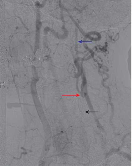
Collateralization in Common Carotid Artery (CCA) Occlusion. An example of collateralization in a patient with left CCA occlusion. The left CCA is occluded (black arrow). The left external carotid artery (ECA) fills via collaterals, and the left carotid bifurcation fills via the ECA (red arrow). The distal left internal carotid artery (ICA) then fills antegrade (blue arrow).
Contributed by S Dulebohn, MD
References
Rerkasem A, Orrapin S, Howard DP, Rerkasem K. Carotid endarterectomy for symptomatic carotid stenosis. The Cochrane database of systematic reviews. 2020 Sep 12:9(9):CD001081. doi: 10.1002/14651858.CD001081.pub4. Epub 2020 Sep 12 [PubMed PMID: 32918282]
Level 1 (high-level) evidenceDoonan RJ, Abdullah A, Steinmetz-Wood S, Mekhaiel S, Steinmetz OK, Obrand DI, Corriveau MM, Mackenzie KS, Gill HL. Carotid Endarterectomy Outcomes in the Elderly: A Canadian Institutional Experience. Annals of vascular surgery. 2019 Aug:59():16-20. doi: 10.1016/j.avsg.2018.12.084. Epub 2019 Feb 22 [PubMed PMID: 30802579]
Morales MM, Anacleto A, Filho CM, Ledesma S, Aldrovani M, Wolosker N. Peak Systolic Velocity for Calcified Plaques Fails to Estimate Carotid Stenosis Degree. Annals of vascular surgery. 2019 Aug:59():1-4. doi: 10.1016/j.avsg.2018.12.086. Epub 2019 Feb 22 [PubMed PMID: 30802575]
Savardekar AR, Narayan V, Patra DP, Spetzler RF, Sun H. Timing of Carotid Endarterectomy for Symptomatic Carotid Stenosis: A Snapshot of Current Trends and Systematic Review of Literature on Changing Paradigm towards Early Surgery. Neurosurgery. 2019 Aug 1:85(2):E214-E225. doi: 10.1093/neuros/nyy557. Epub [PubMed PMID: 30799491]
Level 1 (high-level) evidenceStrömberg S, Gelin J, Osterberg T, Bergström GM, Karlström L, Osterberg K, Swedish Vascular Registry (Swedvasc) Steering Committee. Very urgent carotid endarterectomy confers increased procedural risk. Stroke. 2012 May:43(5):1331-5. doi: 10.1161/STROKEAHA.111.639344. Epub 2012 Mar 15 [PubMed PMID: 22426315]
Level 2 (mid-level) evidenceStrömberg S, Nordanstig A, Bentzel T, Österberg K, Bergström GM. Risk of early recurrent stroke in symptomatic carotid stenosis. European journal of vascular and endovascular surgery : the official journal of the European Society for Vascular Surgery. 2015 Feb:49(2):137-44. doi: 10.1016/j.ejvs.2014.11.004. Epub 2014 Dec 26 [PubMed PMID: 25548062]
Level 2 (mid-level) evidenceBruls S, Van Damme H, Defraigne JO. Timing of carotid endarterectomy: a comprehensive review. Acta chirurgica Belgica. 2012 Jan:112(1):3-7 [PubMed PMID: 22442903]
Morris DR, Ayabe K, Inoue T, Sakai N, Bulbulia R, Halliday A, Goto S. Evidence-Based Carotid Interventions for Stroke Prevention: State-of-the-art Review. Journal of atherosclerosis and thrombosis. 2017 Apr 3:24(4):373-387. doi: 10.5551/jat.38745. Epub 2017 Mar 4 [PubMed PMID: 28260723]
Chang RW, Tucker LY, Rothenberg KA, Lancaster E, Faruqi RM, Kuang HC, Flint AC, Avins AL, Nguyen-Huynh MN. Incidence of Ischemic Stroke in Patients With Asymptomatic Severe Carotid Stenosis Without Surgical Intervention. JAMA. 2022 May 24:327(20):1974-1982. doi: 10.1001/jama.2022.4835. Epub [PubMed PMID: 35608581]
Level 2 (mid-level) evidenceSudheer P, Vibha D, Misra S. Asymptomatic Carotid Stenosis: Several Guidelines with Unclear Answers. Annals of Indian Academy of Neurology. 2022 Mar-Apr:25(2):171-176. doi: 10.4103/aian.aian_566_21. Epub 2022 Jan 13 [PubMed PMID: 35693653]
Goessens BM, Visseren FL, Kappelle LJ, Algra A, van der Graaf Y. Asymptomatic carotid artery stenosis and the risk of new vascular events in patients with manifest arterial disease: the SMART study. Stroke. 2007 May:38(5):1470-5 [PubMed PMID: 17363718]
Arasu R, Arasu A, Muller J. Carotid artery stenosis: An approach to its diagnosis and management. Australian journal of general practice. 2021 Nov:50(11):821-825. doi: 10.31128/AJGP-10-20-5664. Epub [PubMed PMID: 34713283]
Akhtar KH, Metzger DC, Latif F. Carotid Disease and Management. Interventional cardiology clinics. 2025 Apr:14(2):191-204. doi: 10.1016/j.iccl.2024.11.006. Epub 2025 Jan 24 [PubMed PMID: 40049847]
Eckstein HH. European Society for Vascular Surgery Guidelines on the Management of Atherosclerotic Carotid and Vertebral Artery Disease. European journal of vascular and endovascular surgery : the official journal of the European Society for Vascular Surgery. 2018 Jan:55(1):1-2. doi: 10.1016/j.ejvs.2017.06.026. Epub 2017 Aug 26 [PubMed PMID: 28851595]
Aboyans V, Ricco JB, Bartelink MEL, Björck M, Brodmann M, Cohnert T, Collet JP, Czerny M, De Carlo M, Debus S, Espinola-Klein C, Kahan T, Kownator S, Mazzolai L, Naylor AR, Roffi M, Röther J, Sprynger M, Tendera M, Tepe G, Venermo M, Vlachopoulos C, Desormais I, ESC Scientific Document Group. 2017 ESC Guidelines on the Diagnosis and Treatment of Peripheral Arterial Diseases, in collaboration with the European Society for Vascular Surgery (ESVS): Document covering atherosclerotic disease of extracranial carotid and vertebral, mesenteric, renal, upper and lower extremity arteriesEndorsed by: the European Stroke Organization (ESO)The Task Force for the Diagnosis and Treatment of Peripheral Arterial Diseases of the European Society of Cardiology (ESC) and of the European Society for Vascular Surgery (ESVS). European heart journal. 2018 Mar 1:39(9):763-816. doi: 10.1093/eurheartj/ehx095. Epub [PubMed PMID: 28886620]
Meschia JF, Bushnell C, Boden-Albala B, Braun LT, Bravata DM, Chaturvedi S, Creager MA, Eckel RH, Elkind MS, Fornage M, Goldstein LB, Greenberg SM, Horvath SE, Iadecola C, Jauch EC, Moore WS, Wilson JA, American Heart Association Stroke Council, Council on Cardiovascular and Stroke Nursing, Council on Clinical Cardiology, Council on Functional Genomics and Translational Biology, Council on Hypertension. Guidelines for the primary prevention of stroke: a statement for healthcare professionals from the American Heart Association/American Stroke Association. Stroke. 2014 Dec:45(12):3754-832. doi: 10.1161/STR.0000000000000046. Epub 2014 Oct 28 [PubMed PMID: 25355838]
Beckman JA, Ansel GM, Lyden SP, Das TS. Carotid Artery Stenting in Asymptomatic Carotid Artery Stenosis: JACC Review Topic of the Week. Journal of the American College of Cardiology. 2020 Feb 18:75(6):648-656. doi: 10.1016/j.jacc.2019.11.054. Epub [PubMed PMID: 32057380]
Puig N, Solé A, Aguilera-Simon A, Griñán R, Rotllan N, Camps-Renom P, Benitez S. Novel Therapeutic Approaches to Prevent Atherothrombotic Ischemic Stroke in Patients with Carotid Atherosclerosis. International journal of molecular sciences. 2023 Sep 20:24(18):. doi: 10.3390/ijms241814325. Epub 2023 Sep 20 [PubMed PMID: 37762627]
Schwarz F, Bayer-Karpinska A, Poppert H, Buchholz M, Cyran C, Grimm J, Helck A, Nikolaou K, Opherk C, Dichgans M, Saam T. Serial carotid MRI identifies rupture of a vulnerable plaque resulting in amaurosis fugax. Neurology. 2013 Mar 19:80(12):1171-2. doi: 10.1212/WNL.0b013e31828869ad. Epub [PubMed PMID: 23509049]
Saba L, Anzidei M, Marincola BC, Piga M, Raz E, Bassareo PP, Napoli A, Mannelli L, Catalano C, Wintermark M. Imaging of the carotid artery vulnerable plaque. Cardiovascular and interventional radiology. 2014 Jun:37(3):572-85. doi: 10.1007/s00270-013-0711-2. Epub 2013 Aug 3 [PubMed PMID: 23912494]
Saba L, Sanfilippo R, Sannia S, Anzidei M, Montisci R, Mallarini G, Suri JS. Association between carotid artery plaque volume, composition, and ulceration: a retrospective assessment with MDCT. AJR. American journal of roentgenology. 2012 Jul:199(1):151-6. doi: 10.2214/AJR.11.6955. Epub [PubMed PMID: 22733906]
Level 2 (mid-level) evidenceKim HW, Regenhardt RW, D'Amato SA, Nahhas MI, Dmytriw AA, Hirsch JA, Silverman SB, Martinez-Gutierrez JC. Asymptomatic carotid artery stenosis: a summary of current state of evidence for revascularization and emerging high-risk features. Journal of neurointerventional surgery. 2023 Jul:15(7):717-722. doi: 10.1136/jnis-2022-018732. Epub 2022 Sep 9 [PubMed PMID: 36190935]
Saratzis A, Naylor R. 30 Day Outcomes After Carotid Interventions: An Updated Meta-analysis of Randomised Controlled Trials in Asymptomatic Patients. European journal of vascular and endovascular surgery : the official journal of the European Society for Vascular Surgery. 2022 Jan:63(1):157-158. doi: 10.1016/j.ejvs.2021.10.029. Epub 2021 Nov 25 [PubMed PMID: 34838436]
Level 1 (high-level) evidenceNaylor R, Rantner B, Ancetti S, de Borst GJ, De Carlo M, Halliday A, Kakkos SK, Markus HS, McCabe DJH, Sillesen H, van den Berg JC, Vega de Ceniga M, Venermo MA, Vermassen FEG, Esvs Guidelines Committee, Antoniou GA, Bastos Goncalves F, Bjorck M, Chakfe N, Coscas R, Dias NV, Dick F, Hinchliffe RJ, Kolh P, Koncar IB, Lindholt JS, Mees BME, Resch TA, Trimarchi S, Tulamo R, Twine CP, Wanhainen A, Document Reviewers, Bellmunt-Montoya S, Bulbulia R, Darling RC 3rd, Eckstein HH, Giannoukas A, Koelemay MJW, Lindström D, Schermerhorn M, Stone DH. Editor's Choice - European Society for Vascular Surgery (ESVS) 2023 Clinical Practice Guidelines on the Management of Atherosclerotic Carotid and Vertebral Artery Disease. European journal of vascular and endovascular surgery : the official journal of the European Society for Vascular Surgery. 2023 Jan:65(1):7-111. doi: 10.1016/j.ejvs.2022.04.011. Epub 2022 May 20 [PubMed PMID: 35598721]
Level 1 (high-level) evidenceIlluminati G, Ricco JB, Caliò F, Pacilè MA, Miraldi F, Frati G, Macrina F, Toscano M. Short-term results of a randomized trial examining timing of carotid endarterectomy in patients with severe asymptomatic unilateral carotid stenosis undergoing coronary artery bypass grafting. Journal of vascular surgery. 2011 Oct:54(4):993-9; discussion 998-9. doi: 10.1016/j.jvs.2011.03.284. Epub 2011 Jun 23 [PubMed PMID: 21703806]
Level 1 (high-level) evidenceCoelho AP, Lobo M, Nogueira C, Gouveia R, Campos J, Augusto R, Coelho N, Semião AC, Canedo A. Overview of evidence on risk factors and early management of acute carotid stent thrombosis during the last two decades. Journal of vascular surgery. 2019 Mar:69(3):952-964. doi: 10.1016/j.jvs.2018.09.053. Epub [PubMed PMID: 30798846]
Level 3 (low-level) evidenceTexakalidis P, Chaitidis N, Giannopoulos S, Giannopoulos S, Machinis T, Jabbour P, Rivet D, Reavey-Cantwell J, Rangel-Castilla L. Carotid Revascularization in Older Adults: A Systematic Review and Meta-Analysis. World neurosurgery. 2019 Jun:126():656-663.e1. doi: 10.1016/j.wneu.2019.02.030. Epub 2019 Feb 22 [PubMed PMID: 30797928]
Level 1 (high-level) evidenceTokugawa J, Kudo K, Mitsuhashi T, Yanagisawa N, Nojiri S, Hishii M. Surgical Results of Carotid Endarterectomy for Twisted Carotid Bifurcation. World neurosurgery. 2019 Jun:126():e153-e156. doi: 10.1016/j.wneu.2019.01.282. Epub 2019 Feb 19 [PubMed PMID: 30794973]
Wangqin R, Krafft PR, Piper K, Kumar J, Xu K, Mokin M, Ren Z. Management of De Novo Carotid Stenosis and Postintervention Restenosis-Carotid Endarterectomy Versus Carotid Artery Stenting-a Review of Literature. Translational stroke research. 2019 Oct:10(5):460-474. doi: 10.1007/s12975-019-00693-z. Epub 2019 Feb 22 [PubMed PMID: 30793257]
Level 2 (mid-level) evidenceCardounell SZ, Gonzalez L. Carotid Artery Fibromuscular Dysplasia. StatPearls. 2026 Jan:(): [PubMed PMID: 30855787]
Lal BK, Meschia JF, Brott TG, Jones M, Aronow HD, Lackey A, Howard G. Race Differences in High-Grade Carotid Artery Stenosis. Stroke. 2021 Jun:52(6):2053-2059. doi: 10.1161/STROKEAHA.120.032723. Epub 2021 May 4 [PubMed PMID: 33940957]
Feigin VL, Brainin M, Norrving B, Martins S, Sacco RL, Hacke W, Fisher M, Pandian J, Lindsay P. World Stroke Organization (WSO): Global Stroke Fact Sheet 2022. International journal of stroke : official journal of the International Stroke Society. 2022 Jan:17(1):18-29. doi: 10.1177/17474930211065917. Epub [PubMed PMID: 34986727]
Petty GW, Brown RD Jr, Whisnant JP, Sicks JD, O'Fallon WM, Wiebers DO. Ischemic stroke subtypes: a population-based study of incidence and risk factors. Stroke. 1999 Dec:30(12):2513-6 [PubMed PMID: 10582970]
Flaherty ML, Kissela B, Khoury JC, Alwell K, Moomaw CJ, Woo D, Khatri P, Ferioli S, Adeoye O, Broderick JP, Kleindorfer D. Carotid artery stenosis as a cause of stroke. Neuroepidemiology. 2013:40(1):36-41. doi: 10.1159/000341410. Epub 2012 Oct 11 [PubMed PMID: 23075828]
US Preventive Services Task Force, Krist AH, Davidson KW, Mangione CM, Barry MJ, Cabana M, Caughey AB, Donahue K, Doubeni CA, Epling JW Jr, Kubik M, Ogedegbe G, Pbert L, Silverstein M, Simon MA, Tseng CW, Wong JB. Screening for Asymptomatic Carotid Artery Stenosis: US Preventive Services Task Force Recommendation Statement. JAMA. 2021 Feb 2:325(5):476-481. doi: 10.1001/jama.2020.26988. Epub [PubMed PMID: 33528542]
de Weerd M, Greving JP, Hedblad B, Lorenz MW, Mathiesen EB, O'Leary DH, Rosvall M, Sitzer M, Buskens E, Bots ML. Prevalence of asymptomatic carotid artery stenosis in the general population: an individual participant data meta-analysis. Stroke. 2010 Jun:41(6):1294-7. doi: 10.1161/STROKEAHA.110.581058. Epub 2010 Apr 29 [PubMed PMID: 20431077]
Level 1 (high-level) evidenceMoore WS, Barnett HJ, Beebe HG, Bernstein EF, Brener BJ, Brott T, Caplan LR, Day A, Goldstone J, Hobson RW 2nd. Guidelines for carotid endarterectomy. A multidisciplinary consensus statement from the ad hoc Committee, American Heart Association. Stroke. 1995 Jan:26(1):188-201 [PubMed PMID: 7839390]
Level 1 (high-level) evidenceMoore JE Jr, Xu C, Glagov S, Zarins CK, Ku DN. Fluid wall shear stress measurements in a model of the human abdominal aorta: oscillatory behavior and relationship to atherosclerosis. Atherosclerosis. 1994 Oct:110(2):225-40 [PubMed PMID: 7848371]
Ku DN, Giddens DP, Zarins CK, Glagov S. Pulsatile flow and atherosclerosis in the human carotid bifurcation. Positive correlation between plaque location and low oscillating shear stress. Arteriosclerosis (Dallas, Tex.). 1985 May-Jun:5(3):293-302 [PubMed PMID: 3994585]
Saba L, Anzidei M, Sanfilippo R, Montisci R, Lucatelli P, Catalano C, Passariello R, Mallarini G. Imaging of the carotid artery. Atherosclerosis. 2012 Feb:220(2):294-309. doi: 10.1016/j.atherosclerosis.2011.08.048. Epub 2011 Sep 17 [PubMed PMID: 21968317]
Brinjikji W, Huston J 3rd, Rabinstein AA, Kim GM, Lerman A, Lanzino G. Contemporary carotid imaging: from degree of stenosis to plaque vulnerability. Journal of neurosurgery. 2016 Jan:124(1):27-42. doi: 10.3171/2015.1.JNS142452. Epub 2015 Jul 31 [PubMed PMID: 26230478]
Antonopoulos CN, Giosdekos A, Mylonas SN, Liapis CD. Management of internal carotid artery near-occlusion: the need for updated evidence. Annals of translational medicine. 2020 Oct:8(19):1263. doi: 10.21037/atm.2020.03.148. Epub [PubMed PMID: 33178795]
Wan M, Yan L, Xu Z, Hou Z, Kang K, Cui R, Yu Y, Song J, Hui FK, Wang Y, Miao Z, Lou X, Ma N. Symptomatic and Asymptomatic Chronic Carotid Artery Occlusion on High-Resolution MR Vessel Wall Imaging. AJNR. American journal of neuroradiology. 2022 Jan:43(1):110-116. doi: 10.3174/ajnr.A7365. Epub 2021 Dec 2 [PubMed PMID: 34857516]
Gaba K, Bulbulia R. Identifying asymptomatic patients at high-risk for stroke. The Journal of cardiovascular surgery. 2019 Jun:60(3):332-344. doi: 10.23736/S0021-9509.19.10912-3. Epub 2019 Feb 20 [PubMed PMID: 30785251]
Lamanna A, Maingard J, Barras CD, Kok HK, Handelman G, Chandra RV, Thijs V, Brooks DM, Asadi H. Carotid artery stenting: Current state of evidence and future directions. Acta neurologica Scandinavica. 2019 Apr:139(4):318-333. doi: 10.1111/ane.13062. Epub 2019 Feb 6 [PubMed PMID: 30613950]
Level 3 (low-level) evidenceMcGuire LS, See AP, Kwasnicki A, Charbel FT. External carotid artery to internal carotid artery transposition to augment flow for a superficial temporal artery to middle cerebral artery bypass associated with severe external carotid artery stenosis. Acta neurochirurgica. 2021 Dec:163(12):3495-3499. doi: 10.1007/s00701-021-04974-1. Epub 2021 Aug 21 [PubMed PMID: 34420106]
Carreira M, Duarte-Gamas L, Rocha-Neves J, Andrade JP, Fernando-Teixeira J. Management of The Carotid Artery Stenosis in Asymptomatic Patients. Revista portuguesa de cirurgia cardio-toracica e vascular : orgao oficial da Sociedade Portuguesa de Cirurgia Cardio-Toracica e Vascular. 2020 Jul-Sep:27(3):159-166 [PubMed PMID: 33068504]
Raman G, Moorthy D, Hadar N, Dahabreh IJ, O'Donnell TF, Thaler DE, Feldmann E, Lau J, Kitsios GD. Management strategies for asymptomatic carotid stenosis: a systematic review and meta-analysis. Annals of internal medicine. 2013 May 7:158(9):676-685. doi: 10.7326/0003-4819-158-9-201305070-00007. Epub [PubMed PMID: 23648949]
Level 1 (high-level) evidenceYu E, Malik VS, Hu FB. Cardiovascular Disease Prevention by Diet Modification: JACC Health Promotion Series. Journal of the American College of Cardiology. 2018 Aug 21:72(8):914-926. doi: 10.1016/j.jacc.2018.02.085. Epub [PubMed PMID: 30115231]
Kang S, Wu Y, Li X. Effects of statin therapy on the progression of carotid atherosclerosis: a systematic review and meta-analysis. Atherosclerosis. 2004 Dec:177(2):433-42 [PubMed PMID: 15530920]
Level 1 (high-level) evidenceAmarenco P, Bogousslavsky J, Callahan A 3rd, Goldstein LB, Hennerici M, Rudolph AE, Sillesen H, Simunovic L, Szarek M, Welch KM, Zivin JA, Stroke Prevention by Aggressive Reduction in Cholesterol Levels (SPARCL) Investigators. High-dose atorvastatin after stroke or transient ischemic attack. The New England journal of medicine. 2006 Aug 10:355(6):549-59 [PubMed PMID: 16899775]
Level 1 (high-level) evidenceStein JH, Smith SS, Hansen KM, Korcarz CE, Piper ME, Fiore MC, Baker TB. Longitudinal effects of smoking cessation on carotid artery atherosclerosis in contemporary smokers: The Wisconsin Smokers Health Study. Atherosclerosis. 2020 Dec:315():62-67. doi: 10.1016/j.atherosclerosis.2020.11.010. Epub 2020 Nov 15 [PubMed PMID: 33227549]
Brott TG, Hobson RW 2nd, Howard G, Roubin GS, Clark WM, Brooks W, Mackey A, Hill MD, Leimgruber PP, Sheffet AJ, Howard VJ, Moore WS, Voeks JH, Hopkins LN, Cutlip DE, Cohen DJ, Popma JJ, Ferguson RD, Cohen SN, Blackshear JL, Silver FL, Mohr JP, Lal BK, Meschia JF, CREST Investigators. Stenting versus endarterectomy for treatment of carotid-artery stenosis. The New England journal of medicine. 2010 Jul 1:363(1):11-23. doi: 10.1056/NEJMoa0912321. Epub 2010 May 26 [PubMed PMID: 20505173]
Halliday A, Bulbulia R, Bonati LH, Chester J, Cradduck-Bamford A, Peto R, Pan H, ACST-2 Collaborative Group. Second asymptomatic carotid surgery trial (ACST-2): a randomised comparison of carotid artery stenting versus carotid endarterectomy. Lancet (London, England). 2021 Sep 18:398(10305):1065-1073. doi: 10.1016/S0140-6736(21)01910-3. Epub 2021 Aug 29 [PubMed PMID: 34469763]
Level 1 (high-level) evidenceKwolek CJ, Jaff MR, Leal JI, Hopkins LN, Shah RM, Hanover TM, Macdonald S, Cambria RP. Results of the ROADSTER multicenter trial of transcarotid stenting with dynamic flow reversal. Journal of vascular surgery. 2015 Nov:62(5):1227-34. doi: 10.1016/j.jvs.2015.04.460. Epub [PubMed PMID: 26506270]
Level 1 (high-level) evidenceZhu J, Rao A, Ting W, Han D, Tadros R, Finlay D, Phair J, Vouyouka A, Liu H, Marin M, Faries P. Comparison of Transcarotid Artery Revascularization and Transfemoral Carotid Artery Stenting Based on High Risk Anatomic Characteristics. Annals of vascular surgery. 2022 Nov:87():21-30. doi: 10.1016/j.avsg.2022.03.034. Epub 2022 Apr 6 [PubMed PMID: 35398198]
Borger MA, Fremes SE, Weisel RD, Cohen G, Rao V, Lindsay TF, Naylor CD. Coronary bypass and carotid endarterectomy: does a combined approach increase risk? A metaanalysis. The Annals of thoracic surgery. 1999 Jul:68(1):14-20; discussion 21 [PubMed PMID: 10421108]
Manthey S, Spears J, Goldberg S. Coexisting Coronary and Carotid Artery Disease - Which Technique and in Which Order? Case Report and Review of Literature. Clinical Medicine Insights. Cardiology. 2020:14():1179546820951797. doi: 10.1177/1179546820951797. Epub 2020 Aug 27 [PubMed PMID: 32922112]
Level 3 (low-level) evidenceHillis LD, Smith PK, Anderson JL, Bittl JA, Bridges CR, Byrne JG, Cigarroa JE, Disesa VJ, Hiratzka LF, Hutter AM Jr, Jessen ME, Keeley EC, Lahey SJ, Lange RA, London MJ, Mack MJ, Patel MR, Puskas JD, Sabik JF, Selnes O, Shahian DM, Trost JC, Winniford MD, American College of Cardiology Foundation, American Heart Association Task Force on Practice Guidelines, American Association for Thoracic Surgery, Society of Cardiovascular Anesthesiologists, Society of Thoracic Surgeons. 2011 ACCF/AHA Guideline for Coronary Artery Bypass Graft Surgery. A report of the American College of Cardiology Foundation/American Heart Association Task Force on Practice Guidelines. Developed in collaboration with the American Association for Thoracic Surgery, Society of Cardiovascular Anesthesiologists, and Society of Thoracic Surgeons. Journal of the American College of Cardiology. 2011 Dec 6:58(24):e123-210. doi: 10.1016/j.jacc.2011.08.009. Epub 2011 Nov 7 [PubMed PMID: 22070836]
Level 1 (high-level) evidenceAbbott AL. Medical (nonsurgical) intervention alone is now best for prevention of stroke associated with asymptomatic severe carotid stenosis: results of a systematic review and analysis. Stroke. 2009 Oct:40(10):e573-83. doi: 10.1161/STROKEAHA.109.556068. Epub 2009 Aug 20 [PubMed PMID: 19696421]
Level 1 (high-level) evidenceSpence JD, Coates V, Li H, Tamayo A, Muñoz C, Hackam DG, DiCicco M, DesRoches J, Bogiatzi C, Klein J, Madrenas J, Hegele RA. Effects of intensive medical therapy on microemboli and cardiovascular risk in asymptomatic carotid stenosis. Archives of neurology. 2010 Feb:67(2):180-6. doi: 10.1001/archneurol.2009.289. Epub 2009 Dec 14 [PubMed PMID: 20008646]