 Tools
Tools
 Calcaneus Fractures
Introduction
The calcaneus is the largest tarsal bone and serves as the primary weight-bearing structure of the hindfoot, transmitting forces from the talus to the ground while providing attachment for the Achilles tendon and plantar fascia. The subtalar joint between the talus and calcaneus primarily contributes to inversion and eversion (see Image. Labeled Lateral Radiograph of the Hindfoot and Midfoot Bones). Calcaneal fractures account for approximately 60% of all tarsal fractures and are most commonly the result of high-energy axial loading, such as falls from height or motor vehicle collisions. Approximately 70% to 75% are intra-articular fractures involving the posterior facet of the subtalar joint, whereas the remainder are extra-articular injuries. In addition to traumatic fractures, calcaneal stress fractures occur from repetitive submaximal loading and are frequently encountered in runners, military recruits, and individuals with osteoporosis.
The calcaneus articulates superiorly with the talus through the posterior, middle, and anterior subtalar facets and anteriorly with the cuboid. These articulations are essential for subtalar motion, allowing inversion and eversion of the hindfoot. The calcaneus also functions as the insertion site for the Achilles tendon posteriorly and provides origin for the plantar fascia inferiorly. Because of its complex anatomy and limited surrounding soft-tissue envelope, fracture displacement frequently alters hindfoot alignment, reduces calcaneal height, widens the heel, and disrupts normal subtalar biomechanics.
Intra-articular calcaneal fractures are commonly categorized into tongue-type and joint depression-type fracture patterns. Tongue-type fractures involve a fracture line extending through the posterior tuberosity, allowing the Achilles tendon to displace the tuberosity fragment superiorly. This displacement places significant tension on the posterior heel skin, creating an orthopedic emergency because of the risk of skin necrosis.
In contrast, joint depression-type fractures result in depression of the posterior facet with less displacement of the tuberosity. Although they are less likely to threaten the posterior skin, they often produce greater articular incongruity and subtalar joint disruption. Historically, some calcaneal burst fractures were referred to as “lover’s fractures,” a term describing an injury sustained after jumping from a lover’s balcony to avoid detection.[1][2][3]
The natural history of untreated displaced intra-articular fractures is characterized by persistent hindfoot pain, loss of calcaneal height, heel widening, subtalar stiffness, malunion, and progressive post-traumatic arthritis. Calcaneal stress fractures generally follow a more favorable course if recognized early, although continued weight-bearing may result in fracture propagation or displacement. Chronic deformity may lead to altered gait mechanics, peroneal tendon impingement, shoe wear difficulty, and long-term functional impairment.
Management depends on fracture morphology, displacement, patient comorbidities, and soft tissue condition. Nondisplaced fractures and most stress fractures are treated nonoperatively with immobilization, protected or non-weight-bearing, gradual rehabilitation, and progressive return to activity after radiographic evidence of healing. Displaced intra-articular fractures often require open reduction and internal fixation or minimally invasive fixation to restore posterior facet congruity, calcaneal height, width, and hindfoot alignment. Tongue-type fractures with threatened posterior skin require urgent reduction and fixation to prevent soft tissue compromise, whereas definitive fixation of joint depression fractures is typically delayed until soft tissue swelling resolves.
Prognosis is influenced by fracture severity, quality of reduction, associated soft tissue injury, and patient-specific factors such as smoking, diabetes mellitus, and vascular disease. Extra-articular fractures and stress fractures generally have excellent outcomes with appropriate treatment. Conversely, displaced intra-articular fractures frequently require prolonged rehabilitation, with recovery extending 12 to 18 months. Despite optimal management, some patients develop chronic pain, subtalar arthritis, stiffness, or require secondary subtalar arthrodesis. Nevertheless, early diagnosis, meticulous soft tissue management, anatomic restoration of hindfoot alignment, and structured rehabilitation remain the primary determinants of favorable long-term functional outcomes.
Etiology
Register For Free And Read The Full Article
 Search engine and full access to all medical articles
- 10 free questions in your specialty
- Free CME/CE Activities
- Free daily question in your email
- Save favorite articles to your dashboard
- Emails offering discounts
Learn more about a Subscription to StatPearls Point-of-Care
Etiology
Calcaneal fractures most often result from high-energy axial loading of the heel, such as falls from height or motor vehicle collisions, in which the talus wedges into the calcaneus, producing intra-articular or comminuted fractures of the calcaneal body. Lower-energy mechanisms, including twisting injuries, direct blows, or inversion events, can produce extra-articular patterns, such as anterior process, sustentacular, or tuberosity fractures, without the classic burst morphology. Calcaneal stress fractures arise from repetitive submaximal loading that exceeds the bone’s ability to remodel and are seen in runners, military recruits, and patients with risk factors such as osteoporosis, corticosteroid use, or sudden increases in training. These stress injuries include fatigue fractures in normal bone exposed to excessive cyclic loading and insufficiency fractures in weakened bone exposed to normal loading and typically present more subtly than acute, high-energy trauma.[4][5]
Epidemiology
Calcaneus fractures are the most common fractures of the tarsal bones, accounting for approximately 60% of all tarsal fractures and 1% to 2% of all fractures. They most frequently occur in young, working-age adults, with a peak incidence between 30 and 50 years of age. Men are affected significantly more often than women, largely due to greater occupational and recreational exposure to high-energy trauma. The predominant mechanism of injury is axial loading from falls from height, followed by motor vehicle collisions, making these injuries particularly common among construction workers, roofers, and industrial laborers. Approximately 70% to 75% of calcaneal fractures are intra-articular, involving the posterior facet of the subtalar joint, while the remaining fractures are extra-articular.
Because calcaneal fractures typically result from high-energy mechanisms, they are frequently associated with concomitant injuries. Lumbar spine compression fractures, lower extremity fractures, pelvic injuries, and contralateral calcaneal fractures occur in a substantial proportion of patients, necessitating a thorough trauma evaluation. Bilateral calcaneal fractures are reported in approximately 5% to 10% of cases. Smoking, diabetes mellitus, peripheral vascular disease, and obesity are associated with increased complication rates and poorer functional outcomes. Although calcaneal fractures represent a relatively small percentage of all fractures, they account for a disproportionate amount of long-term disability, prolonged work absenteeism, and socioeconomic burden due to persistent pain, post-traumatic arthritis, and limitations in mobility.[6]
Pathophysiology
Calcaneus fractures most commonly result from high-energy axial loading, such as a fall from height or motor vehicle collision, in which compressive forces are transmitted through the talus into the calcaneus. This force causes trabecular and cortical bone failure, frequently resulting in intra-articular fractures involving the posterior facet of the subtalar joint. The degree of fracture comminution and displacement is determined by the magnitude of the applied force, bone quality, and foot position at the time of injury.
Disruption of the subtalar joint alters hindfoot biomechanics, leading to loss of calcaneal height, widening of the heel, decreased Böhler's angle, and increased varus or valgus malalignment. These structural changes impair normal load transmission across the hindfoot and compromise gait mechanics. The initial injury also produces significant soft tissue trauma, edema, and hemorrhage, which contribute to the high rate of wound complications associated with surgical management.
Gunshot wounds and other ballistic injuries produce more unpredictable fracture patterns, although such injuries are uncommon. Avulsion fractures require significant twisting or shearing forces due to the strength of ligamentous and tendinous attachments to the calcaneus. The tibial artery and nerve course along the medial aspect of the calcaneal body and are considered relatively protected by the sustentaculum tali. Therefore, neurovascular injury is uncommon in calcaneal fractures.[7][8]
History and Physical
A thorough history and physical examination are essential for diagnosing calcaneus fractures and identifying associated injuries. Most calcaneal fractures result from high-energy axial loading mechanisms, such as falls from height or motor vehicle collisions, although low-energy mechanisms may occur in patients with osteoporosis or other metabolic bone disorders. Patients typically report immediate onset of severe heel pain, inability or difficulty bearing weight, and substantial swelling following injury. The mechanism of injury should be carefully elicited, as high-energy trauma is frequently associated with concomitant injuries to the lumbar spine, pelvis, lower extremities, and contralateral calcaneus. A complete trauma evaluation and tertiary physical exam is therefore warranted in patients suspected of calcaneus fractures.
The physical examination begins with inspecting the affected foot for swelling, ecchymosis, deformity, and fracture blisters. Plantar ecchymosis extending across the sole (Mondor sign) is highly suggestive of a calcaneal fracture. The heel should be assessed for widening, loss of normal contour, varus or valgus malalignment, and shortening. Palpation typically reveals tenderness over the calcaneal tuberosity, subtalar joint, and lateral wall. Active and passive ankle and subtalar range of motion are often limited because of pain. A comprehensive neurovascular examination should be performed, documenting the posterior tibial and dorsalis pedis pulses and the sensory function of the tibial, sural, superficial peroneal, deep peroneal, and saphenous nerves. The overlying soft tissues require careful assessment, as swelling, blistered fractures, and skin compromise significantly influence treatment timing.
Evaluation differs depending on whether the injury represents a tongue-type or joint depression-type fracture. Tongue-type fractures require urgent identification because the superiorly displaced posterior tuberosity fragment is pulled proximally by the Achilles tendon, creating significant pressure on the thin posterior heel skin. Even minimal skin blanching or tenting constitutes an orthopedic emergency due to the risk of rapid skin necrosis and full-thickness soft-tissue loss. The posterior heel should therefore be inspected meticulously for blanching, threatened skin, or impending ulceration. Immediate plantarflexion splinting may reduce tension on the skin while definitive treatment is arranged.
In contrast, joint depression-type fractures rarely produce posterior skin compromise but typically demonstrate greater disruption of the posterior facet of the subtalar joint. Patients often present with diffuse hindfoot swelling, lateral heel widening, and tenderness over the subtalar joint. Clinical examination should focus on identifying hindfoot malalignment, lateral wall blowout with peroneal tendon impingement, and associated injuries to adjacent joints. Recognition of the fracture pattern during the initial evaluation is critical because tongue-type fractures often require urgent operative intervention to prevent soft-tissue complications, whereas joint-depression-type fractures are generally managed after soft-tissue swelling has sufficiently resolved.
Evaluation
Evaluating a suspected calcaneus fracture begins with a complete neurovascular examination and assessment of lower extremity tendon function. Loss of the ipsilateral dorsalis pedis or posterior tibial pulse compared with the contralateral limb should raise concern for arterial injury and prompt further investigation with angiography or Doppler ultrasonography. Initial radiographic assessment includes anteroposterior, lateral, and oblique views of the foot and ankle (see Image. Lateral Right Ankle Radiograph Exhibiting Calcaneal Fracture). A Harris view may be obtained to demonstrate the calcaneus in axial orientation.[9]
Noncontrast computed tomography (CT) is the gold standard for evaluating traumatic calcaneal injuries, providing detailed characterization for classification and preoperative planning, and is indicated when clinical suspicion remains high despite normal radiographic findings (2-3 mm cuts are recommended). The Mondor sign, representing a plantar hematoma extending along the sole on CT, is considered pathognomonic for a calcaneal fracture. Stress fractures, such as those associated with running, are more appropriately evaluated with bone scintigraphy or magnetic resonance imaging.
The Böhler angle may be decreased on plain radiographs, defined by the angle between a line from the highest point of the tuberosity to the highest point of the posterior facet and a second line from the highest point of the anterior process to the highest point of the posterior facet. Normal values range from 20° to 40°. The critical angle of Gissane may be increased, defined by the angle between a line along the anterior downward slope of the calcaneus and a line along the superior upward slope. Normal values range from 130° to 145°. A normal Böhler angle and critical angle of Gissane do not exclude the occurrence of a fracture. Abnormalities in either measurement should prompt CT for further fracture classification and evaluation.
Calcaneal fractures are broadly classified into extra-articular and intra-articular types. Extra-articular fractures account for approximately 25% of cases and typically represent avulsion injuries involving the calcaneal tuberosity at the Achilles tendon insertion, the anterior process at the bifurcate ligament, or the sustentaculum tali. Intra-articular fractures account for the remaining 75% of calcaneal fractures and result from axial loading in which the talus acts as a wedge or hammer, compressing the calcaneus at the angle of Gissane and producing articular disruption.[10][11]
Two principal classification systems are used to describe calcaneal fractures. The Essex-Lopresti classification describes fracture patterns based on axial loading mechanisms and primary fracture lines.[12]
- Joint depression type is characterized by a single vertical fracture line through the angle of Gissane, separating the anterior and posterior portions of the calcaneus (see Image. Intra-articular Calcaneus Fracture with Subtalar Joint Depression).
- Tongue-type demonstrates a similar vertical fracture line, with an additional horizontal fracture line extending posteriorly, resulting in a superior posterior fragment and potential superior rotation of the tuberosity fragment.
The Sanders classification is based on coronal and axial CT findings of the posterior facet.[13]
- Type I fractures: nondisplaced or minimally displaced fractures with a single fracture line
- Type II fractures: 2 fracture fragments of the posterior facet and are subdivided into types A, B, and C based on medial-to-lateral fracture line location
- Type III fractures: 3 posterior facet fragments with a central depressed fragment and are subdivided into types AB, AC, and BC based on fracture line configuration
- Type IV fractures: severely comminuted injuries with 4 or more fracture fragments
Treatment / Management
Initial Management
Initial treatment includes aggressive wound care with antibiotics as indicated for contaminated wounds. Analgesics are administered for pain control. Ice application and elevation are recommended to reduce swelling. Immobilization is typically achieved with splinting, most commonly using bulky Jones-type splints. Patients selected for outpatient management require a strict non–weight-bearing status at discharge.[14]
Treatment of Closed Fractures
Most extra-articular fractures are managed conservatively with 10 to 12 weeks of immobilization in a cast or splint. Calcaneal tuberosity avulsion fractures, displaced sustentaculum tali fractures, and large calcaneal body fractures may require operative management. Selected intra-articular fractures may be managed nonoperatively depending on severity and patient co-morbidities.
Operative management includes open reduction and internal fixation, percutaneous pinning, and, in severe cases, arthrodesis. Nondisplaced Sanders type I fractures are usually treated with closed, conservative management. If contemplated, surgical treatment aims to restore calcaneal height and width by reestablishing calcaneal anatomy, including normalization of the Böhler and Gissane angles and restoration of subtalar joint congruity. Hindfoot mechanical alignment is reconstituted to restore functional biomechanics.
Surgical Management
Surgical intervention is typically recommended for the following indications:
- Displaced tongue-type fractures
- Joint depression fractures with articular comminution or anterior process involvement
- Fractures with the Böhler angle <5° on initial presentation
- Dislocated fractures
- Anterior process fractures involving >25% of the calcaneocuboid articulation
- Calcaneal body fractures with significant varus or valgus malalignment, lateral impingement, loss of calcaneal height, or significant posterior tuberosity translation [15] (B2)
When surgical management is recommended, the primary goals include restoring calcaneal morphology and reestablishing articular congruency. The decision to proceed with surgery requires fully informed consent, given the substantial risks associated with operative intervention, and should incorporate shared decision-making with the patient. Operative management of calcaneal fractures is associated with a high risk of complications.
Surgical treatment should be delayed until the return of the so-called “wrinkle sign,” typically occurring 5 to 10 days following injury. All serous and hemorrhagic blisters must be fully epithelialized prior to intervention. Sanders et al report that soft tissue swelling may require up to 21 days to resolve, and that surgical intervention should not proceed until adequate soft tissue recovery has occurred.[16](B2)
Treatment of open fractures
Open calcaneal fractures require emergent irrigation, debridement, and meticulous wound management. Outcomes in open calcaneal fractures are generally poorer compared with closed fractures. A 2004 case series by Aldridge et al reviewed outcomes of open calcaneectomy for calcaneal fractures and reported higher-than-expected quality-of-life measures despite a substantial complication rate. Operative treatment was associated with increased complications compared with closed fractures. However, the complication rate observed in the study was lower than previously reported in the literature.[17] Aldridge et al recommended against definitive fixation at the time of initial irrigation and debridement.(B2)
Previous literature by Siebert et al included 36 open calcaneal fractures, with complications including the need for soft tissue coverage in 23 cases, development of osteomyelitis in 9 cases, amputation in 5 cases, and arthrodesis in 1 case. The cohort also demonstrated poor postoperative quality-of-life metrics. The poorest patient-reported outcomes and highest complication rates were observed in Grade III open fractures.[18] Surgical treatment has historically been categorized into 2 main approaches: the open extensile lateral technique and minimally invasive methods. Emerging techniques for primary arthrodesis are also described. Tourniquet use is recommended for all surgical approaches.(B2)
Extensile lateral approach
The extensile lateral technique has served as the primary surgical strategy for the operative management of calcaneal fractures for many years. This approach provides adequate intraoperative visualization and facilitates manipulation of fracture fragments to restore alignment and articular congruity. The extensile lateral approach is performed by creating full-thickness soft-tissue and periosteal flaps, as previously described by Gould and later modified by Benirschke and Sangeorzan.[19] Lateral calcaneal structures and articular surfaces are well visualized using this method. Temporary retraction is typically achieved through placement of Kirschner wires into the talus, fibula, and cuboid.
Fracture lines are opened during this stage and may include temporary removal of the lateral wall. Manual traction is used to restore the height and length of the tuberosity, with placement of a Steinmann pin into the tuberosity to assist in this restoration. Varus deformity requires correction at this stage. Temporary reduction may be maintained through placement of Kirschner wires from the tuberosity fragment into the “constant” fragment of the medial sustentaculum.
The articular surface of the lateral posterior facet requires anatomic reduction under direct visualization and may be stabilized with temporary fixation. Lag screw fixation typically provides compression across the articular surface to facilitate healing, as described by Sanders et al. Care is required to avoid overpenetration of the medial cortex, particularly of the constant fragment, which may result in flexor hallucis longus tendon entrapment and fixed hallux flexion.
Reduction and alignment should be confirmed with fluoroscopy prior to exchange for definitive fixation. Wound closure is performed in layers, with many authors recommending drain placement. Closure is commonly performed using an interrupted Allgöwer–Donati suture to reduce tension across the incision. Level I evidence supports the use of the Allgöwer–Donati suture over the vertical mattress technique in lower extremity trauma, with improved tissue perfusion at the incision site.[20] A recent meta-analysis also demonstrated reduced suture time and drainage duration, without an increased risk of complications, compared with vertical mattress closure.[21](A1)
Minimally invasive approach
The goals of the minimally invasive technique, also known as the sinus tarsi approach, include minimizing soft tissue injury while allowing fracture reduction and stabilization. Indications include Sanders types II and III fractures, comorbidities that increase soft tissue complication risk, such as diabetes mellitus, smoking, obesity, and peripheral vascular disease, and fractures with minimal posterior facet comminution. The sinus tarsi approach is performed through a 2- to 4-cm incision along a line from the tip of the fibula toward the base of the fourth metatarsal, allowing direct visualization of the posterior facet and anterolateral fragment of the lateral wall.
A low-profile plate is typically used following reduction of the joint surface and is positioned along the joint line. Adjunctive percutaneous screws are often placed to restore calcaneal morphology, including height, width, and hindfoot alignment. A Schanz pin is inserted through a percutaneous incision to facilitate manipulation of the calcaneal tuberosity. Alignment is assessed intraoperatively under fluoroscopy.
Absence of an extensile L-shaped flap reduces the risk of fibular (peroneal) tendon irritation and sural nerve complications. The same incision may be utilized for subsequent subtalar arthrodesis if required.[22] Some authors contend that anatomic reduction, fracture disimpaction, and fracture repositioning are more difficult to achieve with limited exposures. Conversion to an extensile lateral approach is generally not recommended once a minimally invasive approach is selected, underscoring the need for careful preoperative selection of the appropriate surgical approach for each patient.(B3)
A recent meta-analysis compared minimally invasive techniques with the standard extensile lateral approach. The analysis included 2179 participants with a mean follow-up of 22.41 months. No statistically significant differences were observed in postoperative calcaneal width, calcaneal length, deep infection rates, or Gissane angle between approaches. Differences favoring minimally invasive techniques were observed in wound complications, superficial infection, nerve injury, Visual Analog Scale (VAS) pain scores, American Orthopaedic Foot and Ankle Society (AOFAS) scores, calcaneal height, and postoperative Böhler's angle.
Findings contribute to the growing body of evidence supporting smaller incision techniques in selected patient populations.[23] No comparison of preoperative fracture morphology was included, and many authors note that more severe fracture patterns often require extensile approaches and are associated with higher complication rates regardless of technique. Kline et al retrospectively reviewed a cohort of 112 calcaneal fractures, with 79 treated using the extensile lateral approach and 33 managed with minimally invasive techniques.(A1)
Allocation was based on surgeon preference; group demographics were comparable. The extensile lateral group included 53% Sanders type II fractures and 47% Sanders type III fractures. The minimally invasive group included 61% Sanders type II fractures and 39% Sanders type III fractures. Both groups demonstrated a 100% union rate and similar radiographic outcomes with respect to Böhler's angle and the Gissane critical angle. The wound complication rate was 29% in the extensile lateral group, with 9% of total cases requiring reoperation for complications. The minimally invasive group demonstrated a 6% wound complication rate, with no cases requiring additional surgery.
Kline et al concluded that minimally invasive techniques may reduce complication rates while maintaining comparable radiographic outcomes.[24] Similar findings have been reported in other studies, suggesting that the sinus tarsi approach achieves comparable radiographic and functional outcomes with fewer overall wound complications.[25] Some caution is warranted with this approach.(A1)
As with many orthopedic techniques, a defined learning curve exists for the sinus tarsi approach. Results from a study of 66 calcaneal fractures at a level I trauma center demonstrated a 29% reduction in surgical complications per year of surgeon experience and an approximately 9% reduction in complications per case. The likelihood of achieving good or excellent radiographic reduction also increased significantly with each additional year and each additional case performed by the surgeon.[26]
Primary arthrodesis
Displaced intra-articular calcaneal fractures carry a high risk of posttraumatic arthritis. Open arthrodesis for Sanders types III and IV fractures has demonstrated favorable outcomes in terms of return-to-work rates and fusion rates. To minimize the risk of wound complications, techniques such as percutaneous reduction combined with posterior subtalar arthrodesis have demonstrated high fusion rates with low complication rates, while still allowing restoration of calcaneal morphology.[27] Another study using a sinus tarsi approach similarly reported high fusion rates and a relatively high return-to-work rate in a workers’ compensation population.[28]
Special Cases
Certain calcaneal fracture patterns require deviation from standard treatment algorithms due to unique biomechanical or soft tissue considerations. These special cases necessitate individualized management strategies based on fracture morphology and associated risk factors. Key examples are explained below.
Severely displaced closed calcaneal fractures with heel varus deformity
Some authors have adopted a 2-stage surgical approach for severely displaced closed calcaneal fractures to mitigate soft tissue compromise. Fracture displacement and loss of the Böhler angle result in a shortened and widened calcaneus with associated heel varus deformity. A small series by Githens et al evaluated medial external fixator placement to improve soft-tissue healing, facilitate early restoration of calcaneal morphology, and reduce wound-related complications by retensioning the soft-tissue envelope during operative management.
The series included 21 fractures and demonstrated restoration of calcaneal length, height, and axial alignment with external fixation. The Böhler angle was not reliably restored with external fixation and ligamentotaxis alone. No deep or superficial wound infections requiring surgical intervention were reported, although the authors cautioned against drawing definitive conclusions given the small sample size, limited statistical power, and absence of a control group.
Tongue-type fractures
Tongue-type fractures produce variable displacement of the calcaneal tuberosity fragment. Achilles tendon traction may result in significant superior and dorsal displacement, with posterior heel skin tenting and an increased risk of soft-tissue breakdown. Emergent reduction is recommended to relieve skin tenting. Temporizing management typically includes a removable posterior splint in plantarflexion with close skin monitoring until definitive treatment.
Delayed reduction may result in skin breakdown with severe sequelae. Urgent fracture reduction should be performed at the first sign of skin compromise. A review of 139 tongue-type fractures at a single institution over a 5-year period reported a 21% rate of soft tissue compromise, with a higher risk associated with greater initial displacement. No subsequent soft-tissue complications were reported among patients with threatened soft tissue at presentation who underwent emergent operative reduction and fixation.[29](B2)
Severely comminuted fractures
Some surgeons favor primary subtalar arthrodesis, as it may reduce the need for subsequent procedures, associated costs, and time away from work.[30] However, other studies have demonstrated that patients who develop posttraumatic arthritis after initial open reduction and internal fixation with intraoperative restoration of calcaneal morphology may experience improved outcomes compared to patients who develop subtalar arthritis secondary to malunion following nonoperative management.[31](A1)
Differential Diagnosis
Differential diagnosis of calcaneal fractures includes severe ankle sprain, Achilles tendon rupture, talar fractures, subtalar dislocation, and calcaneocuboid joint injury. Stress fractures of the calcaneus may mimic overuse injuries, such as plantar fasciitis or heel contusion. Clinical overlap with soft tissue injuries and subtalar pathology may delay diagnosis without appropriate imaging evaluation.
Prognosis
A recent randomized controlled trial evaluated operative versus nonoperative treatment of closed, displaced intra-articular calcaneus fractures. The study included 151 patients, with 73 assigned to the operative arm and 78 assigned to the nonoperative arm. The operative group underwent treatment using the extensile lateral approach (see Extensile lateral approach). The nonoperative group received initial immobilization followed by gentle mobilization as tolerated.
Results demonstrated a higher complication rate in the operative group, including infection and hardware removal in 11% of patients. No significant differences in health outcomes, range of motion, heel width, walking speed, or gait parameters were observed between groups. Recovery was prolonged in both groups, with most functional improvement plateauing at approximately 18 months. Persistent adverse effects were reported by most participants at 2 years. The study concluded with recommendations against operative intervention for closed, displaced intra-articular calcaneus fractures.[32]
A 2017 meta-analysis that included 18 trials involving 1467 patients demonstrated significantly improved anatomic outcomes with operative management, including restoration of the Böhler angle, calcaneal height, and calcaneal width, as well as a higher likelihood of return to work. The analysis confirmed a higher complication rate associated with operative intervention. Operative treatment of displaced intra-articular calcaneal fractures was associated with improved anatomic restoration, functional outcomes, and return-to-work rates.[33]
The Sanders classification demonstrates sustained prognostic value. A cohort of 108 operatively treated calcaneal fractures (Sanders types II and III) was evaluated at a minimum of 10 years, with a mean follow-up of 15.22 years. Postoperative CT imaging demonstrated anatomic posterior facet reduction in 95% of fractures, with no articular step-off and no reductions exceeding 5 mm of malalignment. Within this cohort of well-reduced fractures, Sanders type III fractures were 4 times more likely to require subsequent subtalar arthrodesis compared with type II fractures. Long-term functional outcomes included mild pain and persistent limitations in activities of daily living.
Certain populations demonstrate more favorable outcomes. These groups include women, younger adults, patients engaged in occupations involving lower physical workloads, and individuals without workers’ compensation claims. A higher initial Böhler angle is also associated with improved outcomes, with a Böhler angle less than 0° correlating with poorer functional results. A Böhler angle greater than 15° at presentation is associated with improved functional outcomes regardless of operative or nonoperative treatment.[34] Several factors have been identified as predictors of poorer functional outcomes, including polytrauma, alcohol abuse, psychiatric illness, unemployment at the time of injury, and the development of posttraumatic osteoarthritis.[35]
Complications
Concomitant injuries must be considered during clinical assessment due to the severe nature and high-energy mechanisms required to sustain calcaneal fractures.[36][37] Studies demonstrate that an estimated 70% of patients with calcaneal fractures present with additional injuries. A thorough evaluation of the entire spine should be performed whenever a calcaneal fracture is identified, particularly in cases involving falls from height.
Impact forces transmit through the lower extremity and into the axial skeleton, occasionally resulting in vertebral fractures. Compartment syndrome of the foot is a rare but severe complication of calcaneal fractures and may occur in up to 10% of cases. A high index of suspicion is required in patients presenting with escalating pain during initial assessment or following treatment.
Osteomyelitis, postoperative wound infection, malunion, and subtalar arthritis are potential complications following calcaneal fractures and their surgical management. Infection and wound breakdown are the most common and clinically significant complications associated with the extensile lateral approach. Reported rates of wound complications and infection reach up to 37% and 20%, respectively, following operative intervention.[38]
Subtalar osteoarthritis may develop following either surgical or nonsurgical treatment, with an increasing proportion of patients with nonoperatively managed displaced intra-articular calcaneal fractures requiring delayed subtalar fusion due to progressive subtalar arthritis. Another study demonstrated that nonoperative management was up to 6 times more likely to result in late subtalar fusion secondary to symptomatic subtalar arthritis.[39] This condition represents posttraumatic arthritis. Loss of subtalar motion is also common following a calcaneal fracture.
Sural nerve injury occurs in up to 15% of operatively treated cases and is more commonly associated with the extensile lateral approach. Risk reduction has been reported with the use of a more inferiorly based L-shaped incision.[40] Chronic pain is another frequent complication, often related to posttraumatic subtalar arthritis, malalignment, or stiffness following injury.
Peroneal tendon instability may result from displaced intra-articular calcaneal fractures.[41] Mechanisms include direct tendon injury or impingement by displaced fracture fragments. Up to 40% displacement of the peroneal tendons has been reported on CT imaging in calcaneal fractures.[42] A more recent CT-based study further evaluated the prevalence of peroneal tendon instability.
Assessment included fracture morphology, associated lateral malleolar fracture, tendon dislocation, calcaneal wall displacement, fracture-dislocation, superior fibular retinacular avulsion (fleck sign), and retromalleolar groove morphology. Imaging findings were compared with intraoperative stress testing of the superior peroneal retinaculum to determine the presence of true instability. The study's results reported a 16.7% rate of instability, with calcaneal fracture-dislocation identified as the only preoperative CT factor significantly associated with instability (99.1%).[43]
Furthermore, significant calcaneal height loss, widening, and hindfoot varus may result in subfibular impingement due to osseous or soft tissue abnormalities. Subfibular impingement may present as lateral heel pain, particularly with hindfoot eversion. Techniques have been described involving percutaneous calcaneal osteotomy and fibular tendon decompression to address subfibular impingement following calcaneal malunion.[44]
Postoperative and Rehabilitation Care
Postoperative and rehabilitation care following calcaneus fractures is directed toward protecting fracture healing, minimizing soft tissue complications, restoring hindfoot function, and facilitating a gradual return to weight-bearing activities. Rehabilitation protocols vary according to fracture severity, fixation method, soft tissue condition, and whether treatment is operative or nonoperative. Following operative fixation, the affected extremity is immobilized in a well-padded splint with strict elevation during the immediate postoperative period to reduce edema and protect the surgical incision.
Careful monitoring of wound healing is essential, as calcaneal fractures are associated with a high incidence of wound complications due to the limited soft tissue envelope surrounding the heel. Sutures are generally removed 2 to 3 weeks after surgery once satisfactory wound healing has occurred. Patients remain non–weight-bearing for approximately 8 to 12 weeks, although the duration may vary depending on fracture pattern, fixation stability, and radiographic evidence of healing. Patients are commonly prescribed prophylactic anticoagulation to reduce the risk of deep venous thrombosis and pulmonary embolism.
Early rehabilitation focuses on edema control, pain management, and preservation of joint mobility. Gentle active and passive ankle range-of-motion exercises are typically initiated within the first 2 to 3 weeks after surgery, once the incision has healed adequately. Early subtalar joint mobilization is encouraged when fixation stability permits, as prolonged immobilization increases the risk of postoperative stiffness.
Progressive strengthening exercises targeting the gastrocnemius-soleus complex, intrinsic foot musculature, and peroneal muscles are introduced as healing progresses. Weight-bearing is advanced gradually following radiographic confirmation of fracture union, initially in a controlled walking boot, then to supportive footwear. Balance training, gait retraining, and proprioceptive exercises are incorporated to restore functional stability and normalize walking mechanics.
Patients managed nonoperatively, typically those with nondisplaced extra-articular fractures or minimally displaced intra-articular fractures, follow similar rehabilitation principles without surgical wound considerations. Initial treatment consists of immobilization in a splint, cast, or removable walking boot with strict limb elevation and edema control. Patients remain non–weight-bearing for approximately 6 to 8 weeks until clinical and radiographic evidence of healing is demonstrated. As pain and swelling improve, ankle and subtalar range-of-motion exercises are initiated to minimize stiffness while maintaining fracture stability. Progressive strengthening, gait training, and gradual advancement to weight-bearing follow radiographic evidence of union.
Regardless of treatment method, rehabilitation emphasizes restoration of hindfoot mobility, muscular strength, and functional balance while avoiding premature loading that may compromise fracture healing. Patients should be counseled that recovery following calcaneus fractures is often prolonged, with maximal functional improvement frequently requiring 12 to 18 months. Long-term follow-up is important to monitor for complications including subtalar stiffness, chronic pain, post-traumatic arthritis, malunion, and difficulty returning to high-demand occupational or recreational activities.
Deterrence and Patient Education
Primary prevention of calcaneal fractures focuses on reducing high-energy trauma through workplace safety measures, fall prevention, protective equipment use, and adherence to motor vehicle safety practices. Measures that can help prevent stress-related calcaneal fractures include gradual training progression, use of appropriate footwear, and avoidance of repetitive overloading. Secondary prevention emphasizes smoking cessation, optimization of bone health, glycemic control in patients with diabetes mellitus, adherence to weight-bearing restrictions, and structured rehabilitation to reduce complications, recurrent injury, chronic pain, and long-term functional impairment.
Pearls and Other Issues
Calcaneal fractures are severe injuries with potential for long-term functional impairment. Initial evaluation includes a complete neurovascular and musculoskeletal examination, supported by a focused history. Splinting, ice application, and elevation help optimize soft tissues prior to definitive management. Accurate diagnosis requires orthogonal radiographs with additional specialized views, while CT is the gold standard for comprehensive fracture assessment.
Given the high-energy mechanisms commonly associated with calcaneal fractures, additional examination and imaging of other anatomical regions, such as the spine, should be considered based on clinical history and associated injury patterns. Patient counseling should address both operative and nonoperative treatment options, including associated risks and benefits. Fracture morphology and patient-specific factors must be considered. Certain patterns necessitate operative management, while specific patient populations face potentially poorer surgical outcomes.
Treatment goals in the setting of operative management include anatomic reduction of the articular surfaces and restoration of calcaneal morphology. The appropriate surgical approach and fixation strategy should be selected to achieve these objectives. Patients should be counseled regarding expected outcomes following surgery, including the risk of developing post-traumatic subtalar osteoarthritis, even with adequate reduction.
Enhancing Healthcare Team Outcomes
Optimal management of calcaneal fractures requires a coordinated, multidisciplinary approach to maximize functional outcomes, minimize complications, and ensure patient-centered care. Orthopedic surgeons are responsible for establishing the diagnosis, assessing fracture characteristics and soft tissue status, determining operative versus nonoperative management, and performing definitive fixation when indicated. Clinical decision-making should incorporate patient-specific factors, including medical comorbidities, functional demands, smoking status, and vascular health, while emphasizing informed, shared decision-making.
Advanced practice clinicians (eg, physician assistants and nurse practitioners) play a critical role in perioperative management by performing serial neurovascular and soft-tissue examinations, coordinating diagnostic testing, reinforcing weight-bearing restrictions, and facilitating timely follow-up. Nurses contribute by frequently assessing pain, swelling, wound integrity, and neurovascular status, and by providing education on limb elevation, cast or splint care, medication adherence, and recognition of complications such as compartment syndrome, wound infection, or deep venous thrombosis.
Pharmacists optimize multimodal pain management, monitor for adverse medication interactions, ensure appropriate venous thromboembolism prophylaxis when indicated, and counsel patients regarding safe opioid use and medication compliance. Physical therapists guide progressive rehabilitation by restoring ankle and subtalar motion, improving strength, correcting gait abnormalities, and facilitating a safe transition to weight-bearing as fracture healing allows. Occupational therapists assist patients with activities of daily living, adaptive equipment, and strategies to maintain independence during prolonged periods of non-weight-bearing. Social workers and case managers coordinate discharge planning, arrange home health services when necessary, and address barriers to rehabilitation, transportation, and financial resources.
Effective interprofessional communication is essential throughout treatment. Timely exchange of clinical information regarding soft tissue status, imaging findings, operative plans, rehabilitation milestones, and complications ensures continuity of care and reduces preventable errors. Standardized documentation and collaborative treatment planning facilitate consistent messaging among team members and improve patient understanding of postoperative expectations.
Ethical principles including patient autonomy, beneficence, nonmaleficence, and justice should guide management decisions. Healthcare professionals must provide balanced counseling regarding treatment options, expected outcomes, and potential complications while respecting patient preferences and functional goals. Through collaborative care coordination, evidence-based practice, and effective communication, the multidisciplinary team can optimize fracture healing, improve functional recovery, enhance patient safety, and reduce the incidence of long-term disability following calcaneal fractures.
Media
(Click Image to Enlarge)
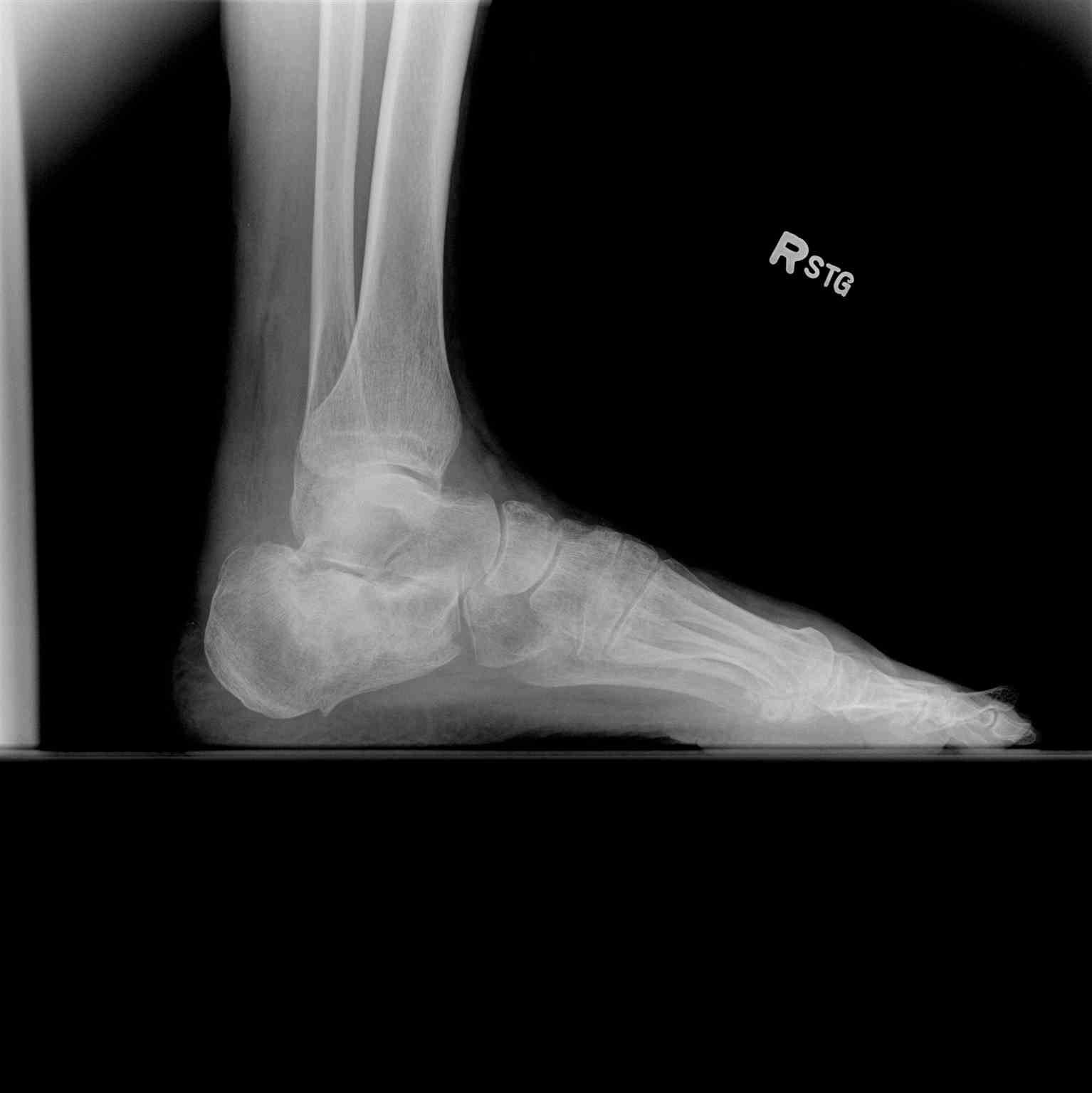
Lateral Right Ankle Radiograph Exhibiting Calcaneal Fracture. This weight-bearing lateral radiograph of the right ankle demonstrates a fracture of the calcaneus with articular involvement of the subtalar joint. Sclerosis and subtle cortical disruption are visible along the superior aspect of the calcaneal body.
Contributed by S Dulebohn, MD
(Click Image to Enlarge)
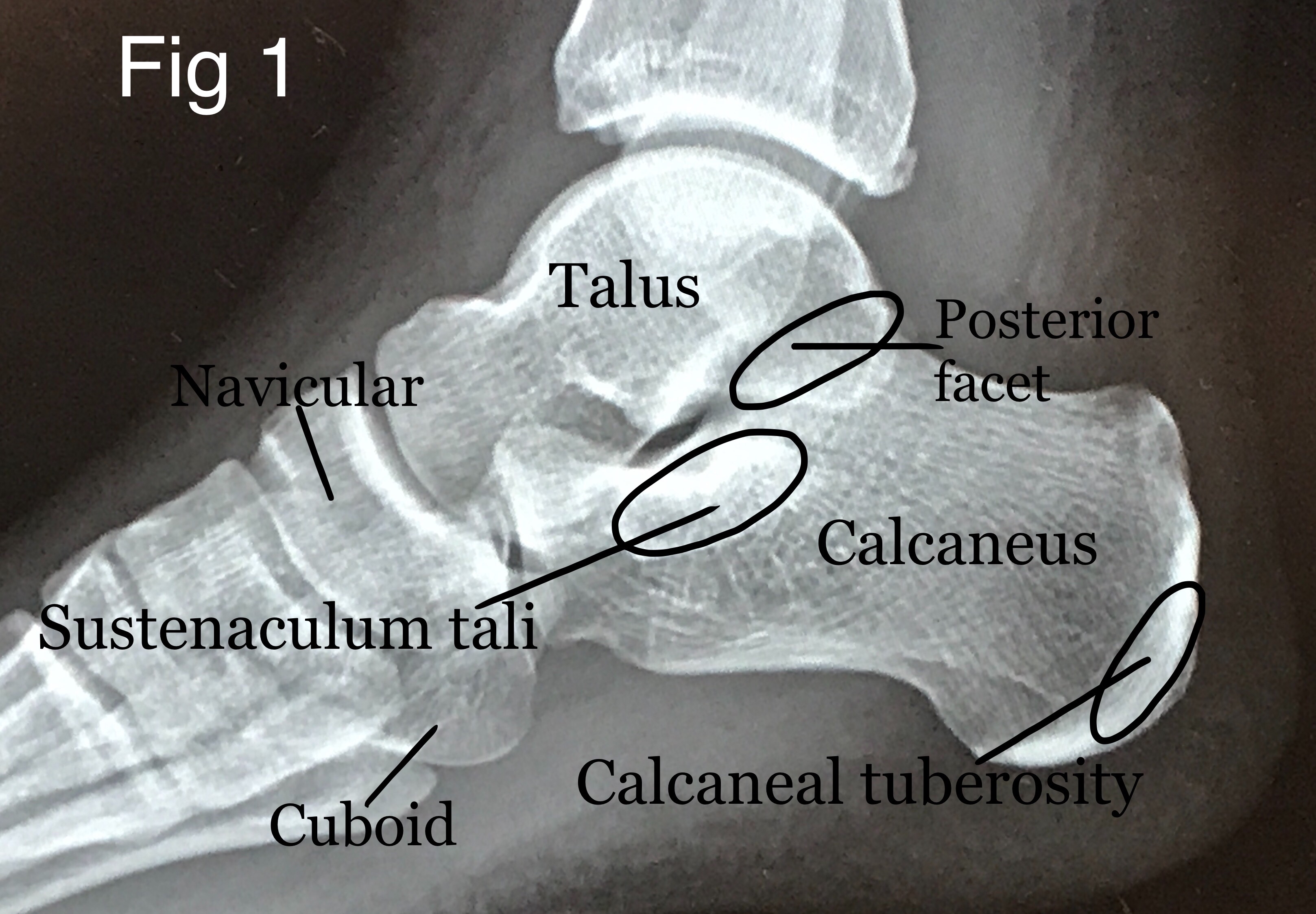
Labeled Lateral Radiograph of the Hindfoot and Midfoot Bones. This annotated lateral radiograph outlines the osseous structures and articulation sites of the right foot. The diagram highlights the subregions of the calcaneus, specifically emphasizing the sustentaculum tali, the posterior facet of the subtalar joint, and the posterior calcaneal tuberosity. The adjacent tarsal bones, including the talus, navicular, and cuboid, are also labeled for anatomical context.
Contributed by David R Davis MD
(Click Image to Enlarge)
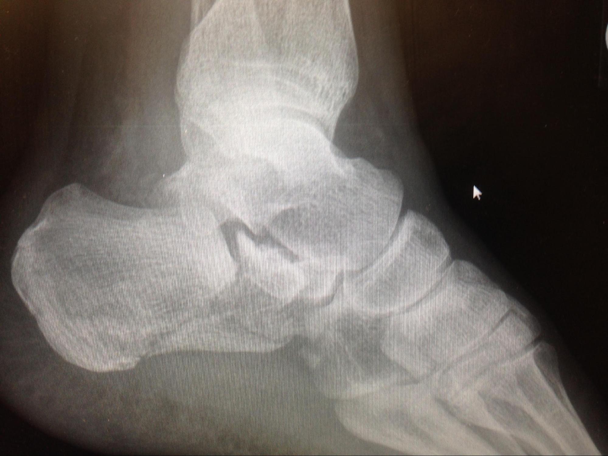
Intraarticular Calcaneus Fracture with Subtalar Joint Depression. This lateral radiograph demonstrates a severe, comminuted calcaneus fracture extending into the posterior facet of the subtalar joint. Marked cortical collapse and depression of the articular surface are visible, accompanied by an altered trabecular pattern within the calcaneal body.
Contributed by Mark A. Dreyer, DPM, FACFAS
References
Adams MR, Koury KL, Mistry JB, Braaksma W, Hwang JS, Firoozabadi R. Plantar Medial Avulsion Fragment Associated With Tongue-Type Calcaneus Fractures. Foot & ankle international. 2019 Jun:40(6):634-640. doi: 10.1177/1071100719830948. Epub 2019 Mar 6 [PubMed PMID: 30841752]
Bibbo C, Siddiqui N, Fink J, Powers J, Ehrlich DA, Kovach SJ. Wound Coverage Options for Soft Tissue Defects Following Calcaneal Fracture Management (Operative/Surgical). Clinics in podiatric medicine and surgery. 2019 Apr:36(2):323-337. doi: 10.1016/j.cpm.2018.10.012. Epub 2018 Dec 12 [PubMed PMID: 30784540]
Cottom JM, Douthett SM, McConnell KK. Intraoperative Reduction Techniques for Surgical Management of Displaced Intra-Articular Calcaneal Fractures. Clinics in podiatric medicine and surgery. 2019 Apr:36(2):269-277. doi: 10.1016/j.cpm.2018.10.008. Epub 2019 Jan 24 [PubMed PMID: 30784536]
Hordyk PJ, Fuerbringer BA, Roukis TS. Clinical Management of Acute, Closed Displaced Intra-Articular Calcaneal Fractures. Clinics in podiatric medicine and surgery. 2019 Apr:36(2):163-171. doi: 10.1016/j.cpm.2018.10.001. Epub 2018 Dec 12 [PubMed PMID: 30784528]
Schepers T. Sinus Tarsi Approach with Screws-Only Fixation for Displaced Intra-Articular Calcaneal Fractures. Clinics in podiatric medicine and surgery. 2019 Apr:36(2):211-224. doi: 10.1016/j.cpm.2018.10.004. Epub 2019 Jan 24 [PubMed PMID: 30784532]
Mitchell MJ, McKinley JC, Robinson CM. The epidemiology of calcaneal fractures. Foot (Edinburgh, Scotland). 2009 Dec:19(4):197-200. doi: 10.1016/j.foot.2009.05.001. Epub [PubMed PMID: 20307476]
Herrera-Pérez M, Oller-Boix A, Valderrabano V, González-Casamayor S, Gutiérrez-Morales MJ, Guerra-Ferraz A, País-Brito JL. [Calcaneal fractures: controversies and consensus]. Acta ortopedica mexicana. 2018 May-Jun:32(3):172-181 [PubMed PMID: 30521710]
Level 3 (low-level) evidenceDiacon AL, Kimmel LA, Hau RC, Gabbe BJ, Edwards ER. Outcomes of midfoot and hindfoot fractures in multitrauma patients. Injury. 2019 Feb:50(2):558-563. doi: 10.1016/j.injury.2018.11.021. Epub 2018 Nov 12 [PubMed PMID: 30448328]
Buckley RE, Tough S. Displaced intra-articular calcaneal fractures. The Journal of the American Academy of Orthopaedic Surgeons. 2004 May-Jun:12(3):172-8 [PubMed PMID: 15161170]
Jiménez-Almonte JH, King JD, Luo TD, Aneja A, Moghadamian E. Classifications in Brief: Sanders Classification of Intraarticular Fractures of the Calcaneus. Clinical orthopaedics and related research. 2019 Feb:477(2):467-471. doi: 10.1097/CORR.0000000000000539. Epub [PubMed PMID: 30664605]
Shih JT, Kuo CL, Yeh TT, Shen HC, Pan RY, Wu CC. Modified Essex-Lopresti procedure with percutaneous calcaneoplasty for comminuted intra-articular calcaneal fractures: a retrospective case analysis. BMC musculoskeletal disorders. 2018 Mar 9:19(1):77. doi: 10.1186/s12891-018-1995-9. Epub 2018 Mar 9 [PubMed PMID: 29523122]
Level 2 (mid-level) evidenceESSEX-LOPRESTI P. The mechanism, reduction technique, and results in fractures of the os calcis. The British journal of surgery. 1952 Mar:39(157):395-419 [PubMed PMID: 14925322]
Sanders R, Fortin P, DiPasquale T, Walling A. Operative treatment in 120 displaced intraarticular calcaneal fractures. Results using a prognostic computed tomography scan classification. Clinical orthopaedics and related research. 1993 May:(290):87-95 [PubMed PMID: 8472475]
Sop JL, Sop A. Open Fracture Management. StatPearls. 2026 Jan:(): [PubMed PMID: 28846249]
Githens M, Shatsky J, Agel J, Bransford RJ, Benirschke SK. Medial external fixation for staged treatment of closed calcaneus fractures: Surgical technique and case series. Journal of orthopaedic surgery (Hong Kong). 2017 Sep-Dec:25(3):2309499017727915. doi: 10.1177/2309499017727915. Epub [PubMed PMID: 28844198]
Level 2 (mid-level) evidenceSanders R, Vaupel ZM, Erdogan M, Downes K. Operative treatment of displaced intraarticular calcaneal fractures: long-term (10-20 Years) results in 108 fractures using a prognostic CT classification. Journal of orthopaedic trauma. 2014 Oct:28(10):551-63. doi: 10.1097/BOT.0000000000000169. Epub [PubMed PMID: 25243849]
Level 2 (mid-level) evidenceAldridge JM 3rd, Easley M, Nunley JA. Open calcaneal fractures: results of operative treatment. Journal of orthopaedic trauma. 2004 Jan:18(1):7-11 [PubMed PMID: 14676550]
Level 2 (mid-level) evidenceSiebert CH, Hansen M, Wolter D. Follow-up evaluation of open intra-articular fractures of the calcaneus. Archives of orthopaedic and trauma surgery. 1998:117(8):442-7 [PubMed PMID: 9801778]
Level 2 (mid-level) evidenceBenirschke SK, Sangeorzan BJ. Extensive intraarticular fractures of the foot. Surgical management of calcaneal fractures. Clinical orthopaedics and related research. 1993 Jul:(292):128-34 [PubMed PMID: 8519099]
Shannon SF, Houdek MT, Wyles CC, Yuan BJ, Cross WW 3rd, Cass JR, Sems SA. Allgöwer-Donati Versus Vertical Mattress Suture Technique Impact on Perfusion in Ankle Fracture Surgery: A Randomized Clinical Trial Using Intraoperative Angiography. Journal of orthopaedic trauma. 2017 Feb:31(2):97-102. doi: 10.1097/BOT.0000000000000731. Epub [PubMed PMID: 28129268]
Level 1 (high-level) evidenceLai HX, Peng LP, Wu LH, Ye W. A meta-analysis of Allgöwer-Donati versus interrupted vertical mattress suturing in preventing postoperative incisional complications of calcaneal fracture surgery: A systematic review and meta-analysis. Joint diseases and related surgery. 2026 May 1:37(2):390-401. doi: 10.52312/jdrs.2026.2500. Epub 2026 Mar 20 [PubMed PMID: 41906834]
Level 1 (high-level) evidenceHsu AR, Anderson RB, Cohen BE. Advances in Surgical Management of Intra-articular Calcaneus Fractures. The Journal of the American Academy of Orthopaedic Surgeons. 2015 Jul:23(7):399-407. doi: 10.5435/JAAOS-D-14-00287. Epub [PubMed PMID: 26111874]
Level 3 (low-level) evidenceSeat A, Seat C. Lateral Extensile Approach Versus Minimal Incision Approach for Open Reduction and Internal Fixation of Displaced Intra-articular Calcaneal Fractures: A Meta-analysis. The Journal of foot and ankle surgery : official publication of the American College of Foot and Ankle Surgeons. 2020 Mar-Apr:59(2):356-366. doi: 10.1053/j.jfas.2019.08.007. Epub [PubMed PMID: 32131003]
Level 1 (high-level) evidenceKline AJ, Anderson RB, Davis WH, Jones CP, Cohen BE. Minimally invasive technique versus an extensile lateral approach for intra-articular calcaneal fractures. Foot & ankle international. 2013 Jun:34(6):773-80. doi: 10.1177/1071100713477607. Epub 2013 Mar 4 [PubMed PMID: 23460669]
Level 2 (mid-level) evidenceLi LH, Guo YZ, Wang H, Sang QH, Zhang JZ, Liu Z, Sun TS. Less wound complications of a sinus tarsi approach compared to an extended lateral approach for the treatment of displaced intraarticular calcaneal fracture: A randomized clinical trial in 64 patients. Medicine. 2016 Sep:95(36):e4628. doi: 10.1097/MD.0000000000004628. Epub [PubMed PMID: 27603354]
Level 1 (high-level) evidenceJoseph NM, McCaskey M, Bhatt FR, Watson DT, Sanders RW, Mir HR. Surgeon Learning Curve for the Sinus Tarsi Approach to Intra-articular Calcaneus Fractures: Improvement With Experience. Journal of orthopaedic trauma. 2023 Dec 1:37(12):644-649. doi: 10.1097/BOT.0000000000002682. Epub [PubMed PMID: 37559211]
Anand S, Belmonte AD, Grzeskiewicz E, Groth A, Martin KD. Radiographic Results of Percutaneous Reduction of Calcaneal Fractures and Posterior Arthroscopic Subtalar Arthrodesis (C-PASTA). Foot & ankle international. 2025 Jul:46(7):679-687. doi: 10.1177/10711007251333367. Epub 2025 Apr 24 [PubMed PMID: 40275601]
Bloomer AK, McKnight RR, Johnson NR, Macknet DM, Wally MK, Yu Z, Seymour RB, Hsu JR. Screws-Only Primary Subtalar Arthrodesis for Calcaneus Fractures. Foot & ankle international. 2022 Apr:43(4):509-519. doi: 10.1177/10711007211058689. Epub 2022 Jan 4 [PubMed PMID: 34996306]
Gardner MJ, Nork SE, Barei DP, Kramer PA, Sangeorzan BJ, Benirschke SK. Secondary soft tissue compromise in tongue-type calcaneus fractures. Journal of orthopaedic trauma. 2008 Aug:22(7):439-45. doi: 10.1097/BOT.0b013e31817ace7e. Epub [PubMed PMID: 18670282]
Level 2 (mid-level) evidenceBuckley R, Leighton R, Sanders D, Poon J, Coles CP, Stephen D, Paolucci EO. Open reduction and internal fixation compared with ORIF and primary subtalar arthrodesis for treatment of Sanders type IV calcaneal fractures: a randomized multicenter trial. Journal of orthopaedic trauma. 2014 Oct:28(10):577-83. doi: 10.1097/BOT.0000000000000191. Epub [PubMed PMID: 24983433]
Level 1 (high-level) evidenceRadnay CS, Clare MP, Sanders RW. Subtalar fusion after displaced intra-articular calcaneal fractures: does initial operative treatment matter? Surgical technique. The Journal of bone and joint surgery. American volume. 2010 Mar:92 Suppl 1 Pt 1():32-43. doi: 10.2106/JBJS.I.01267. Epub [PubMed PMID: 20194342]
Griffin D, Parsons N, Shaw E, Kulikov Y, Hutchinson C, Thorogood M, Lamb SE, UK Heel Fracture Trial Investigators. Operative versus non-operative treatment for closed, displaced, intra-articular fractures of the calcaneus: randomised controlled trial. BMJ (Clinical research ed.). 2014 Jul 24:349():g4483. doi: 10.1136/bmj.g4483. Epub 2014 Jul 24 [PubMed PMID: 25059747]
Level 3 (low-level) evidenceWei N, Yuwen P, Liu W, Zhu Y, Chang W, Feng C, Chen W. Operative versus nonoperative treatment of displaced intra-articular calcaneal fractures: A meta-analysis of current evidence base. Medicine. 2017 Dec:96(49):e9027. doi: 10.1097/MD.0000000000009027. Epub [PubMed PMID: 29245290]
Level 1 (high-level) evidenceBuckley R, Tough S, McCormack R, Pate G, Leighton R, Petrie D, Galpin R. Operative compared with nonoperative treatment of displaced intra-articular calcaneal fractures: a prospective, randomized, controlled multicenter trial. The Journal of bone and joint surgery. American volume. 2002 Oct:84(10):1733-44 [PubMed PMID: 12377902]
Level 1 (high-level) evidenceSimske NM, Hermelin MJ, Vallier HA. Impact of Psychosocial and Economic Factors on Functional Outcomes After Operative Calcaneus Fractures. Journal of orthopaedic trauma. 2021 Nov 1:35(11):e423-e428. doi: 10.1097/BOT.0000000000002082. Epub [PubMed PMID: 33967225]
Spierings KE, Min M, Nooijen LE, Swords MP, Schepers T. Managing the open calcaneal fracture: A systematic review. Foot and ankle surgery : official journal of the European Society of Foot and Ankle Surgeons. 2019 Dec:25(6):707-713. doi: 10.1016/j.fas.2018.10.005. Epub 2018 Nov 5 [PubMed PMID: 30467055]
Level 1 (high-level) evidenceZhang Z, Wang Z, Zhang Y, Qiu X, Chen Y. Risk factors for increased postoperative drainage of calcaneal fractures after open reduction and internal fixation: An observational study. Medicine. 2018 Aug:97(32):e11818. doi: 10.1097/MD.0000000000011818. Epub [PubMed PMID: 30095652]
Level 2 (mid-level) evidenceAllegra PR, Rivera S, Desai SS, Aiyer A, Kaplan J, Gross CE. Intra-articular Calcaneus Fractures: Current Concepts Review. Foot & ankle orthopaedics. 2020 Jul:5(3):2473011420927334. doi: 10.1177/2473011420927334. Epub 2020 Jul 28 [PubMed PMID: 35097384]
Csizy M, Buckley R, Tough S, Leighton R, Smith J, McCormack R, Pate G, Petrie D, Galpin R. Displaced intra-articular calcaneal fractures: variables predicting late subtalar fusion. Journal of orthopaedic trauma. 2003 Feb:17(2):106-12 [PubMed PMID: 12571499]
Level 2 (mid-level) evidenceEastwood DM, Langkamer VG, Atkins RM. Intra-articular fractures of the calcaneum. Part II: Open reduction and internal fixation by the extended lateral transcalcaneal approach. The Journal of bone and joint surgery. British volume. 1993 Mar:75(2):189-95 [PubMed PMID: 8444935]
Toussaint RJ, Lin D, Ehrlichman LK, Ellington JK, Strasser N, Kwon JY. Peroneal tendon displacement accompanying intra-articular calcaneal fractures. The Journal of bone and joint surgery. American volume. 2014 Feb 19:96(4):310-5. doi: 10.2106/JBJS.L.01378. Epub [PubMed PMID: 24553887]
Level 2 (mid-level) evidenceOhashi K, Sanghvi T, El-Khoury GY, Ahn JM, Bennett DL, Geijer M, Inaoka T, Berbaum K. Diagnostic accuracy of 3D color volume-rendered CT images for peroneal tendon dislocation in patients with acute calcaneal fractures. Acta radiologica (Stockholm, Sweden : 1987). 2015 Feb:56(2):190-5. doi: 10.1177/0284185114522224. Epub 2014 Feb 3 [PubMed PMID: 24493866]
Vosoughi AR, Afaridi E, Solooki S, Shayan Z, Rammelt S. Prevalence and Predictors of Peroneal Tendon Instability Accompanying Calcaneal Fractures. Foot & ankle international. 2023 Sep:44(9):825-833. doi: 10.1177/10711007231175666. Epub 2023 Sep 2 [PubMed PMID: 37658714]
Chu KM, Lui TH. Endoscopic Lateral Calcaneal Ostectomy and Peroneal Tendon Decompression With the Patient in the Prone Position as Management of Subfibular Impingement After Calcaneal Fracture. Arthroscopy techniques. 2019 Oct:8(10):e1069-e1073. doi: 10.1016/j.eats.2019.05.021. Epub 2019 Sep 19 [PubMed PMID: 31921576]