 Tools
Tools
 Breast Reconstruction Free Flaps
Introduction
Breast cancer is the leading type of cancer diagnosis in women. In 2022, 670,000 deaths were attributed to breast cancer in women, and 2.3 million new breast cancer cases were diagnosed. Despite its incidence, it is commonly a treatable disease, with 90% 5-year and 83% 10-year survival rates. Mastectomy is a common measure for both the treatment and prophylaxis of breast cancer.[1]
Although mastectomies have been performed for many years, reconstruction has only been a consideration more recently. The first reported case of breast reconstruction was in 1887, when Aristide Verneuil used a pedicle from the opposite breast. This was closely followed by Vincent Czerny, who used a lipoma to reconstruct a lumpectomy defect.
Iginio Tansini first performed a latissimus dorsi flap in 1906, although most advocated against reconstruction during this period, as they felt it inhibited cancer care. Not until the 1950s did breast reconstruction become an option again, with surgeons like Dr Gilles performing tubed pedicled flaps.[2] The invention of silicone implants in the 1960s started the age of prosthetic breast reconstruction.
Free flaps were not used until the late 1970s when Holmstrom published the use of a “free abdominoplasty flap” for breast reconstruction. However, microsurgery was not commonplace during that time. Autologous reconstruction really took off when Dr Hartrampf published his method for pedicled transverse rectus abdominis myocutaneous (TRAM) flap in 1982.
The pedicled TRAM evolved into the free TRAM as microsurgery became more common, and the deep inferior epigastric artery offered improved blood supply compared to the superior epigastric artery (the basis of the pedicled flap). This process has further evolved into the free muscle-sparing TRAM (MS-TRAM), the deep inferior epigastric perforator (DIEP) flap, and the use of other free flaps for breast reconstruction.[2][3][4][5]
Breast reconstructions are common operations. The 2 most common methods for breast reconstruction are implant-based and free autologous tissue transfer. In a review of the Breast-Q, patients who underwent autologous-based reconstruction had higher postoperative scores.[6]
Since there are so many survivors who go on to live a long life, quality of life issues are paramount. Plastic surgeons can contribute to the quality of life issues of these patients by performing breast reconstruction. In addition, all insurance carriers must cover breast reconstruction under the Women’s Health and Cancer Rights Act of 1998.[7]
Patients undergoing autologous reconstruction reported a higher quality of life and improved outcomes than their implant-based counterparts.[3][8] Advantages of autologous reconstruction include high postoperative satisfaction and long-lasting results, with natural aging, ptosis, responsiveness to changes in body weight, improved aesthetics, and body contouring at the donor site. Autologous reconstructions do not have the major disadvantages of implants, like capsular contracture and the risk of device failure.
A unilateral autologous reconstruction is easier to match to the remaining natural breast. Also, some patients will not have enough skin after a mastectomy and will not be candidates for reconstruction with implants alone. Because autologous reconstructions more closely resemble the preoperative form, they are now considered the gold standard. The DIEP flap is typically the flap of choice, if available, secondary to the improved aesthetic result at the donor site and minimal donor-site morbidity. Other donor sites are also suitable depending on the patient’s habitus and reconstructive needs.[9][10][11][12]
Anatomy and Physiology
Register For Free And Read The Full Article
 Search engine and full access to all medical articles
- 10 free questions in your specialty
- Free CME/CE Activities
- Free daily question in your email
- Save favorite articles to your dashboard
- Emails offering discounts
Learn more about a Subscription to StatPearls Point-of-Care
Anatomy and Physiology
Anatomy varies widely depending on the operation chosen. Therefore, this will include the most common choices for free flap breast reconstruction, though it is not exhaustive. See the images below for the anatomy.
Recipient Site
Patients who are having free flap breast reconstruction have or are undergoing a mastectomy. Therefore, the recipient's vessels need to be located near the reconstruction site. Historically, the thoracodorsal arteries were utilized, although the internal mammary arteries are now more commonly used. The results are comparable regardless of the site chosen.
The thoracodorsal arteries are similarly sized to the internal mammary arteries at 1 to 2.5 mm in diameter. These arteries are already exposed in patients who have undergone an axillary dissection but are prone to injury in those who require a delayed axillary dissection. The internal mammary arteries have a higher flow than the thoracodorsal arteries.
Also, they can be used for anastomosis in both anterograde and retrograde directions and are best used at the third to fifth intercostal spaces. Occasionally, perforators from the internal mammary artery are large enough to use. Internal mammary arteries require less pedicle length to be reached and are advantageous for small flaps with short pedicles.[3][4][8][13][11]
Below is the list of the common donor sites/flaps:
- Abdomen
- There are several iterations of abdominal flaps that can be utilized, from most invasive to least invasive: the free TRAM, ms-TRAM, DIEP, and superficial inferior epigastric artery (SIEA) flap. These flaps are beneficial to harvest, as many patients have excess abdominal tissue and gain the added benefit of abdominal contouring from harvesting the transverse skin island. The TRAM derives from the rectus muscle and is a Mathes and Nahai type III flap with 2 dominant pedicles. TRAM, ms-TRAM (ms-1 and 2), and DIEP (ms-3) all depend on how much muscle is removed.
- The free TRAM takes the entire muscle (ms-0); ms-1 leaves either a medial or a lateral component; ms-2 leaves both medial and lateral components; ms-3 (DIEP) is a true perforator flap and leaves all muscle intact. The muscle is split to free the vessels. The typical anatomy of the rectus muscle consists of two rows of perforators, although variants with 1 or 3 rows also exist.
- The advantage of taking more muscle is improved blood supply, but it comes at the cost of increased abdominal wall morbidity. A large amount of tissue is harvestable, and the pedicle is about 12 cm in length and 2.0 mm in diameter. The SIEA is considered ms-4 because it only removes the skin and fat from the fascia, based on the superficial vessel, and does not violate the muscle or fascia. However, only a hemi-abdomen can be used with this flap, which often lacks a vessel large enough to use.[9][10][14][15]
- When the abdominal tissue is not available or sufficient, secondary flap options are a possible choice. This option varies between buttock and thigh flaps, including the superior gluteal artery perforator (SGAP), inferior gluteal artery perforator (IGAP), transverse upper gracilis (TUG), and PAP (profundal artery perforator).[14] Some surgeons prefer to use the thigh, as the tissue is more pliable, it does not require an intraoperative position change, and the donor site scar is less obtrusive.
- Medial thigh
- There are 2 options from the medial thigh: the TUG flap and the PAP flap. The TUG flap produces a relatively small breast but is an easy flap to elevate. This flap utilizes the area that would otherwise be discarded during a medial thigh lift.
- Still, it keeps the gracilis, and thus the vascular pedicle (the ascending branch of the medial circumflex femoral artery) with it. Since the TUG flap is based on the gracilis, it is a Mathes and Nahai type II flap. The transversely oriented skin paddle can be up to 25 x 10 cm in size, with a small pedicle (6 cm in length and 1.6 mm vessels). This flap is ideal for small-breasted women who do not have an abdominal donor site or do not want an abdominal scar.[4][8]
- The profunda artery perforator flap, or PAP flap, has recently been described as an improvement over the TUG flap. This flap has the advantages of a longer pedicle length, a better donor-site scar, a larger skin paddle, no muscle harvest, and a decreased risk of lymphedema.
- However, preoperative imaging is necessary to determine the pedicle location and properly design the skin paddle. This is more posteriorly located than the TUG donor site and can make a flap of about 6 to 7 cm by 18 to 20 cm. The pedicle is about 10 cm in length and usually has a diameter of 2.2 mm. These flaps can be coned to reshape the breast and tend to look more natural than gluteal tissue.[8][10][14][16]
- Gluteal flaps
- The gluteal flaps were actually described before the thigh-based flaps, but are used less commonly because they require repositioning for harvest and inset. Gluteal tissue is firmer and more difficult to shape and inset. They can be based on either the superior (SGAP) or inferior gluteal arteries (IGAP). The SGAP skin island measures 10 to 12 × 25 to 32 cm with a 6- to 8-cm pedicle. The IGAP is usually about 8 × 18 cm, and the pedicle is about 8 to 11 cm. The inferiorly based flap avoids the divot created by the SGAP donor site but risks exposing the sciatic nerve if the muscle is taken.[3][8][17][18]
- Lateral thigh flap
- A recently described flap is the lateral thigh or subcutaneous tensor fascia lata perforator flap, based on perforators originating from the ascending branch of the lateral circumflex artery. The perforators are in the septum between the tensor fascia lata and the gluteus medius muscles. Flap dimensions range from 6 to 9 cm x 18 to 22 cm, with a 6- to 8-cm pedicle length and a 2-mm diameter.[9]
Indications
The main indication of free flap breast reconstruction is the acquired absence of the breast(s), typically from a mastectomy for breast cancer or prophylactic mastectomy for BRCA or another genetic predisposition for breast cancer, but also congenital breast deformity. Although breast-sparing options exist, many women still choose or need a mastectomy to treat their cancer. The goals are to restore the breast dimensions, contour, and consistency.[3][8][9]
Contraindications
There are no specific contraindications for breast reconstruction, presuming the patient is medically able to tolerate a general anesthetic for the duration of the chosen case. However, there are risk factors that might make reconstruction unsafe or undesirable. Age is not a risk factor for these procedures.
Those aged older than 65 are at increased risk for a hernia after abdominally based surgery and thrombosis. Obesity with a body mass index (BMI) over 30 is associated with increased overall complications, donor site complications, partial flap loss, fat necrosis, and recipient site complications. BMI over 40 is at high risk for flap failure and should prompt caution before performing the reconstruction. American Society of Anesthesiologists (ASA) class III and increased operative times are also risk factors for complications.
Certain flaps are contraindicated in individual patients depending on their habitus and anatomy; for example, contraindications to DIEP include a prior abdominoplasty. Collagen vascular diseases and thrombotic diseases increase the risk of free flap failure. Patients on chemotherapy and steroids have an increased risk of delayed wound healing.
Notably, an elective free flap is contraindicated during pregnancy.[8][15] Smoking is a relative contraindication, with some surgeons refusing to perform the procedure until the patient ceases smoking to prevent wound complications and healing issues.[19] Radiation needs to be taken into consideration as well. While autologous methods are the ideal reconstruction in patients who require postmastectomy radiotherapy, radiation to the flap can decrease the aesthetics and increase the risk of partial flap loss and fibrosis.[8]
Equipment
The equipment required is similar to that of a standard operation, with the addition of specific microsurgical equipment. Surgeons require some magnification, either with loupes, a microscope, or both. Microsurgical equipment includes microsutures (8-0 to 10-0), jeweler’s forceps, microscissors, a vessel dilator, a microneedle holder, microvascular clamps, microsurgical hemoclips, and a micro-instrument wipe.
Medications include heparin saline (100 u/mL), papaverine, and possibly tissue plasminogen activator (TPA). A venous coupler is often necessary for vein anastomosis. Also, standard equipment for monitoring the flap is mandatory; typically, this is a Doppler probe, but an internal Doppler and other devices can be used depending on surgeon preferences.
Personnel
The personnel needed for free flap breast reconstruction are similar to those for other surgeries. A microsurgeon and an assistant are required, as are an anesthesiologist, a circulating nurse, and a scrub tech. Also, the floor nurses need training to monitor the flap appropriately.
Preparation
A thorough history and physical is required. Risks and comorbidities are reviewed and modified when appropriate; this requires discussing the plan with anesthesia, including avoiding bands, intravenous catheters, and blood pressure cuffs on donor extremities; avoiding pressors and vasoactive drugs during the case; and ensuring anticoagulants are available.
Technique or Treatment
Surgical techniques depend on the flap chosen. In brief, there are 2 parts of the operation: flap elevation and preparation of the recipient's vessels, which 2 surgeons can perform simultaneously. In all instances, the patient gets a usual surgical prep and sterile drape in the supine position unless otherwise stated.
In all cases, surgical staff will mark the patient in the preoperative holding area. Standard markings include sternal midline and standard breast markings for the mastectomy (ellipse, Wise pattern, vertical reduction pattern, nipple-sparing incisions), depending on patient habitus, tumor characteristics, and surgeon preference. The donor site is also marked: an elliptical abdominoplasty-style incision for DIEP, TRAM, and ms-TRAM flaps, medial thigh lift for TUG (which can be modified based on where the imaging shows the perforator for a PAP), near the superior or inferior gluteal creases for SGAP/IGAP, and an ellipse around the lateral thigh perforator (LTP) for the LTP flap (see Image. Anatomy of Donor and Recipient Sites for Breast Reconstruction).
The flap is elevated on its perforator for all flaps, dissected down the pedicle, and kept in situ until the recipient site is prepared. Once the vessels are ready (internal mammary in most cases), the pedicle is ligated distally, and the flap is removed from the site. The flap is flushed with heparinized saline, and the vessels are cleaned, separated, and prepared for anastomosis.
The recipient vessels are also prepared, distally ligated, and flushed with heparinized saline. Arterial inflow is checked, and the vessels are temporarily clamped with Acklund clamps to keep a dry surgical field. The flap is temporarily fixed in a position to allow inset while keeping the flap secure, preventing motion or tension on the vessels during the anastomosis.
The vessel path must also be assessed to ensure the vessels are straight and that nothing will kink them after anastomosis. There are many techniques to anastomose vessels. One preferred technique is a coupler for the vein and an interrupted hand-sewn anastomosis with 8-0 or 9-0 nylon sutures for the artery. The flap is inset, the breast is shaped with sutures, and the drains are placed within the cavity.
Flap Elevation
- Abdominally based flaps
- The abdomen is marked as described for use with either the whole abdomen or a hemi-abdomen, depending on the size of the required flap. One preferred method is to make the superior incision first, down to the fascia, and elevate the upper abdominal skin, which is transposed, so the surgeon knows where to make the lower incision to close the abdomen. The inferior incision is then made, with care to find and dissect free the superficial inferior epigastric vein.
- Next, (if needed), the midline incision and umbilical incisions are made. The flap is elevated off the abdominal wall from lateral to medial. Depending on the type of flap chosen, the whole rectus muscle, part of it, or just the perforator(s) chosen are dissected free. These are isolated from the deep inferior epigastric artery and vein and taken as described above. The abdomen is closed over drains by transposing the superior abdominal skin down and closing in layers. The umbilicus is inset into the abdominal flap.
- TUG
- The skin flap marking is as above, and the patient is in the supine frog-leg position. The flap should center over the gracilis and the pedicle, which is about 10 cm from the pubis. The skin is incised, leaving it attached to the gracilis muscle and staying superficial over the lymphatics. Once the flap is isolated on the gracilis, the adductor longus is retracted, and the pedicle is dissected out to the origin. When the pedicle is ready, the gracilis is divided, the vessels are taken, and the flap is transferred.
- PAP
- Again, the patient is marked as described and centered on the perforator identified by imaging. The patients are in a supine, frog-leg position. The incision is started medially down to the deep fascia. The flap is elevated in a subfascial plane posterior to the adductor longus, extending until the perforators are identified (usually at the level of the adductor magnus fascia). The perforator dissection proceeds through the adductor magnus up until its origin at the profunda vessels. The flap is taken and transferred as above.
- SGAP/IGAP
- The patient is preoperatively marked for the flap chosen. The patient is positioned prone or in lateral decubitus for flap harvest. The flap is marked based on anatomic landmarks (SGAP perforators are one-third of the distance along a line from the ASIS to the greater trochanter, and IGAP perforators are two-thirds of the distance along a line from the anterior superior iliac spine [ASIS] to the gluteal fold).
- The incision is made with a bevel so that extra gluteal fat can be included superiorly and inferiorly. The dissection is taken down to the level of the muscle, and the dissection proceeds distally to proximally until the perforator(s) is(are) found. A perforator of at least 1 mm is selected and dissected through the muscle to the pedicle of either the superior or inferior gluteal artery. The vessel is taken at a level with sufficient length and caliber for a microsurgical anastomosis.
- Lateral thigh
- Again, the patient is in the supine position, and the flap markings are drawn preoperatively. The dissection through the skin proceeds, and then the flap is elevated from medial to lateral until the perforators are reached. A line is drawn from the ASIS to the lateral patella; this is the anterior border of the flap, and another line is drawn from the pubic bone perpendicular to it.
- Perforators are usually located along the horizontal line posterior to the vertical line. During the dissection, the lateral femoral cutaneous nerve is identified and preserved. The perforators are found and dissected into the septum between the tensor fascia lata and the gluteus medius up to the ascending branch of the lateral circumflex femoral artery. The pedicle is taken, and the flap transferred.
Preparation of the Recipient's Vessels
Many surgeons use the internal mammary vessels as the preferred recipient vessels for our anastomosis. Either through the old scar or the mastectomy incision, the space between the third and fourth ribs is palpated. If the space is large, a rib-sparing approach is possible. If not, the cartilage of the third rib is removed. The pectoralis muscle is split, leaving a groove for the vessels and the intercostal muscles to be excised. The vessels are dissected free for the length of the space.[16][17][18]
Complications
All surgeries have the risk of complications, and this is no exception. The most common complications are wound-related and include infections, seromas, hematomas, skin flap necrosis, and delayed healing. In some series, the percentage of wound complications reaches 50%.
Complications with microsurgery may also occur, with loss-of-flap rates due to venous or arterial thrombosis ranging from 1% to 4% and fat necrosis from 5% to 40%. If the mammary arteries are chosen, there is a small risk of pneumothorax. Donor site complications depend on the flap chosen.
For abdominally based flaps, complications include abdominal bulge, hernia, and weakness. Thigh flaps have a high rate of breakdown, sensory disturbance to the thigh, and risk of lymphedema. Gluteal flaps are at risk of sciatic nerve exposure or insufficient padding and may also have wound-healing complications.[10][20][21]
Clinical Significance
Autologous breast reconstruction using free tissue flaps is a clinically important reconstructive option for patients undergoing mastectomy because it restores breast volume and contour using the patient’s own tissue while avoiding dependence on an internal implant or external prosthesis. The availability of multiple donor sites, including the abdomen, thigh, buttock, and back, expands reconstructive options and may allow more patients to pursue mastectomy with an individualized reconstruction plan. For some patients, breast reconstruction can facilitate acceptance of mastectomy as a treatment or risk-reduction strategy and may reduce the need for ongoing mammographic surveillance of the reconstructed breast, although continued oncologic follow-up remains essential.
Loss of a breast can have substantial psychological, emotional, and body-image consequences. Free flap reconstruction may improve self-confidence, body image, sexual well-being, and overall mental health by helping patients regain a sense of physical wholeness after mastectomy.[3][8] Because autologous reconstruction can provide durable, natural-appearing results, it is particularly valuable for patients who prefer to avoid prosthetic reconstruction or who have prior radiation exposure, inadequate soft-tissue coverage, or implant-related complications. Appropriate patient selection, shared decision-making, and counseling regarding operative complexity, donor-site morbidity, recovery, and potential complications are essential to optimize functional, psychosocial, and reconstructive outcomes.
Enhancing Healthcare Team Outcomes
Breast reconstruction with free tissue flaps requires coordinated multidisciplinary care to optimize flap survival, minimize complications, and support patient-centered recovery. Plastic and reconstructive surgeons must perform careful patient selection, assess donor-site and recipient-vessel anatomy, plan microsurgical reconstruction, and promptly recognize vascular compromise. Advanced practitioners assist with perioperative assessment, patient education, postoperative monitoring, and follow-up coordination.
Anesthesiologists support flap perfusion through meticulous hemodynamic management, normothermia, appropriate fluid administration, and, when possible, avoidance of vasoconstrictive agents or hypoperfusion. Nurses are essential for frequent flap assessments, including evaluation of color, temperature, capillary refill, turgor, Doppler signals, and drainage, as well as early escalation of concerning findings to the surgical team.
Effective communication among surgeons, anesthesiologists, nurses, pharmacists, physical and occupational therapists, wound care specialists, and oncology teams is critical throughout preoperative planning, surgery, and recovery. Pharmacists optimize multimodal analgesia, venous thromboembolism prophylaxis, antimicrobial therapy, and medication reconciliation while monitoring for adverse effects. Physical and occupational therapists facilitate safe mobilization, shoulder range-of-motion recovery, and functional rehabilitation while protecting the donor site during healing. Structured handoffs, standardized flap-monitoring protocols, early recognition of thrombosis or hematoma, and coordinated discharge planning improve patient safety, reduce delays in re-exploration, and support long-term reconstructive outcomes, quality of life, and continuity of oncologic care.
Media
(Click Image to Enlarge)
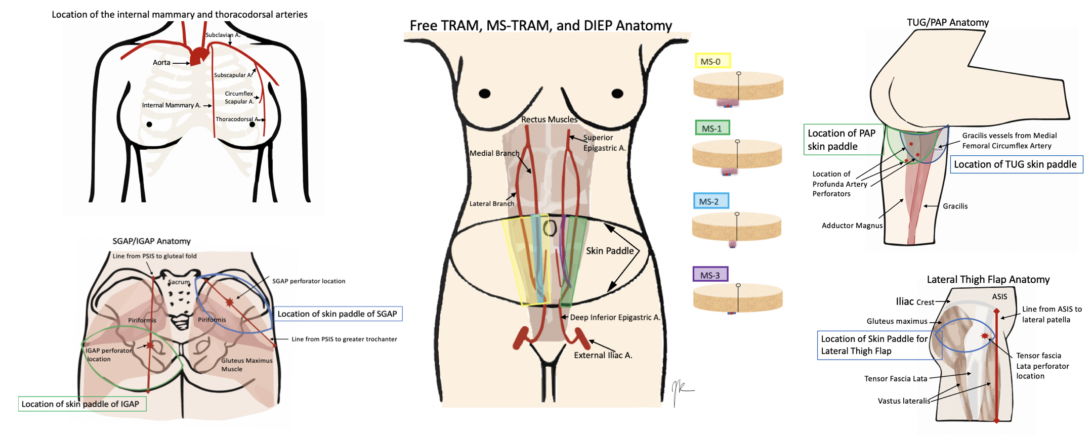
Anatomy of Donor and Recipient Sites for Breast Reconstruction.
Contributed by J Rose, DO
References
Kim J, Harper A, McCormack V, Sung H, Houssami N, Morgan E, Mutebi M, Garvey G, Soerjomataram I, Fidler-Benaoudia MM. Global patterns and trends in breast cancer incidence and mortality across 185 countries. Nature medicine. 2025 Apr:31(4):1154-1162. doi: 10.1038/s41591-025-03502-3. Epub 2025 Feb 24 [PubMed PMID: 39994475]
Rozen WM, Rajkomar AK, Anavekar NS, Ashton MW. Post-mastectomy breast reconstruction: a history in evolution. Clinical breast cancer. 2009 Aug:9(3):145-54. doi: 10.3816/CBC.2009.n.024. Epub [PubMed PMID: 19661037]
Serletti JM, Fosnot J, Nelson JA, Disa JJ, Bucky LP. Breast reconstruction after breast cancer. Plastic and reconstructive surgery. 2011 Jun:127(6):124e-135e. doi: 10.1097/PRS.0b013e318213a2e6. Epub [PubMed PMID: 21617423]
Arnez ZM, Pogorelec D, Planinsek F, Ahcan U. Breast reconstruction by the free transverse gracilis (TUG) flap. British journal of plastic surgery. 2004 Jan:57(1):20-6 [PubMed PMID: 14672674]
Level 3 (low-level) evidenceHartrampf CR, Scheflan M, Black PW. Breast reconstruction with a transverse abdominal island flap. Plastic and reconstructive surgery. 1982 Feb:69(2):216-25 [PubMed PMID: 6459602]
Level 3 (low-level) evidenceSeth I, Seth N, Bulloch G, Rozen WM, Hunter-Smith DJ. Systematic Review of Breast-Q: A Tool to Evaluate Post-Mastectomy Breast Reconstruction. Breast cancer (Dove Medical Press). 2021:13():711-724. doi: 10.2147/BCTT.S256393. Epub 2021 Dec 16 [PubMed PMID: 34938118]
Level 1 (high-level) evidenceWilkins EG, Alderman AK. Breast reconstruction practices in north america: current trends and future priorities. Seminars in plastic surgery. 2004 May:18(2):149-55. doi: 10.1055/s-2004-829049. Epub [PubMed PMID: 20574493]
Macadam SA, Bovill ES, Buchel EW, Lennox PA. Evidence-Based Medicine: Autologous Breast Reconstruction. Plastic and reconstructive surgery. 2017 Jan:139(1):204e-229e. doi: 10.1097/PRS.0000000000002855. Epub [PubMed PMID: 28027256]
Tuinder SMH, Beugels J, Lataster A, de Haan MW, Piatkowski A, Saint-Cyr M, van der Hulst RRWJ, Allen RJ. The Lateral Thigh Perforator Flap for Autologous Breast Reconstruction: A Prospective Analysis of 138 Flaps. Plastic and reconstructive surgery. 2018 Feb:141(2):257-268. doi: 10.1097/PRS.0000000000004072. Epub [PubMed PMID: 29019861]
Patel NG, Ramakrishnan V. Microsurgical Tissue Transfer in Breast Reconstruction. Clinics in plastic surgery. 2017 Apr:44(2):345-359. doi: 10.1016/j.cps.2016.12.002. Epub 2017 Feb 13 [PubMed PMID: 28340667]
Cho MJ, Schroeder M, Flores Garcia J, Royfman A, Moreira A. The Current State of the Art in Autologous Breast Reconstruction: A Review and Modern/Future Approaches. Journal of clinical medicine. 2025 Feb 25:14(5):. doi: 10.3390/jcm14051543. Epub 2025 Feb 25 [PubMed PMID: 40095465]
Heidekrueger PI, Fritschen U, Moellhoff N, Germann G, Giunta RE, Zeman F, Prantl L. Impact of body mass index on free DIEP flap breast reconstruction: A multicenter cohort study. Journal of plastic, reconstructive & aesthetic surgery : JPRAS. 2021 Aug:74(8):1718-1724. doi: 10.1016/j.bjps.2020.12.043. Epub 2021 Jan 16 [PubMed PMID: 33461890]
Todd AR, Genereux O, Schrag C, Hatchell A, Matthews J. Improved Operative Efficiency and Surgical Times in Autologous Breast Reconstruction: A 15-year Single-center Retrospective Review. Plastic and reconstructive surgery. Global open. 2023 Sep:11(9):e5231. doi: 10.1097/GOX.0000000000005231. Epub 2023 Sep 15 [PubMed PMID: 38152707]
Level 2 (mid-level) evidenceHealy C, Allen RJ Sr. The evolution of perforator flap breast reconstruction: twenty years after the first DIEP flap. Journal of reconstructive microsurgery. 2014 Feb:30(2):121-5. doi: 10.1055/s-0033-1357272. Epub 2013 Oct 25 [PubMed PMID: 24163223]
Howard MA, Mehrara B. Emerging trends in microsurgical breast reconstruction: deep inferior epigastric artery perforator (DIEP) and the superior gluteal artery perforator (SGAP) flaps. International journal of surgery (London, England). 2005:3(1):53-60 [PubMed PMID: 17462259]
Allen RJ Jr, Lee ZH, Mayo JL, Levine J, Ahn C, Allen RJ Sr. The Profunda Artery Perforator Flap Experience for Breast Reconstruction. Plastic and reconstructive surgery. 2016 Nov:138(5):968-975. doi: 10.1097/PRS.0000000000002619. Epub [PubMed PMID: 27391834]
Allen RJ, Tucker C Jr. Superior gluteal artery perforator free flap for breast reconstruction. Plastic and reconstructive surgery. 1995 Jun:95(7):1207-12 [PubMed PMID: 7761507]
Level 3 (low-level) evidenceAllen RJ, Levine JL, Granzow JW. The in-the-crease inferior gluteal artery perforator flap for breast reconstruction. Plastic and reconstructive surgery. 2006 Aug:118(2):333-9 [PubMed PMID: 16874198]
Muller-Sloof E, de Laat HEW, Hummelink SLM, Peters JWB, Ulrich DJO. The effect of postoperative closed incision negative pressure therapy on the incidence of donor site wound dehiscence in breast reconstruction patients: DEhiscence PREvention Study (DEPRES), pilot randomized controlled trial. Journal of tissue viability. 2018 Nov:27(4):262-266. doi: 10.1016/j.jtv.2018.08.005. Epub 2018 Aug 14 [PubMed PMID: 30126630]
Level 3 (low-level) evidenceVega S, Smartt JM Jr, Jiang S, Selber JC, Brooks CJM, Herrera HR, Serletti JM. 500 Consecutive patients with free TRAM flap breast reconstruction: a single surgeon's experience. Plastic and reconstructive surgery. 2008 Aug:122(2):329-339. doi: 10.1097/PRS.0b013e31817f45cb. Epub [PubMed PMID: 18626347]
Level 2 (mid-level) evidenceMrad MA, Al Qurashi AA, Shah Mardan QNM, Alqarni MD, Alhenaki GA, Alghamdi MS, Fathi AB, Alobaidi HA, Alnamlah AA, Aljehani SK, Daghistani G, Alsharif TH. Predictors of Complications after Breast Reconstruction Surgery: A Systematic Review and Meta-analysis. Plastic and reconstructive surgery. Global open. 2022 Dec:10(12):e4693. doi: 10.1097/GOX.0000000000004693. Epub 2022 Dec 13 [PubMed PMID: 36583164]
Level 1 (high-level) evidence