 Tools
Tools
 Breast Abscess
Introduction
A breast abscess represents a specific type of infection characterized by an organized collection of inflammatory exudate, or pus. Breast infections can be categorized as lactational or nonlactational, corresponding broadly to postpartum and nonpostpartum disease. The lactational and nonlactational classification will be utilized here to describe breast abscesses and their clinical characteristics.
Most breast infections occur in females aged 18 to 50 years, although breast abscesses can also develop in adolescents, neonates, and males. Nonlactational breast abscesses carry a substantial risk of recurrence, defined as the need for repeat drainage within 6 months. Reported recurrence rates range from 25% to 50%.[1][2][3] Particularly among nonlactating individuals who present with signs and symptoms suggestive of a breast abscess, clinicians should evaluate for serious underlying pathology, including malignancy and previously undiagnosed chronic conditions (eg, diabetes).
Etiology
Register For Free And Read The Full Article
 Search engine and full access to all medical articles
- 10 free questions in your specialty
- Free CME/CE Activities
- Free daily question in your email
- Save favorite articles to your dashboard
- Emails offering discounts
Learn more about a Subscription to StatPearls Point-of-Care
Etiology
Lactational breast abscesses develop as a progression of infectious mastitis. Although breast skin, tissue, and milk were previously considered sterile, each harbors a diverse microbiome whose clinical significance is still being investigated.[4] Tissue trauma, including nipple or skin damage and mechanical trauma associated with breastfeeding, combined with inflammation and disruption of the breast microbiome, or “mammary dysbiosis,” can contribute to breast infection and subsequent abscess formation.[5] Risk factors associated with progression from lactational mastitis to breast abscess include more than 42 days postpartum, symptom onset lasting longer than 2 days, milk or pus cultures positive for methicillin-resistant Staphylococcus aureus (MRSA), a history of massage performed by nonprofessionals, lesions involving the nipple-areolar complex, and, paradoxically, a temperature below 38.5 °C, which may indicate delayed presentation.[6]
Nonlactational breast abscesses can develop secondary to ductal abnormalities and predisposing factors, eg, diabetes or smoking. Nipple piercing is associated with subareolar breast abscess formation. Smoking also demonstrates a strong association with recurrent breast abscesses.[7][3]
Microbiology
Bacterial species isolated from lactational breast abscesses are most often skin-colonizing flora, including Staphylococcus aureus (and other Staphylococcus species, including S epidermidis, S lugdunensis, and S hominis) and Streptococcus species, including S mitis, S salivarius, S pyogenes, and S agalactiae.[5] MRSA is becoming increasingly common.[8] Staphylococcus and Streptococcus species are also commonly found in healthy breast microbiomes. In the setting of an abscess, an overgrowth or imbalance is implicated.
Staphylococcus aureus is also the most commonly implicated pathogen for nonlactational breast abscess. Other common pathogens in nonlactational breast abscesses include Streptococcus species, anaerobic bacteria, and mixed flora.[ABS. Pesce et al. Abscess/Infections/Periareolar Mastitis. 2021][7][8] Recurrent abscesses show higher rates of mixed bacterial and anaerobic infections compared to primary abscesses.[3]
In one retrospective cohort study conducted in Ireland, gram-positive bacteria were identified in 71% of lactational breast abscesses, MRSA in 11.8%, and anaerobes in 17.7%. For nonlactational breast abscesses, gram-positive bacteria were identified in 58.2% of cases, MRSA in 7.7%, anaerobes in 25.3%, gram-negative bacteria in 7.7%, and Candida in 1.1%.[9]
Epidemiology
Breast abscesses occur most frequently among premenopausal females aged 18 to 50 years.[1] However, neonates, adolescents, and males can also develop breast abscesses.[10][11][12]
Lactational Breast Abscess
Lactational mastitis affects an estimated 1% to 24% of lactating people.[1] Among individuals with lactational mastitis, approximately 3% to 11% may progress to breast abscess formation.[5] The estimated overall global incidence of breast abscess among lactating individuals ranges from 0.1% to 3%.[13] Most lactational breast abscesses develop between 3 and 8 weeks postpartum.[14]
Reported risk factors for breast abscess include primiparity, maternal age older than 30 years, gestation exceeding 41 weeks, breastfeeding difficulties, nipple trauma, and employment outside the home.[14][15] High-quality studies examining risk factors for lactational mastitis remain limited, with even fewer studies specifically addressing breast abscess risk factors. Because lactational breast abscesses most commonly represent progression from mastitis, the two conditions may share several risk factors. A systematic review identified nipple damage or nipple pain as the factor most consistently associated with lactational mastitis.[16] Additional risk factors for lactational mastitis include nipple cream use, nipple shield use, wearing a tight bra, a history of lactational mastitis, breastfeeding difficulties, hyperlactation, and breast pump use.[16][17] Studies assessing age, education, income, occupation, and access to private care have produced mixed results.[16]
A retrospective cohort study of childbirth-associated breast infections, including mastitis, abscess, and nipple infection, evaluated cases in the United States over 10 years. Most breast abscesses occurred among individuals aged 22 to 30 years (50.4%), followed by those aged 31 years and older (24.8%), 18 to 21 years (18.7%), and 12 to 17 years (6.1%).[18] Within this study, Black women had a disproportionate share of admissions for breast abscess compared with mastitis, accounting for 60.0% versus 37.9%, respectively. Among white women, the corresponding proportions were 33.9% versus 65.1%.[18] Breast abscess incidence decreased as income quartile increased. Among patients presenting with breast abscess, 51.1% had Medicaid insurance and 40.0% had private insurance.[18]
A retrospective study from China comparing single lactational breast abscesses with multiple abscesses (defined as 2 or more) identified several factors associated with multiple abscesses. These factors included mastitis occurring before 6 weeks postpartum, separation from the infant, nonexclusive breastfeeding, an inverted nipple, nipple pain during breastfeeding, breast massage performed by nonmedical staff, and fever, redness, and swelling of the breast skin.[19]
Nonlactational Breast Abscess
A study evaluating risk factors for breast abscess, with 93% of cases classified as nonlactational, reported a mean patient age of 38 years. Univariate analysis identified smoking as a significant risk factor, with an odds ratio of 8.0, along with diabetes mellitus with an odds ratio of 5.7, nipple piercing with an odds ratio of 10.2, and obesity with an odds ratio of 3.6.[7] Multivariate analysis demonstrated that smoking remained a significant risk factor, with an odds ratio of 6.15, while nipple piercing remained significantly associated with subareolar breast abscess, with an odds ratio of 20.26. Nonlactational breast abscesses have a higher recurrence rate than lactational abscesses, with reported rates of 53% to 57%.[3][7][3] Increasing age, smoking, and surgical treatment have been associated with recurrence.[7]
In a study of 89 patients diagnosed with a primary breast abscess requiring surgical intervention, 14% of cases were lactational, and 86% were nonlactational. Compared with the general population, patients with primary breast abscesses included a disproportionately high percentage of African American individuals (64% vs. 12%), individuals with obesity defined by a body mass index greater than 30 (43% vs. 22%), and tobacco smokers (45% vs. 23%).[3]
Risk factors may vary according to geographic region. A small study of nonlactational breast abscesses in India found that most cases occurred among females aged 31 to 50 years. Associated risk factors included tobacco consumption, which demonstrated the strongest association, along with diabetes mellitus, tuberculosis, and HIV.[European Journal of Cardiovascular Medicine. Breast Abscess in Non-lactating Women- a Clinical Study in a Tertiary Care Centre. 2025]
Pathophysiology
Breast anatomy provides an essential foundation for understanding the pathophysiology of breast abscesses. The breast consists of glandular tissue, fibrous connective tissue, and adipose tissue, supported by an extensive network of blood vessels, lymphatic vessels, and nerves. The glandular system contains 15 to 20 lobes arranged in a radial, spoke-like pattern extending from the nipple. Each lobe contains 20 to 40 lobules. The terminal duct lobular unit, composed of lobules and their associated ducts, is the functional unit responsible for milk production. Within the terminal duct lobular unit, small milk-producing sacs called alveoli or acini contain cuboidal epithelial cells that produce milk proteins and lipids. The lobes produce milk, while the ducts transport milk. Each lobe drains into a lactiferous duct that widens to form lactiferous sinuses that converge at the nipple (see Images. Breast Sagittal View and Terminal Duct Lobular Unit). During lactation, the lactiferous sinuses function as milk reservoirs.[20] The mammary gland remains dynamic and hormonally responsive throughout life, undergoing changes associated with puberty, the menstrual cycle, pregnancy, breastfeeding, cessation of breastfeeding, menopause, and aging.[21]
Contrary to earlier beliefs, available evidence does not establish milk stasis as the root cause of breast abscess formation. Excessive expression of the lactating breast in an effort to completely “empty” milk can promote overproduction and ultimately worsen breast inflammation, as supply-and-demand dynamics regulate milk production. Although nipple trauma commonly accompanies lactational mastitis, available data remain confounded, and evidence indicates that breast infection does not result solely from retrograde bacterial infection.[5][22]
Breast inflammation encompasses a spectrum of disorders that can progress through several stages. Ductal lumen narrowing, often referred to colloquially as “plugging,” represents an early step in the pathogenesis. Rather than a single duct becoming physically “plugged” with milk, ductal narrowing reflects focal induration and congestion of breast tissue. Edema and hyperemia associated with hyperlactation and mammary dysbiosis can contribute to this process. Ductal narrowing can progress to inflammatory mastitis, which produces erythema, edema, and pain in a segmental distribution involving the ducts, alveoli, and surrounding connective tissue. Inflammatory mastitis can subsequently progress to bacterial mastitis, during which bacterial infection promotes neutrophil recruitment and local tissue injury. Ductal narrowing and inflammatory mastitis can also produce a phlegmon, representing a fluid collection that deep massage may exacerbate.[5][23] An abscess develops when a fluid collection associated with bacterial mastitis or a phlegmon becomes infected. Breast abscesses generally present as well-defined, palpable collections of infected fluid or pus and may become encapsulated by fibrin.[5][23]
Alterations in the breast microbiome may also contribute to the pathogenesis of breast abscesses. During healthy lactation, numerous bacterial species can inhabit the mammary gland ecosystem. The breast microbiome reflects interactions between maternal skin flora and infant oral flora and can also be influenced by factors, eg, mode of delivery (cesarean versus vaginal). Mammary dysbiosis, characterized by an imbalance in the breast microbiome, together with local inflammation, can promote progression from mastitis to abscess formation.[24]
Nonlactating breasts also contain a microbiome, although this state has received less extensive research. The pathogenesis of nonlactational breast abscess remains less clearly established. Underlying immunocompromising conditions, including diabetes and smoking, frequently contribute to disease development. Similar inflammatory mechanisms can occur, beginning with ductal inflammation, progressing to fluid collection, and culminating in infection. Skin trauma, including trauma associated with nipple piercings, may also contribute. Nonlactational abscesses more commonly occur in subareolar or periareolar locations compared to peripheral breast tissue.[25]
Smoking contributes to breast abscess pathogenesis through both immunosuppressive effects and the potential for tobacco toxins to damage lactiferous ducts. Smoking has a strong association with periductal mastitis accompanied by squamous metaplasia of the lactiferous ducts, a condition known as Zuska disease. During this process, the normal cuboidal epithelium of the terminal ducts undergoes squamous metaplasia, resulting in keratin accumulation and subsequent luminal obstruction.[26] Bacterial infection can subsequently develop, and fistula formation may occur. This pathophysiologic process may help explain the strong association between smoking and breast abscess formation, although additional research remains necessary.[3]
Histopathology
Histopathology can be especially useful in evaluating a nonlactating breast abscess, though biopsy is not always necessary. Importantly, histopathologic examination can rule out underlying malignancy and may uncover an undiagnosed underlying condition, eg, idiopathic granulomatous mastitis.[25][27] If a biopsy of a breast abscess is performed, histopathology typically demonstrates a mixed, predominantly neutrophilic inflammatory infiltrate (see Image. Breast Abscess Inflammatory Infiltration). In lactation, lactational changes, including dilated ducts and secretory epithelium, can be seen. During the healing process, histopathologic findings of neutrophilic inflammation are replaced by chronic inflammatory changes and granulation tissue. In nonlactational subareolar abscesses, dilated ducts with squamous metaplasia of the lactiferous ducts with keratin plugging may be seen.[28]
History and Physical
Clinical History
Patients with a breast abscess typically present with progressive unilateral breast pain, erythema, induration, warmth, and fluctuance, with a palpable mass also possible. Systemic manifestations may include malaise, fever, chills, body aches, fatigue, nausea, and vomiting.
A thorough history should document symptom duration, the presence of pain or a palpable mass, skin thickening, changes in symptoms over time, and the location, color, and amount of any nipple discharge. Clinicians should determine whether the patient is breastfeeding and, if so, for how long, as well as whether the patient is pregnant or could be pregnant. The history should also identify previous breast infections, prior treatments, eg, incision and drainage or aspiration, and any cosmetic or plastic surgery procedures. A comprehensive medical, surgical, and social history should include assessment of diabetes and tobacco use. Breast cancer risk factors, including family history, should also be identified.[29]
Physical Examination
Following the history, clinicians should perform a focused physical examination. Visual inspection should assess breast size and symmetry, skin changes (eg, erythema, edema, or peau d’orange), areas of skin bulging or retraction, changes involving the nipple-areolar complex, and visible nipple discharge (see Images. Breast Abscess and Peau D'Orange). Palpation should include both breasts and the axillary and supraclavicular lymph nodes. Breast examinations most commonly occur with the patient supine, although some experts recommend palpation in both seated and supine positions. Clinicians should characterize any palpable mass by documenting its size, distance from the nipple-areolar complex, clock-position location, mobility, and presence of any erythema, fluctuance, or tenderness. A mass, for example, may be documented as a “3-centimeter (cm), well-circumscribed fluctuant mass in the left breast at 12 o’clock with limited mobility, 2 cm from the nipple-areolar complex, exquisitely tender to palpation with surrounding erythema.”[29] Vital signs should also be recorded, with particular attention to fever and tachycardia.
Evaluation
Initial Diagnostic Testing
The physical examination serves as the cornerstone of breast abscess diagnosis. A complete blood count (CBC) may be obtained to assess for leukocytosis. When frank drainage occurs, clinicians should obtain cultures to guide antibiotic selection and subsequent treatment. Ultrasound represents the preferred imaging modality for evaluating a suspected breast abscess. Breast ultrasound can distinguish mastitis or cellulitis from a drainable abscess. Ultrasound can also help guide drainage by defining the abscess size and location and identifying any loculations. On ultrasound, breast abscesses may appear as hypoechoic fluid collections containing internal debris, septations, and irregular borders.
Breast Abscess Biopsy
In selected cases, needle aspiration can confirm a breast abscess, with subsequent analysis of the aspirated fluid. When a suspected breast abscess fails to improve within 7 to 10 days of empiric treatment, clinicians should pursue additional diagnostic evaluation to exclude malignancy. A palpable mass or skin changes concerning for inflammatory breast cancer, including peau d’orange, warrant core needle biopsy. For patients without a palpable mass, diagnostic mammography should be considered to evaluate for an underlying malignancy, particularly among patients older than 30 years. Depending on clinical and imaging findings, tissue diagnosis may involve fine-needle aspiration, core needle biopsy, or excisional biopsy.[29] For nonlactational breast abscesses, clinicians should consider screening for diabetes and HIV according to the individual clinical scenario.
Treatment / Management
As with abscesses elsewhere on the body, local source control is the mainstay of treatment for breast abscesses.[Surgical Oncology Insight. Society of Surgical Oncology Medical Student and Trainee Primer for Breast Surgical Oncology. 2025] While antibiotics are generally sufficient to treat bacterial mastitis, and may be sufficient for small abscesses identified early, antibiotic penetration into a large abscess is limited.[30] Historically, incision and drainage has been the treatment of choice. Recent evidence supports needle aspiration as a viable treatment option.[30][31](A1)
A 2023 meta-analysis evaluated 9 randomized controlled trials involving 703 patients and compared needle aspiration with incision and drainage for the treatment of breast abscesses. Of the 9 trials, 2 included both lactational and nonlactational abscesses, while 7 included lactational abscesses exclusively. The analysis found no significant differences in cure or recurrence rates between treatment groups. Ultrasound guidance during needle aspiration also did not significantly affect treatment outcomes. However, needle aspiration provided several advantages, including a shorter healing time of 11.02 fewer days, a lower rate of breastfeeding interruption (relative risk, 0.28), a lower incidence of fistula formation (relative risk, 0.21), and greater satisfaction with breast appearance (relative risk, 1.51).[31] Across the studies, mean abscess diameter ranged from 3 cm to 7.9 cm, with some trials reporting maximum abscess diameters of 3 cm or 5 cm.(A1)
Factors associated with needle aspiration failure include multilocular abscesses and abscesses larger than 5 cm. Abscesses smaller than 3 cm have a high likelihood of healing after a single aspiration. Some abscesses require multiple aspirations, ranging from 1 to 5 procedures, or subsequent incision and drainage.[31] A retrospective study associated smoking and nipple piercing with the need for repeat procedures.[32] Several clinical trials evaluating breast abscess management remain ongoing (see Table 1).[ClinicalTrials.gov. Breast Abscess](A1)
Although needle aspiration provides an appropriate treatment option for many patients, incision and drainage should be considered for large-volume or multicompartmental abscesses and for abscesses that fail needle aspiration.[31] Expert consensus from the American Society of Breast Surgeons, the Society of Breast Imaging, and the College of American Pathologists supports aspiration for lactational fluid collections smaller than 3 cm and does not recommend serial aspiration. For lactational abscesses or fluid collections larger than 3 cm, or when initial aspiration fails, the consensus recommends a stab incision using an 11-blade. Loculations can be disrupted with an instrument, eg, a hemostat. Gravity drainage with a one-fourth-inch Penrose, Foley, or seroma catheter for 3 to 5 days is recommended, and the drained material should be cultured. The incision site may leak milk but should gradually close. Patients should continue breastfeeding or pumping at physiologic volumes that do not exceed the infant’s intake. Pumping should be avoided only when the process would traumatize the drainage site. Patients should avoid squeezing the drainage site or attempting to express its contents. Persistent milk fistula remains uncommon, with a complication rate below 2%.[26](A1)
For nonlactational subareolar breast abscesses measuring 3 to 5 cm, an initial aspiration attempt is recommended. Abscesses associated with thin or attenuated overlying skin, those larger than 5 cm, and those that fail serial aspiration should undergo incision and drainage. Drain placement or packing generally remains unnecessary in these circumstances. When a drain is placed, the previously described drainage guidance applies.[26] When readily available, ultrasound should be used for diagnosis and drainage guidance. If ultrasound cannot be obtained, clinicians can perform aspiration or drainage without imaging.[26]
Needle aspiration and many incision-and-drainage procedures can be performed under local anesthesia in an outpatient setting. Patients who cannot tolerate an office-based procedure, those with very large abscesses or heavily loculated collections, and those with abscesses containing solid components may require incision and drainage in the operating room. When overlying skin necrosis occurs, the affected skin should be excised. The incision should remain as small as feasible while providing adequate drainage and should be positioned as far from the nipple-areolar complex as possible. Wound packing is not recommended. Drain placement promotes continued drainage and prevents premature closure of the skin incision before the abscess has adequately drained.[26]
Healing time varies according to abscess size and patient-specific factors. Small abscesses may heal within days to a week, whereas larger abscesses may require 4 to 6 weeks for complete healing. Diabetes and smoking can prolong the healing process. For recurrent abscesses, particularly subareolar nonlactational breast abscesses, surgical excision of the affected lactiferous ducts may be considered.[33] Squamous metaplasia can promote keratin accumulation and recurrent infection when ductal obstruction develops. Total duct excision generally provides curative treatment and can be considered after at least one recurrence or whenever a persistent fistula develops.[26][ABS. Pesce and Yao. Abscess/Infections/Periareolar Mastitis. 2021]
Antibiotics
Culture and Gram stain results obtained during aspiration or incision and drainage should guide antimicrobial selection, particularly for recurrent abscesses. For lactational abscesses and abscesses occurring during pregnancy, dicloxacillin or cephalexin is generally the first-line empiric option. Patients with MRSA risk factors or beta-lactam hypersensitivity can receive trimethoprim-sulfamethoxazole or clindamycin. Nonlactational breast abscesses more frequently involve mixed and gram-negative organisms, making amoxicillin-clavulanate or metronidazole combined with dicloxacillin or cephalexin appropriate options.[30] Antibiotic treatment following needle aspiration or incision and drainage commonly continues for 10 to 14 days.
Trimethoprim-sulfamethoxazole should be avoided in patients breastfeeding infants younger than 8 weeks, premature infants, jaundiced infants, ill infants, or infants with glucose-6-phosphate dehydrogenase (G6PD) deficiency because of the risk of hyperbilirubinemia and kernicterus. Clinicians generally avoid trimethoprim-sulfamethoxazole during pregnancy, particularly during the first trimester and near delivery. Hemodynamically unstable patients or those with sepsis requiring hospitalization generally receive intravenous vancomycin as first-line therapy. Other intravenous options include clindamycin, while linezolid may be considered for refractory infections. The attached antibiotic choice flowchart provides commonly used doses. An obstetrician-gynecologist should be consulted whenever concerns arise regarding antibiotic safety during pregnancy or lactation.
Pain Control and Breastfeeding Recommendations
Pain management should include consideration of nonsteroidal anti-inflammatory drugs (NSAIDs) and prescription narcotics when clinically appropriate. Cold therapy remains preferable to heat for reducing breast inflammation. Patients should continue breastfeeding from the affected breast whenever possible. Breastfeeding or pumping should occur at physiologic volumes and should not exceed the amount of milk consumed by the infant.
Table 1. Ongoing Clinical Trials Regarding Treatment and Management of Breast Abscess
| Title | Clinical Trial ID | Location | Design | Outcomes | Status |
| Comparison of Efficacy in Treating Breast Abscess With Systemic Antibiotics Against Local Instillation of Antibiotics Along With Ultrasound Guided Aspiration | NCT06594276 | Gujranwala, Punjab Province, Pakistan |
|
|
Recruiting |
| A Phase 2 Study of Methylene Blue Photodynamic Therapy for Treatment of Breast Abscesses | NCT07179003 | Rochester, New York |
|
|
Not yet recruiting |
| Comparison of Multiple Percutaneous Needle Aspiration Versus Incision and Drainage in Small Breast Abscesses: Pain, Cosmesis, and Early Breastfeeding Outcomes | NCT06951373 | Bahawalpur, Punjab Province, Pakistan |
|
|
Completed |
| Outcomes of a Novel Technique of Mini-incision and Self-Express (MISE) for Breast Abscess | NCT05762016 | Singapore, Singapore |
|
|
Not listed |
Differential Diagnosis
The most important differential diagnosis to rule out is malignancy, including inflammatory breast cancer, which is rare but extremely aggressive. This is especially true for nonlactating breast abscesses, those with atypical presentations, and those not responding to treatment for abscess (see Table 2).
Table 2. Differential Diagnoses of Breast Abscesses
| Diagnosis | Description |
| Benign breast mass | Conditions, eg, fat necrosis, cyst, fibroadenoma, fibrocystic changes, lipoma, and hematoma. |
| Lactating adenoma |
Painless, movable lump; benign, the most common breast mass found in pregnancy or lactation. |
| Malignant breast mass | Conditions, eg, ductal carcinoma in situ (DCIS), invasive ductal carcinoma, and invasive lobular carcinoma. |
| Inflammatory breast cancer [34] | Rare, aggressive cancer that causes rapid skin changes (within days to weeks), including erythema, edema, and thickening (peau d'orange). |
| Paget's disease of the breast | Rare malignancy affecting the skin of the nipple areolar complex, most often associated with underlying DCIS or invasive ductal carcinoma. |
| Cellulitis | Bacterial infection affecting the dermis and subcutaneous tissue, leading to a rapid onset of erythema, edema, tenderness, and pain. |
| Mastitis [35] |
Inflammation of the breast, often unilateral, presenting with tenderness, erythema, and edema in a wedge-shaped distribution. Can be infectious or noninfectious; may be associated with systemic flu-like symptoms. |
|
Galactocele [36] |
Benign milk-filled cyst (most often during lactation or postpartum); painless, firm, mobile, round lump. |
| Physiologic breast engorgement | Symptoms peak between days 3 and 5 postpartum. |
| Mammary duct ectasia [37] |
Benign, commonly occurring in perimenopausal/menopausal women. Lactiferous ducts widen, thicken, and become inflamed. This condition can cause nipple discharge, pain, tenderness, and sometimes nipple inversion. |
| Granulomatous mastitis [38] |
A rare benign chronic inflammation of the breast, which can cause a firm, painful mass. Core needle biopsy should be completed and may demonstrate noncaseating granulomas, multinucleated giant cells, and plasma cell infiltrate. Can lead to abscess or fistula, often drains spontaneously, can mimic cancer (may also cause reactive axillary lymph nodes), and is often sterile (idiopathic). May be associated with Corynebacterium infection, which leads to cystic neutrophilic granulomatous mastitis (treated with doxycycline). Granulomatous mastitis is treated with intralesional corticosteroid injection. For refractory cases, systemic corticosteroids may be needed, potentially followed by immunosuppressants like methotrexate or azathioprine.[26] |
| Tuberculosis (TB) mastitis [39] |
A rare presentation of tuberculosis, affecting endemic regions including parts of Asia (particularly India) and Africa. Can mimic a pyogenic breast abscess or breast carcinoma. Diagnosed by fine needle aspiration cytology or biopsy and is treated with anti-TB drugs in combination with aspiration or drainage. |
| Necrotizing fasciitis of the breast [40] |
A rare but life-threatening condition that is rapidly progressive, causes systemic toxicity, and requires urgent surgical debridement. |
| Other skin conditions | Conditions, eg, eczema, psoriasis, contact dermatitis, hidradenitis suppurativa, sebaceous cyst, and candidal infection. |
Prognosis
The majority of isolated breast abscess cases have good outcomes. Healing time can range from 1 to 6 weeks after treatment, with larger abscesses taking longer to heal. Most breast abscesses can be managed in the outpatient setting; however, up to 20% of patients may need hospitalization, most often for intravenous antibiotics.[41][42] The average hospital length of stay is 2 to 3 days.[18] Many individuals can continue breastfeeding. In one study, about one-third stopped breastfeeding within 3 months, the average duration was 5 months, and 40% breastfed for 6 months or longer.[41]
Lactational breast abscesses generally have higher cure rates and lower risk of recurrence. Some studies report no recurrence of lactational breast abscess.[3] In one study of breast abscess recurrence, only 7% were lactational.[7] Conversely, recurrence rates in nonlactational breast abscesses can be as high as 40% to 57%.[3][7] The most significant risk factor in multivariate analyses is tobacco smoking, with an odds ratio of 14.73 in one study.[3][7] Patients with recurrence are more likely to have mixed bacterial and anaerobic infections.[3] Interestingly, in some studies, patients with recurrence were less likely to have MRSA infections.[3][7] Up to 50% of individuals with nonlactational breast abscess, especially subareolar, will require surgical excision of affected ducts, which is usually curative.[43]
Complications
Complications associated with breast abscesses include:
- Breast asymmetry
- Fistula
- Impact on future lactation/breastfeeding
- Pain
- Recurrence, need for additional procedure
- Retraction of nipple-areolar complex
- Scarring, cosmetic deformity
- Sepsis
Incision and drainage may result in unfavorable cosmetic outcomes, including scarring and breast asymmetry. Fistula formation can occur during the healing process and may cause milk to drain through the skin. In severe cases, tissue injury associated with infection or surgical intervention may compromise future lactation capacity. Patients who qualify for needle aspiration may experience a shorter healing time, lower incidence of fistula formation, less interruption of breastfeeding, and greater satisfaction with breast appearance compared with patients managed with incision and drainage.[31] (Please refer to the Treatment/Management section for more information.)
Breast abscesses rarely progress to severe or life-threatening infections, including sepsis, although increased risk is associated with delayed treatment and immunosuppression. Persistent symptoms after 4 weeks of empiric treatment warrant evaluation for malignancy with core needle biopsy.
Breast Abscess and Malignancy
A 10-year retrospective study found biopsy-proven malignancy in 4.4% of breast abscess cases, underscoring the importance of assessing breast cancer risk and ensuring appropriate screening.[44] Although available evidence has not established a causal relationship, several epidemiologic studies have identified a modest association between mastitis or breast abscess and subsequent breast cancer risk. The association appeared stronger among postmenopausal individuals. These studies did not account for all recognized confounding factors, including smoking, diet, and parity.[45][46][47][48] Chronic inflammation leading to genomic instability and subsequent mutations represents a possible mechanism underlying the observed association between mastitis or breast abscess and breast cancer. Additional research remains necessary to clarify the relationship between breast mastitis, breast abscess, and subsequent malignancy risk.
Postoperative and Rehabilitation Care
Following abscess drainage, the patient should keep the incision site clean and dry to support uncomplicated healing and reduce the risk of secondary infection. Any packing or dressings should be changed daily according to the treatment plan. When a drain has been placed, clinicians generally remove it after 3 to 5 days, depending on drainage and clinical progress. Over-the-counter NSAIDs (eg, ibuprofen) and acetaminophen generally provide adequate pain relief. Cold applications are preferred over heat for comfort because cold can reduce inflammation. Heat, breast massage, and attempts at “complete emptying” of the breast are no longer recommended because these measures may increase inflammation and exacerbate symptoms.
Patients who are breastfeeding should generally continue breastfeeding, including from the affected breast, when clinically feasible. Direct breastfeeding remains preferable to pumping when possible. Patients experiencing pain may benefit from taking frequent breaks during breastfeeding while maintaining physiologic milk removal. For dry or cracked nipples, a barrier moisturizer may provide protection and promote healing. Lanolin-based ointment represents a commonly used option and remains safe during breastfeeding. Bras should provide comfortable support without excessive tightness and should feature soft fabric and a wire-free design to minimize pressure and additional breast irritation.
Consultations
Patients with breast abscesses may present in outpatient, urgent care, or emergency settings, depending on symptom severity and access to timely evaluation. General surgery or breast surgery teams typically perform abscess drainage procedures when surgical expertise is required. Radiology consultation can assist when clinical findings do not clearly establish the presence of a drainable collection or when imaging raises concern for an underlying malignancy. Ultrasound-guided assessment can help define the collection and support procedural planning.
For lactating patients, lactation consultants can provide individualized education on supportive care, breastfeeding techniques, physiologic milk removal, and, when appropriate, continuation of breastfeeding. Obstetrician-gynecologists may participate in the care of pregnant or postpartum patients, particularly when treatment decisions involve breastfeeding considerations or medication safety during pregnancy and lactation. Pathologists evaluate biopsy or cytology specimens when tissue or cellular evaluation becomes necessary, particularly when malignancy remains a concern. Infectious disease specialists may contribute to the management of refractory, recurrent, atypical, or treatment-resistant infections and can assist with interpretation of microbiologic results and optimization of antimicrobial therapy.
Deterrence and Patient Education
Lactational Abscesses
Early recognition and appropriate management of lactational mastitis represent key strategies for preventing progression to breast abscess. Patients should receive education regarding effective breastfeeding technique, including achieving a good latch, using an appropriately sized pump flange, avoiding tight bras and clothing, allowing nipples to air-dry, and positioning the infant’s chin toward areas of breast inflammation when feasible.[23] Breastfeeding should generally continue during inflammation when tolerated, with feeding at physiologic volumes. Patients should understand that excessive pumping to empty the breast can promote hyperlactation and worsen inflammation. Deep or vigorous breast massage should also be avoided because tissue trauma can contribute to progression toward phlegmon or abscess.
Patients with early mastitis should be counseled to use rest, ice, and NSAIDs within 1 to 2 days of symptom onset and seek reassessment if symptoms fail to gradually improve over several days to 1 week or worsen sooner. When bacterial mastitis is suspected or if symptoms do not gradually improve over several days to 1 week, antibiotics should be initiated for 10 days to reduce the risk of progression to abscess.[23] Patients should receive clear instructions regarding warning signs, including increasing pain, erythema, swelling, warmth, fluctuance, a persistent palpable mass, fever, chills, or systemic illness. Patients with suspected abscess should seek prompt medical evaluation, as ultrasound evaluation and drainage treatment may be necessary. Education should also reinforce avoidance of squeezing, aggressive expression, or nonprofessional massage of affected tissue.
For patients with hyperlactation, clinicians and lactation professionals can provide guidance regarding block feeding and other strategies that restore balance between milk supply and infant demand.[23] Traditional guidance regarding complete breast emptying to prevent milk stasis can lead to excessive pumping and hyperlactation, which can actually exacerbate symptoms and contribute to abscess formation.[26] Therefore, lactation consultants and nurses can provide updated education to support continued breastfeeding, optimize technique, address pump-related trauma, and reinforce individualized prevention strategies. Medication to suppress lactation is generally unnecessary; however, if needed, oral cabergoline 0.5 mg every 3 days for 3 doses may be considered in selected circumstances under appropriate clinical supervision.[26] Patients undergoing abscess drainage should receive counseling regarding wound care, expected healing, pain control, breastfeeding continuation when appropriate, and symptoms of recurrent infection or complications, eg, fistula formation.
Nonlactational Abscesses
Prevention of nonlactational breast abscess requires identification and management of modifiable risk factors and underlying disease. Patients should receive counseling regarding tobacco cessation because smoking strongly increases the risk of recurrent subareolar abscess and ductal disease. Clinicians should evaluate and manage conditions, eg, diabetes and HIV, when clinically indicated. Patients should also understand the association between nipple piercing and subareolar abscess, and receive counseling on appropriate piercing care and jewelry removal when indicated, particularly in the setting of squamous metaplasia.[26]
Persistent or recurrent abscesses, nonresolving masses, or concerning skin changes require timely follow-up and evaluation for malignancy. Shared decision-making, clear return precautions, coordinated follow-up, and communication among an interdisciplinary team of primary care clinicians, breast specialists, surgeons, nurses, pharmacists, and lactation professionals can reduce recurrence, prevent complications, and promote safe, patient-centered care.
Pearls and Other Issues
Patients may have recurrent or chronic infections, which can lead to chronic pain and scarring. In some cases of recurrence of periductal abscess with squamous metaplasia, a duct excision is indicated. A high index of suspicion for inflammatory breast cancer should be present for patients with a nonresolving breast abscess. Patients should also be screened for new-onset diabetes.
Patients with breast abscess resolution should still have age-appropriate breast cancer screening (mammography), starting at age 40 years for average-risk individuals and earlier for those with family history and increased breast cancer risk. Individualized breast cancer risk can be calculated using validated risk assessment tools, eg, the IBIS (Tyrer-Cuzick) model (accessible online: https://ibis.ikonopedia.com/).
Neonatal mastitis can occur in infants, typically younger than 2 months old, and is twice as common in females; approximately 50% of these cases progress to breast abscess formation. These are treated with antibiotics and drainage as with adults.[49] Other causes of breast abscesses include infected sebaceous cysts, hidradenitis suppurativa, and granulomatous mastitis, which have specific evaluation and treatment.
Enhancing Healthcare Team Outcomes
Breast abscess represents a localized collection of infected inflammatory exudate that may develop from lactational mastitis or nonlactational ductal inflammation. Patients commonly present with unilateral breast pain, erythema, warmth, induration, fluctuance, and a palpable mass, with systemic symptoms occurring in some cases. Ultrasound remains the preferred imaging modality for confirming a drainable collection and guiding aspiration. Management centers on source control, culture-directed antimicrobial therapy, and appropriate follow-up. Needle aspiration offers favorable healing, breastfeeding, fistula, and cosmetic outcomes in selected patients, whereas larger, multiloculated, or aspiration-refractory abscesses may require incision and drainage. Persistent or recurrent disease warrants evaluation for malignancy and underlying conditions such as diabetes or smoking-related ductal disease.
Interprofessional collaboration promotes timely, patient-centered care and reduces preventable complications. Emergency and primary care physicians and advanced practitioners evaluate presentations, initiate diagnostic testing, assess risk factors, and coordinate treatment and referral. Radiologists distinguish mastitis from drainable collections and identify concerning findings that may warrant biopsy. General or breast surgeons perform aspiration or incision and drainage when indicated. Nurses provide monitoring, wound care, education, and symptom assessment, while lactation consultants support continued physiologic breastfeeding and address lactation concerns. Obstetrician-gynecologists assist with pregnancy and postpartum management, and pharmacists optimize antimicrobial selection, duration, and medication safety during pregnancy or lactation. Infectious disease specialists may support refractory or chronic infections, while pathologists evaluate biopsy specimens for malignancy. Clear communication, shared decision-making, timely referral, coordinated follow-up, and management of modifiable risk factors such as smoking and diabetes can support outpatient treatment, reduce hospitalization and healthcare costs, preserve breastfeeding, and improve safety.
Media
(Click Image to Enlarge)
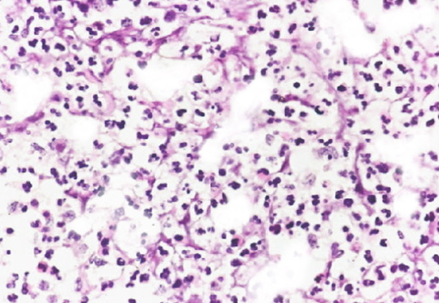
Breast Abscess Inflammatory Infiltration. Histopathology typically demonstrates a mixed, predominantly neutrophilic inflammatory infiltrate.
Contributed by Emma Gregory
(Click Image to Enlarge)
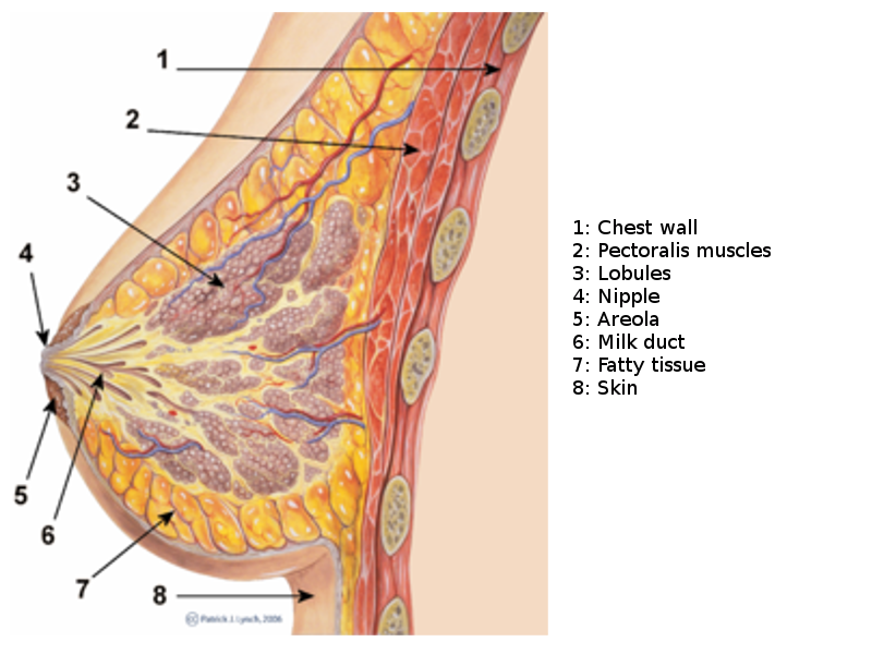
Breast Sagittal View. This illustration shows the chest wall, pectoralis, lobules, nipple, areola, milk duct, fatty tissue, and skin.
PJ Lynch and Morgoth666, Public Domain, via Wikimedia Commons
(Click Image to Enlarge)
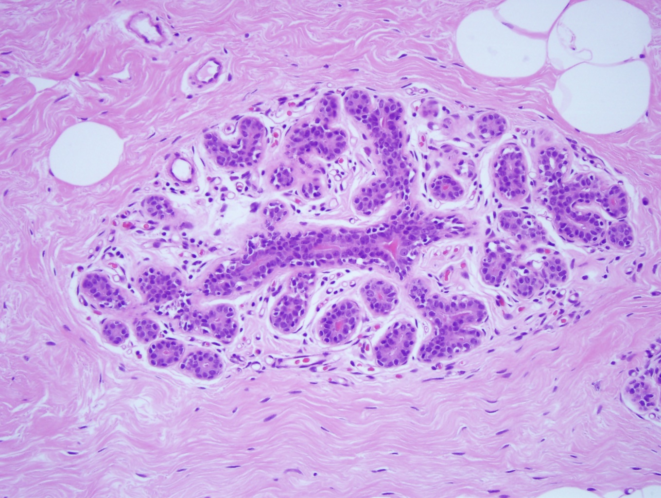
Terminal Duct Lobular Unit. Microscopic anatomy of normal female breast tissue showing a terminal duct lobular unit (×10). The ducts and lobules lumens are open as the epithelial cells do not distend them. Note that the epithelial nuclei overlap, are small and are round, and the nucleoli are inconspicuous and consistent with benign breast tissue.
Contributed by M Khan, DO
(Click Image to Enlarge)
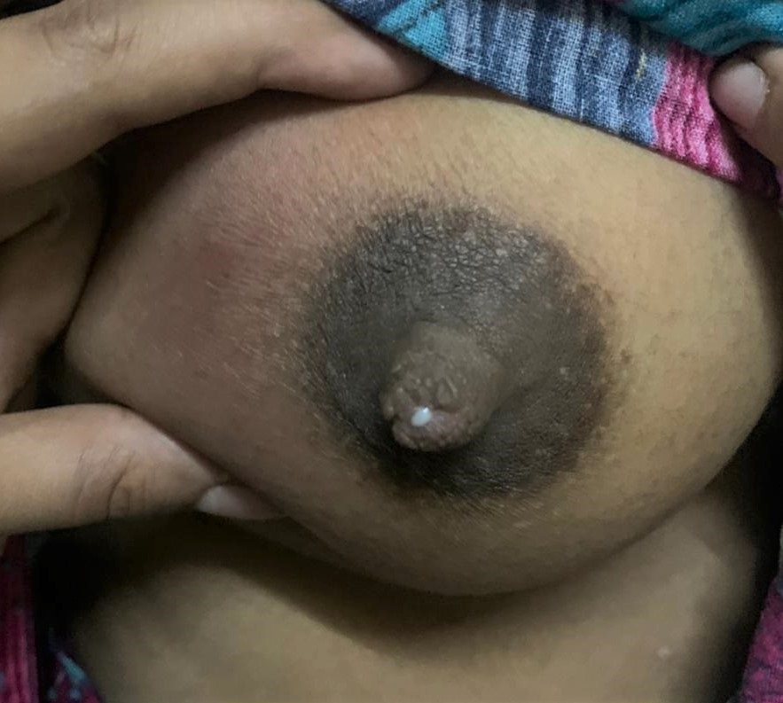
Breast Abscess. Right breast abscess in a lactating mother.
Contributed by P Gada, MBBS
(Click Image to Enlarge)
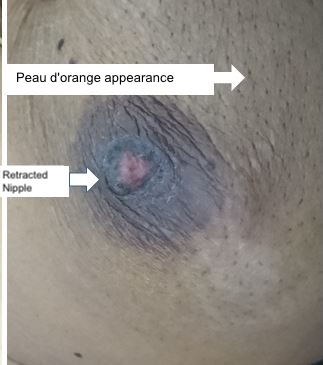
Peau D'Orange. Peau d'orange appearance of the breast.
Contributed by S Munakomi, MD
(Click Image to Enlarge)
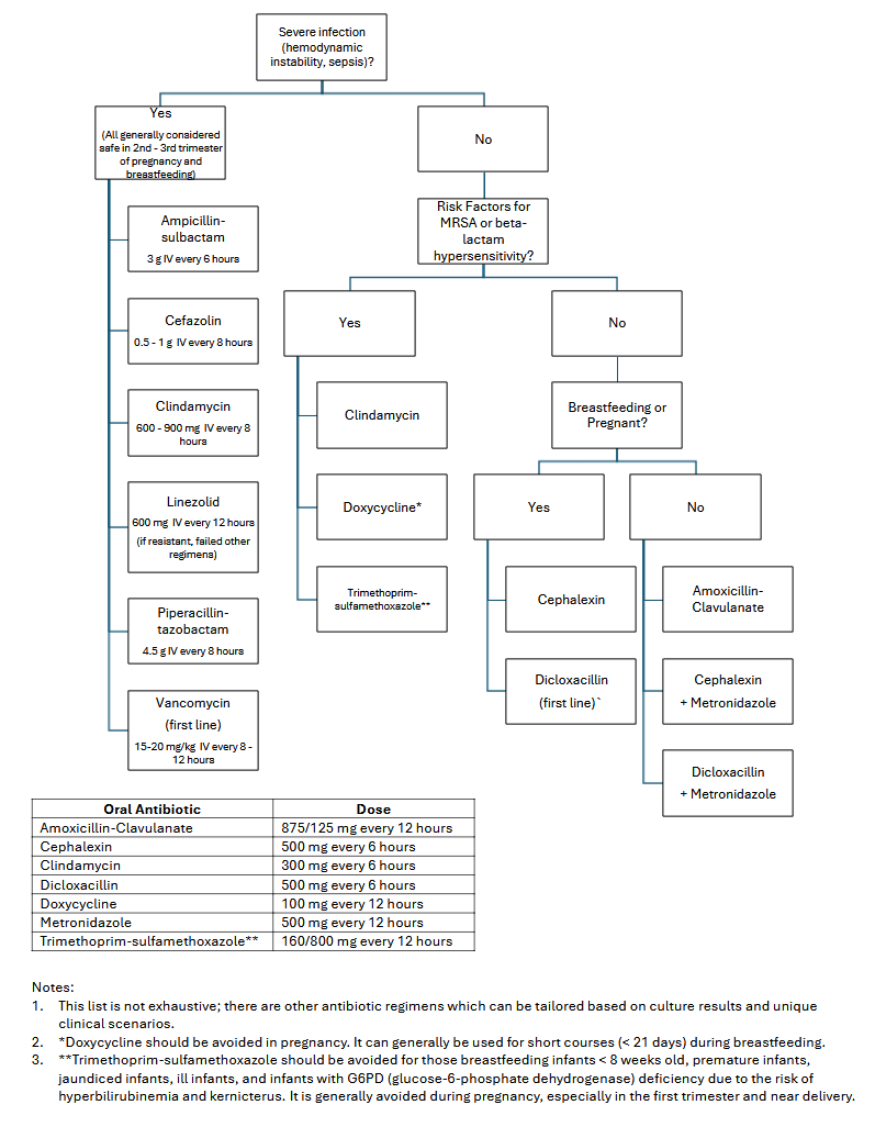
Breast Abscess Antibiotic Flowchart.
Contributed by HG Peifer, MD, MPH
References
Ammann AM, Pratt CG, Lewis JD, Ahmad SA, Shaughnessy E, Heelan AA. Breast infections: A review of current literature. American journal of surgery. 2024 Feb:228():78-82. doi: 10.1016/j.amjsurg.2023.10.040. Epub 2023 Oct 20 [PubMed PMID: 37949727]
Trop I, Dugas A, David J, El Khoury M, Boileau JF, Larouche N, Lalonde L. Breast abscesses: evidence-based algorithms for diagnosis, management, and follow-up. Radiographics : a review publication of the Radiological Society of North America, Inc. 2011 Oct:31(6):1683-99. doi: 10.1148/rg.316115521. Epub [PubMed PMID: 21997989]
Bharat A, Gao F, Aft RL, Gillanders WE, Eberlein TJ, Margenthaler JA. Predictors of primary breast abscesses and recurrence. World journal of surgery. 2009 Dec:33(12):2582-6. doi: 10.1007/s00268-009-0170-8. Epub [PubMed PMID: 19669231]
Urbaniak C, Cummins J, Brackstone M, Macklaim JM, Gloor GB, Baban CK, Scott L, O'Hanlon DM, Burton JP, Francis KP, Tangney M, Reid G. Microbiota of human breast tissue. Applied and environmental microbiology. 2014 May:80(10):3007-14. doi: 10.1128/AEM.00242-14. Epub 2014 Mar 7 [PubMed PMID: 24610844]
Mitchell KB, Johnson HM, Rodríguez JM, Eglash A, Scherzinger C, Zakarija-Grkovic I, Cash KW, Berens P, Miller B, Academy of Breastfeeding Medicine. Academy of Breastfeeding Medicine Clinical Protocol #36: The Mastitis Spectrum, Revised 2022. Breastfeeding medicine : the official journal of the Academy of Breastfeeding Medicine. 2022 May:17(5):360-376. doi: 10.1089/bfm.2022.29207.kbm. Epub [PubMed PMID: 35576513]
Li D, Li J, Yuan Y, Zhou J, Xiao Q, Yang T, Li Y, Jiang L, Gao H. Risk factors and prognosis of acute lactation mastitis developing into a breast abscess: A retrospective longitudinal study in China. PloS one. 2022:17(9):e0273967. doi: 10.1371/journal.pone.0273967. Epub 2022 Sep 1 [PubMed PMID: 36048839]
Level 2 (mid-level) evidenceGollapalli V, Liao J, Dudakovic A, Sugg SL, Scott-Conner CE, Weigel RJ. Risk factors for development and recurrence of primary breast abscesses. Journal of the American College of Surgeons. 2010 Jul:211(1):41-8. doi: 10.1016/j.jamcollsurg.2010.04.007. Epub [PubMed PMID: 20610247]
Moazzez A, Kelso RL, Towfigh S, Sohn H, Berne TV, Mason RJ. Breast abscess bacteriologic features in the era of community-acquired methicillin-resistant Staphylococcus aureus epidemics. Archives of surgery (Chicago, Ill. : 1960). 2007 Sep:142(9):881-4 [PubMed PMID: 17891866]
Russell SP, Neary C, Abd Elwahab S, Powell J, O'Connell N, Power L, Tormey S, Merrigan BA, Lowery AJ. Breast infections - Microbiology and treatment in an era of antibiotic resistance. The surgeon : journal of the Royal Colleges of Surgeons of Edinburgh and Ireland. 2020 Feb:18(1):1-7. doi: 10.1016/j.surge.2019.03.008. Epub 2019 May 7 [PubMed PMID: 31076276]
Jean Bertrand KA, Rose NK, Franck LG, Célestin BA, Ibrahim T, N'gouan Constance BU. Mastitis and breast abscess in newborns and infants. African journal of paediatric surgery : AJPS. 2022 Oct-Dec:19(4):238-240. doi: 10.4103/ajps.ajps_92_21. Epub [PubMed PMID: 36018205]
Warren R, Degnim AC. Uncommon benign breast abnormalities in adolescents. Seminars in plastic surgery. 2013 Feb:27(1):26-8. doi: 10.1055/s-0033-1343993. Epub [PubMed PMID: 24872736]
Alzayyat R, Bokhari D, Almuhanna A, Al-Maghrebi D. Male breast abscess: A rare entity. Radiology case reports. 2024 Mar:19(3):1122-1127. doi: 10.1016/j.radcr.2023.12.014. Epub 2023 Dec 29 [PubMed PMID: 38259713]
Level 3 (low-level) evidenceBoakes E, Woods A, Johnson N, Kadoglou N. Breast Infection: A Review of Diagnosis and Management Practices. European journal of breast health. 2018 Jul:14(3):136-143. doi: 10.5152/ejbh.2018.3871. Epub 2018 Jul 1 [PubMed PMID: 30123878]
Kvist LJ, Rydhstroem H. Factors related to breast abscess after delivery: a population-based study. BJOG : an international journal of obstetrics and gynaecology. 2005 Aug:112(8):1070-4 [PubMed PMID: 16045520]
Branch-Elliman W, Golen TH, Gold HS, Yassa DS, Baldini LM, Wright SB. Risk factors for Staphylococcus aureus postpartum breast abscess. Clinical infectious diseases : an official publication of the Infectious Diseases Society of America. 2012 Jan 1:54(1):71-7. doi: 10.1093/cid/cir751. Epub 2011 Nov 3 [PubMed PMID: 22052894]
Wilson E, Woodd SL, Benova L. Incidence of and Risk Factors for Lactational Mastitis: A Systematic Review. Journal of human lactation : official journal of International Lactation Consultant Association. 2020 Nov:36(4):673-686. doi: 10.1177/0890334420907898. Epub 2020 Apr 14 [PubMed PMID: 32286139]
Level 1 (high-level) evidenceLouis-Jacques AF, Berwick M, Mitchell KB. Risk Factors, Symptoms, and Treatment of Lactational Mastitis. JAMA. 2023 Feb 21:329(7):588-589. doi: 10.1001/jama.2023.0004. Epub [PubMed PMID: 36701134]
Koziol KJ, Smiley A, Latifi R, Castaldi MT. Predictive Risk Factors for Childbirth-Associated Breast Infections in the United States: A 10-Year Perspective. International journal of environmental research and public health. 2023 Jul 10:20(14):. doi: 10.3390/ijerph20146333. Epub 2023 Jul 10 [PubMed PMID: 37510566]
Level 3 (low-level) evidenceDing ST, Gao YJ, Zhang Y, He XP. Analysis of risk factors leading to multiple breast abscesses during lactation. Medicine. 2024 Mar 1:103(9):e37367. doi: 10.1097/MD.0000000000037367. Epub [PubMed PMID: 38428874]
Alex A, Bhandary E, McGuire KP. Anatomy and Physiology of the Breast during Pregnancy and Lactation. Advances in experimental medicine and biology. 2020:1252():3-7. doi: 10.1007/978-3-030-41596-9_1. Epub [PubMed PMID: 32816256]
Level 3 (low-level) evidenceSanten RJ, Mansel R. Benign breast disorders. The New England journal of medicine. 2005 Jul 21:353(3):275-85 [PubMed PMID: 16034013]
Kim SY, Yi DY. Analysis of the human breast milk microbiome and bacterial extracellular vesicles in healthy mothers. Experimental & molecular medicine. 2020 Aug:52(8):1288-1297. doi: 10.1038/s12276-020-0470-5. Epub 2020 Aug 3 [PubMed PMID: 32747701]
Morcomb EF, Dargel CM, Anderson SA. Mastitis: Rapid Evidence Review. American family physician. 2024 Aug:110(2):174-182 [PubMed PMID: 39172675]
Fernández L, Pannaraj PS, Rautava S, Rodríguez JM. The Microbiota of the Human Mammary Ecosystem. Frontiers in cellular and infection microbiology. 2020:10():586667. doi: 10.3389/fcimb.2020.586667. Epub 2020 Nov 20 [PubMed PMID: 33330129]
Abdelhadi MS, Bukharie HA. Breast infections in non-lactating women. Journal of family & community medicine. 2005 Sep:12(3):133-7 [PubMed PMID: 23012091]
Mitchell KB, Valente SA, Snider HC, Fowler AM, Allison KH, Pass HA, Boolbol S, Johnson NM, Boughey JC, Yao K. American Society of Breast Surgeons, Society of Breast Imaging, and College of American Pathology 2025 Guidelines for the Management of Infectious and Inflammatory Lesions of the Breast. JAMA surgery. 2026 Jun 1:161(6):637-645. doi: 10.1001/jamasurg.2026.0613. Epub [PubMed PMID: 41920556]
Brennan ME, Morgan M, Heilat GB, Kanesalingam K. Granulomatous lobular mastitis: Clinical update and case study. Australian journal of general practice. 2020 Jan-Feb:49(1-2):44-47. doi: 10.31128/AJGP-08-19-5042. Epub [PubMed PMID: 32008263]
Level 3 (low-level) evidenceD'Alfonso TM, Ginter PS, Shin SJ. A Review of Inflammatory Processes of the Breast with a Focus on Diagnosis in Core Biopsy Samples. Journal of pathology and translational medicine. 2015 Jul:49(4):279-87. doi: 10.4132/jptm.2015.06.11. Epub 2015 Jul 15 [PubMed PMID: 26095437]
. Practice Bulletin No. 164: Diagnosis and Management of Benign Breast Disorders: Correction. Obstetrics and gynecology. 2021 Feb 1:137(2):382. doi: 10.1097/AOG.0000000000004264. Epub [PubMed PMID: 33481521]
Rao R, Ludwig K, Bailey L, Berry TS, Buras R, Degnim A, Fayanju OM, Froman J, Golesorkhi N, Greenburg C, Ma AT, Mautner SK, Krontiras H, Sowden M, Wexelman B, Landercasper J. Select Choices in Benign Breast Disease: An Initiative of the American Society of Breast Surgeons for the American Board of Internal Medicine Choosing Wisely(®) Campaign. Annals of surgical oncology. 2018 Oct:25(10):2795-2800. doi: 10.1245/s10434-018-6584-5. Epub 2018 Jul 2 [PubMed PMID: 29968026]
Zhou F, Li Z, Liu L, Wang F, Yu L, Xiang Y, Zheng C, Huang S, Yu Z. The effectiveness of needle aspiration versus traditional incision and drainage in the treatment of breast abscess: a meta-analysis. Annals of medicine. 2023 Dec:55(1):2224045. doi: 10.1080/07853890.2023.2224045. Epub [PubMed PMID: 37350731]
Level 1 (high-level) evidenceDavid M, Handa P, Castaldi M. Predictors of outcomes in managing breast abscesses-A large retrospective single-center analysis. The breast journal. 2018 Sep:24(5):755-763. doi: 10.1111/tbj.13053. Epub 2018 May 20 [PubMed PMID: 29781232]
Level 2 (mid-level) evidenceGiacalone PL, Rathat G, Fournet S, Rouleau C. Surgical treatment of recurring subareolar abscess using oncoplastic techniques. Journal of visceral surgery. 2010 Dec:147(6):e389-94. doi: 10.1016/j.jviscsurg.2010.10.008. Epub 2010 Nov 19 [PubMed PMID: 21094113]
Chippa V, Barazi H. Inflammatory Breast Cancer. StatPearls. 2026 Jan:(): [PubMed PMID: 33231994]
Blackmon MM, Nguyen H, Vadakekut ES, Mukherji P. Acute Mastitis. StatPearls. 2026 Jan:(): [PubMed PMID: 32491714]
Gada PB, Vadakekut ES, Bakhshi G. Galactocele. StatPearls. 2026 Jan:(): [PubMed PMID: 35201708]
Hamwi MW, Winters R. Mammary Duct Ectasia. StatPearls. 2026 Jan:(): [PubMed PMID: 32491597]
Zhou F, Yu LX, Ma ZB, Yu ZG. Granulomatous lobular mastitis. Chronic diseases and translational medicine. 2016 Mar:2(1):17-21. doi: 10.1016/j.cdtm.2016.02.004. Epub 2016 Apr 22 [PubMed PMID: 29063020]
Tauro LF, Martis JS, George C, Kamath A, Lobo G, Hegde BR. Tuberculous mastitis presenting as breast abscess. Oman medical journal. 2011 Jan:26(1):53-5. doi: 10.5001/omj.2011.14. Epub [PubMed PMID: 22043382]
Benlghazi A, Benali S, Belouad M, Fakri A, El Mangoub F, Kouach J. Uncommon presentation of necrotizing fasciitis affecting the breast in a young breastfeeding patient: Case report. International journal of surgery case reports. 2024 Jan:114():109147. doi: 10.1016/j.ijscr.2023.109147. Epub 2023 Dec 10 [PubMed PMID: 38134617]
Level 3 (low-level) evidencePileri P, Sartani A, Mazzocco MI, Giani S, Rimoldi S, Pietropaolo G, Pertusati A, Vella A, Bazzi L, Cetin I. Management of Breast Abscess during Breastfeeding. International journal of environmental research and public health. 2022 May 9:19(9):. doi: 10.3390/ijerph19095762. Epub 2022 May 9 [PubMed PMID: 35565158]
Courtney A, Clymo J, Parks R, Wilkins A, Brown R, O'Connell R, Dave R, Dillon M, Fatayer H, Gallimore R, Gandhi A, Gardiner M, Harmer V, Hookway L, Irwin G, Ives C, Mathers H, Murray J, O'Leary DP, Patani N, Paterson S, Potter S, Prichard R, Satta G, Teoh TG, Ziprin P, McIntosh S, Boland MR, Leff DR, MAMMA Research Collaborative. Mastitis and Mammary Abscess Management Audit (MAMMA) in the UK and Ireland. The British journal of surgery. 2024 Jan 3:111(1):. doi: 10.1093/bjs/znad333. Epub [PubMed PMID: 37930678]
Lannin DR. Twenty-two year experience with recurring subareolar abscess andlactiferous duct fistula treated by a single breast surgeon. American journal of surgery. 2004 Oct:188(4):407-10 [PubMed PMID: 15474436]
Scott BG, Silberfein EJ, Pham HQ, Feanny MA, Lassinger BK, Welsh FJ, Carrick MM. Rate of malignancies in breast abscesses and argument for ultrasound drainage. American journal of surgery. 2006 Dec:192(6):869-72 [PubMed PMID: 17161110]
Chang CM, Lin MC, Yin WY. Risk of breast cancer in women with non-lactational mastitis. Scientific reports. 2019 Oct 30:9(1):15587. doi: 10.1038/s41598-019-52046-3. Epub 2019 Oct 30 [PubMed PMID: 31666573]
Lambe M, Johansson AL, Altman D, Eloranta S. Mastitis and the risk of breast cancer. Epidemiology (Cambridge, Mass.). 2009 Sep:20(5):747-51 [PubMed PMID: 19688877]
Krishnan VD, Kostev K, Kalder M. Is there an association between mastitis and breast cancer? a retrospective cohort study from Germany. Cancer causes & control : CCC. 2024 Dec:35(12):1517-1523. doi: 10.1007/s10552-024-01909-w. Epub 2024 Aug 29 [PubMed PMID: 39207599]
Level 2 (mid-level) evidencePeters F, Kiesslich A, Pahnke V. Coincidence of nonpuerperal mastitis and noninflammatory breast cancer. European journal of obstetrics, gynecology, and reproductive biology. 2002 Oct 10:105(1):59-63 [PubMed PMID: 12270566]
Rudoy RC, Nelson JD. Breast abscess during the neonatal period. A review. American journal of diseases of children (1960). 1975 Sep:129(9):1031-4 [PubMed PMID: 1103616]