 Tools
Tools
 Boutonniere Deformity
Introduction
A boutonniere deformity is characterized by flexion of the proximal interphalangeal (PIP) joint and hyperextension of the distal interphalangeal (DIP) joint. Etiologies include acute injuries, such as forceful PIP flexion, dorsal lacerations, or full-thickness burns near the PIP joint, as well as chronic processes, including progressive capsular distension in the context of rheumatoid arthritis.[1][2][3][4] All mechanisms produce disruption of the central slip of the digital extensor mechanism, resulting in volar migration of the lateral bands and formation of the characteristic boutonniere deformity (see Image. Boutonniere Deformity Anatomy).
Etiology
Register For Free And Read The Full Article
 Search engine and full access to all medical articles
- 10 free questions in your specialty
- Free CME/CE Activities
- Free daily question in your email
- Save favorite articles to your dashboard
- Emails offering discounts
Learn more about a Subscription to StatPearls Point-of-Care
Etiology
The digital extensor mechanism originates in the dorsal forearm and extends to the metacarpophalangeal (MCP) joint, where it joins the extensor hood. The mechanism courses distally along the digit, with a portion inserting onto the dorsal base of the middle phalanx as the central slip, which facilitates PIP joint extension. Disruption of the central slip at or near its insertion results in a boutonniere deformity. The extensor hood continues distally to insert onto the dorsal base of the distal phalanx as the terminal tendon, which facilitates DIP joint extension. Disruption of the terminal tendon produces a mallet deformity.[5]
Additional relevant structures include the lateral bands, formed from the interossei, which course laterally at the level of the middle phalanx before merging dorsally into a conjoined tendon that becomes the terminal tendon. The transverse retinacular ligament, located volarly near the PIP joint, inserts into the lateral bands to prevent dorsal migration. The triangular ligament, located dorsally, connects the lateral bands to prevent volar subluxation. The lumbricals, intrinsic muscles inserting into the lateral bands, assist with MCP flexion and PIP and DIP extension.
Epidemiology
Blunt force trauma encompasses a heterogeneous group of digital injuries, with central slip disruption and subsequent boutonniere deformity representing a well-documented sequela. Sports-related injuries most commonly arise from football and basketball participation.[6][7] Dorsal lacerations and burn injuries of the finger also produce central slip compromise and boutonniere deformities, although less frequently. Approximately 50% of patients with rheumatoid arthritis eventually develop a boutonniere deformity, which can markedly reduce quality of life by impairing hand functions, such as grasping and pinching.[8][9] Atraumatic, noninflammatory boutonniere deformities of the thumb occur in an estimated 13% of patients, with prevalence increasing among older populations.[10]
Pathophysiology
The primary inciting injury leading to boutonniere deformity is the disruption of the central slip. Causes include avulsion-type injury from forceful PIP flexion during blunt trauma; dorsal laceration or burn over the PIP joint; and PIP capsular distension associated with underlying inflammatory conditions such as rheumatoid arthritis. In inflammatory cases, synovial fluid accumulation promotes synovitis, resulting in the formation of a thickened fibrous layer and progressive distension of the joint capsule. Chronic inflammation progressively erodes articular surfaces and ultimately compromises the central slip.[11][12]
Following central slip disruption, a sequence of pathoanatomical changes develops that ultimately produces a boutonniere deformity. Injury to the central slip compromises the digital extensor mechanism, preventing adequate active extension of the digit at the PIP joint. Altered biomechanics gradually attenuate the triangular ligament, permitting volar subluxation of the lateral bands (see Image. Isolated Lateral View of a Fifth-Digit Boutonniere Deformity).
In the volar position, the lateral bands act as flexors of the PIP joint. Combined with impaired PIP extension due to central slip disruption, this mechanism generates a flexion deformity at the PIP joint. The joint protrudes through the disrupted central slip, analogous to a button through a buttonhole, which gives the deformity its name (“boutonniere,” meaning “buttonhole” in French).[13]
The lumbricals, lacking a balancing force, retract the lateral bands proximally, increasing tension on the terminal tendon and producing hyperextension at the DIP joint. Persistent abnormal digit posture allows contraction of the transverse retinacular ligament, which further exacerbates PIP flexion. These pathoanatomical changes may require several weeks to develop, and a boutonniere deformity may not be immediately apparent after central slip injury.
Extensor mechanism injuries of the hand are classified by anatomical region into 5 discrete zones; this system accounts for the unique anatomy and functional demands of the thumb. Each zone corresponds to a specific segment of the digit or forearm, as follows:
- Zone 1: DIP joint
- Zone 2: Middle phalanx
- Zone 3: PIP joint, including the central slip
- Zone 4: Proximal phalanx
- Zone 5: MCP joint
- Zone 6: Metacarpal bones
- Zone 7: Carpal bones
- Zone 8: Distal forearm
- Zone 9: Proximal forearm
History and Physical
A thorough history for a suspected boutonniere deformity should include the mechanism of injury, chronicity, associated pain or neurologic symptoms, prior treatments, and degree of functional limitation. Past medical history should be reviewed for systemic inflammatory or autoimmune conditions, such as rheumatoid arthritis. Overall disease control and the use of prior or current disease-modifying medications should be documented if present.[14]
Physical examination begins with assessment of overall digit posture, with a boutonniere deformity defined by PIP flexion and DIP hyperextension. In cases of dorsal laceration, the affected area should be thoroughly cleaned and examined in a bloodless field to visualize extensor integrity.[15] Digital range of motion (ROM) should be evaluated actively and passively to determine stiffness, weakness, and correctability of each joint. A complete neurovascular examination should supplement these findings.
Early recognition of central slip disruption prior to the development of a boutonniere deformity may be achieved using the Elson test. The affected digit is placed over the edge of a surface so that the PIP joint flexes to 90°. A force is applied over the middle phalanx to maintain PIP flexion while the patient attempts active extension of the PIP joint against resistance. The test result is positive for central slip injury if PIP extension is weak and the DIP remains rigid. The test result is negative if PIP extension is strong and the DIP remains supple.[16]
Evaluation
Diagnosis of a suspected central slip injury or a boutonniere deformity can be established by clinical evaluation alone, although supplemental imaging studies may be performed (see Image. Fifth-Digit Boutonniere Deformity, Posteroanterior Radiographic View). Radiographs, including anteroposterior, oblique, and lateral views of the affected digit, assess fractures at the base of the middle phalanx, the site of central slip insertion, and evaluate PIP and DIP joint congruency (see Image. Lateral Radiograph of a Fifth-Digit Boutonniere Deformity). Magnetic resonance imaging provides a detailed visualization of the central slip and surrounding soft tissues.[17]
Treatment / Management
The primary goal in the treatment of a central slip injury and a boutonniere deformity is restoration of the full ROM of the affected digit. Treatment options include nonoperative and operative modalities.
Nonoperative Approaches
Patients presenting within 4 weeks of a closed injury are often treated conservatively. Management involves fabrication of a splint or brace that maintains the PIP joint in full extension, facilitating approximation of the central slip to its insertion at the dorsal base of the middle phalanx. Immobilization of the DIP joint is avoided to prevent unnecessary stiffness. The splint should be worn full-time for 6 to 8 weeks to allow central slip healing, with strict avoidance of PIP flexion during this period to prevent disruption of early repair.[18] Daily passive and active flexion and extension of the free DIP joint should be performed. After the initial 6 to 8 weeks, the splint should be worn at night only for an additional 4 to 6 weeks. For patients returning to sports or activities that risk early reinjury, the digit should be taped or otherwise protected during the transition period.
Operative Treatment
Surgical intervention is indicated in cases of central slip injury or a boutonniere deformity with delayed presentation beyond 4 weeks, associated lacerations, displaced avulsion fractures, underlying rheumatoid arthritis, or failure of nonoperative management.[19][20] Primary repair of a lacerated central slip may be performed with sutures. Avulsed central slips may be reattached to the middle phalanx insertion using a suture anchor. Displaced dorsal base fractures of the middle phalanx that are amenable to fixation may undergo closed reduction with percutaneous Kirschner wire (K-wire) stabilization. Fractures unsuitable for closed reduction require open reduction followed by internal fixation with interfragmentary screws or plate and screw constructs.
Chronic deformities with preserved digital ROM following hand therapy or soft tissue releases may undergo central slip reconstruction using a tendon graft or dorsal transposition and suturing of the lateral bands to restore PIP extension. Terminal tendon hyperextension may be corrected via tenotomy in similar chronic deformities. Painful arthritic PIP joints in rheumatoid arthritis may be managed with PIP arthrodesis using K-wires, screws, or plate and screw constructs, which may be accompanied by terminal tendon tenotomy.
Following surgical procedures, the PIP joint should be maintained in full extension for 6 to 8 weeks with temporary K-wire fixation to stabilize the repair or reconstruction. Rehabilitation with a hand therapist and appropriate bracing is integral to management. In patients with rheumatoid arthritis, concurrent control of the underlying inflammatory disease is essential to optimize long-term outcomes and prevent the development of boutonniere deformities in other digits.
Differential Diagnosis
Differential diagnoses for a suspected central slip injury or a boutonniere deformity include conditions that mimic the characteristic PIP flexion and DIP hyperextension. Pseudo-boutonniere presents with PIP flexion without associated DIP hyperextension and typically results from more remote blunt trauma to the PIP joint. Traumatic dislocation of the PIP joint produces a more pronounced deformity.
Dupuytren disease and locked trigger finger can limit finger extension and produce a flexed posture. Skin contracture at the proximal finger may occur following burn injury. Less common causes include contractures resulting from inherited or systemic conditions, such as epidermolysis bullosa or Hansen disease.
Prognosis
Prevention of progression to a boutonniere deformity after central slip disruption significantly improves functional outcomes. Injuries recognized early and treated acutely with PIP extension splinting achieve moderate to good or excellent functional results.[21] Chronic deformities requiring surgical intervention demonstrate more variable outcomes that depend on preoperative PIP joint rigidity and the surgical technique employed. Overall improvement is generally fair to moderate, with outcomes more unpredictable in patients with rheumatoid arthritis.[22][23]
Complications
Complications of central slip injury or a boutonniere deformity, with or without treatment, include chronic or persistent joint stiffness, persistent PIP extensor lag, reinjury or recurrent deformity, posttraumatic arthritis, and chronic digital swelling.[24] Awareness and early management of these complications are critical to optimizing long-term functional outcomes.
Postoperative and Rehabilitation Care
Referral to hand therapy is recommended following completion of immobilization to restore digital ROM and strength.[25] Custom protective splints may be fabricated by the therapist to support recovery. Restoration of full function may require several months to more than a year.[26][27]
Deterrence and Patient Education
Prognosis and outcomes of central slip injuries and boutonniere deformities are often variable, making patient education essential. Instruction should include potential complications, such as chronic ROM limitation, development of early arthritis, and predisposition to reinjury. Healthcare professionals treating athletes, particularly those at risk for digital injuries, such as football and basketball players, should emphasize early evaluation following blunt trauma to the digits.[28] Delayed management can produce long-term sequelae that may restrict future sports participation. Emphasizing maintenance of PIP extension during treatment and ensuring adherence to follow-up protocols supports optimal functional outcomes.
Enhancing Healthcare Team Outcomes
Diagnosis and management of a boutonniere deformity can be complex and require an interprofessional team comprising a hand surgeon, hand therapist, emergency medicine and primary care clinicians, advanced practitioners, and nurses. Clinicians in emergency, urgent care, or primary care settings frequently encounter patients with suspected central slip injuries. Prompt initiation of diagnostic evaluation and recognition of the injury are critical.
Once identified or suspected, referral to a hand surgeon allows confirmation of the diagnosis and initiation of nonoperative or operative treatment. Hand therapists assume a central role during the rehabilitation phase, while advanced practitioners and nurses facilitate care coordination. Collaboration among these healthcare professionals optimizes patient outcomes and minimizes complications associated with boutonniere deformities.
Media
(Click Image to Enlarge)
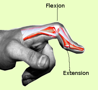
Boutonniere Deformity Anatomy. The diagram illustrates the permanent flexion of the proximal interphalangeal joint. The distal interphalangeal joint simultaneously exhibits hyperextension due to a ruptured central slip of the extensor tendon.
Contributed by S Bhmiji, MD
(Click Image to Enlarge)
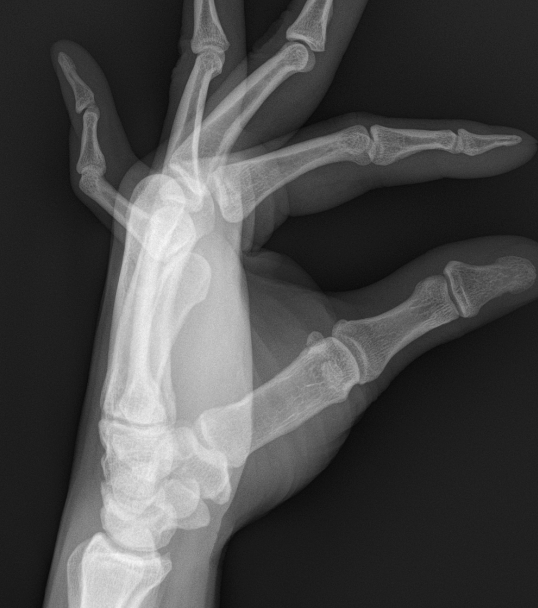
Lateral Radiograph of a Fifth-Digit Boutonniere Deformity. The image shows a boutonniere deformity affecting the extensor mechanism of the fifth digit. This view demonstrates the degree of middle phalanx flexion relative to the proximal phalanx without the overlap of the other fingers.
Contributed by R Flores, MD
(Click Image to Enlarge)
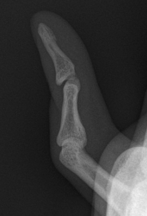
Isolated Lateral View of a Fifth-Digit Boutonniere Deformity. This coned-down perspective removes the radiographic overlap of the other 4 digits, allowing for an unobstructed assessment of the proximal interphalangeal joint. The view highlights a volar subluxation of the middle phalanx, a hallmark of a ruptured central slip.
Contributed by R Flores, MD
(Click Image to Enlarge)
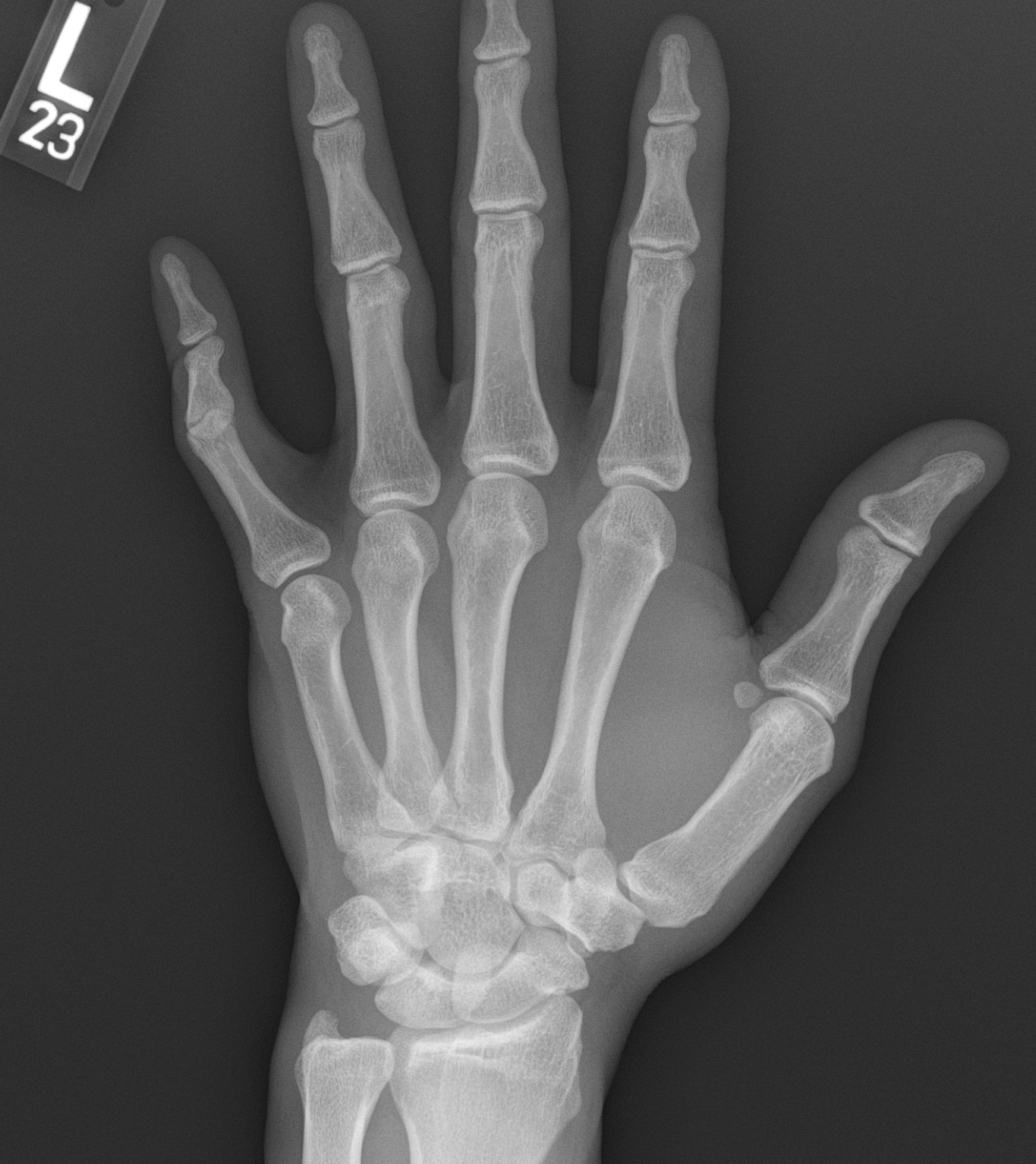
Fifth-Digit Boutonniere Deformity, Posteroanterior Radiographic View. This image provides a primary assessment of the hand's skeletal alignment and joint spaces. In this view, the unique finding is the foreshortening of the 5th digit’s middle phalanx, which occurs because the bone is angled toward the palm in a fixed flexion posture.
Contributed by R Flores, MD
References
Pencle F, Doehrmann R, Waseem M. Fingertip Injuries. StatPearls. 2026 Jan:(): [PubMed PMID: 28613777]
Boussakri H, Azarkane M, Dahmani O, Elidrissi M, Shimi M, Elibrahimi A, Elmrini A. Unusual combination of lesions of the traumatic hand: closed central slip laceration of the extensor and interphalangeal thumb joint's dislocation (a case report). The Pan African medical journal. 2014:18():230. doi: 10.11604/pamj.2014.18.230.3240. Epub 2014 Jul 18 [PubMed PMID: 25426188]
Level 3 (low-level) evidenceSabapathy SR, Bajantri B, Bharathi RR. Management of post burn hand deformities. Indian journal of plastic surgery : official publication of the Association of Plastic Surgeons of India. 2010 Sep:43(Suppl):S72-9. doi: 10.4103/0970-0358.70727. Epub [PubMed PMID: 21321661]
Vedel PN, Tranum-Jensen J, Dahlin LB, Brogren E, Søe NH. [Deformities of the finger joints]. Ugeskrift for laeger. 2017 Nov 27:179(48):. pii: V04170324. Epub [PubMed PMID: 29208202]
Beutel BG, Waseem M. Mallet Finger Injuries. StatPearls. 2026 Jan:(): [PubMed PMID: 29083648]
Bai RJ, Zhang HB, Zhan HL, Qian ZH, Wang NL, Liu Y, Li WT, Yin YM. Sports Injury-Related Fingers and Thumb Deformity Due to Tendon or Ligament Rupture. Chinese medical journal. 2018 May 5:131(9):1051-1058. doi: 10.4103/0366-6999.230721. Epub [PubMed PMID: 29692376]
Level 2 (mid-level) evidenceCarruthers KH, Skie M, Jain M. Jam Injuries of the Finger: Diagnosis and Management of Injuries to the Interphalangeal Joints Across Multiple Sports and Levels of Experience. Sports health. 2016 Sep:8(5):469-78. doi: 10.1177/1941738116658643. Epub 2016 Jul 15 [PubMed PMID: 27421747]
Sood A, Kotamarti VS, Granick MS. Boutonnière Deformity Following Volar Proximal Interphalangeal Joint Dislocation. Eplasty. 2016:16():ic25 [PubMed PMID: 27347279]
G A, C G, C J M, B L, E K. Continuum finger rehabilitation system for rheumatoid arthritis. Annual International Conference of the IEEE Engineering in Medicine and Biology Society. IEEE Engineering in Medicine and Biology Society. Annual International Conference. 2025 Jul:2025():1-6. doi: 10.1109/EMBC58623.2025.11254614. Epub [PubMed PMID: 41337080]
Hara T, Yoneda H, Kurimoto S, Yamamoto M, Iwatsuki K, Hirata H. Thumb boutonnière deformity without rheumatoid arthritis or trauma. The Journal of hand surgery, European volume. 2018 Mar:43(3):324-330. doi: 10.1177/1753193417704610. Epub 2017 May 2 [PubMed PMID: 28462679]
Di Matteo A, Bathon JM, Emery P. Rheumatoid arthritis. Lancet (London, England). 2023 Nov 25:402(10416):2019-2033. doi: 10.1016/S0140-6736(23)01525-8. Epub 2023 Oct 27 [PubMed PMID: 38240831]
Toyama S, Tokunaga D, Tsuchida S, Kushida R, Oda R, Kawahito Y, Takahashi K. Comprehensive assessment of alterations in hand deformities over 11 years in patients with rheumatoid arthritis using cluster analysis and analysis of covariance. Arthritis research & therapy. 2021 Feb 27:23(1):66. doi: 10.1186/s13075-021-02448-4. Epub 2021 Feb 27 [PubMed PMID: 33640011]
Grau L, Baydoun H, Chen K, Sankary ST, Amirouche F, Gonzalez MH. Biomechanics of the Acute Boutonniere Deformity. The Journal of hand surgery. 2018 Jan:43(1):80.e1-80.e6. doi: 10.1016/j.jhsa.2017.07.011. Epub 2017 Sep 7 [PubMed PMID: 28888567]
Sharif K, Sharif A, Jumah F, Oskouian R, Tubbs RS. Rheumatoid arthritis in review: Clinical, anatomical, cellular and molecular points of view. Clinical anatomy (New York, N.Y.). 2018 Mar:31(2):216-223. doi: 10.1002/ca.22980. Epub 2017 Oct 27 [PubMed PMID: 28833647]
Beutel BG, Wang J, Taqi M. Finger Dislocation. StatPearls. 2025 Jan:(): [PubMed PMID: 31855352]
Solitro GF, Echenique DB, Gonzalez MH, Amirouche F. Assessment of the ligamentous stress distribution in the pathomechanics of the boutonniere deformity through a computational 3D model. Journal of orthopaedic surgery and research. 2025 Oct 21:20(1):903. doi: 10.1186/s13018-025-06329-3. Epub 2025 Oct 21 [PubMed PMID: 41121395]
Meadows L, Gorbachova T. MR Imaging of Injury of the Extensor Mechanism of the Fingers. Magnetic resonance imaging clinics of North America. 2026 Feb:34(1):117-128. doi: 10.1016/j.mric.2025.08.007. Epub 2025 Oct 7 [PubMed PMID: 41265971]
McCue FC, Honner R, Johnson MC, Gieck JH. Athletic injuries of the proximal interphalangeal joint requiring surgical treatment. The Journal of bone and joint surgery. American volume. 1970 Jul:52(5):937-56 [PubMed PMID: 5479483]
Fox PM, Chang J. Treating the Proximal Interphalangeal Joint in Swan Neck and Boutonniere Deformities. Hand clinics. 2018 May:34(2):167-176. doi: 10.1016/j.hcl.2017.12.006. Epub [PubMed PMID: 29625636]
Hesse N, Reidler P, Schmitt R. [Sports-related injuries of the thumb and fingers]. Radiologie (Heidelberg, Germany). 2023 Apr:63(4):284-292. doi: 10.1007/s00117-023-01127-6. Epub 2023 Mar 14 [PubMed PMID: 36917239]
Tong Y, Donnelly M, Paksima N. Nonoperative treatment of the Boutonniere deformity: Is there a difference in outcomes? Journal of hand therapy : official journal of the American Society of Hand Therapists. 2025 Oct-Dec:38(4):810-816. doi: 10.1016/j.jht.2025.02.013. Epub 2025 Apr 23 [PubMed PMID: 40274447]
El-Sallakh S, Aly T, Amin O, Hegazi M. Surgical management of chronic boutonniere deformity. Hand surgery : an international journal devoted to hand and upper limb surgery and related research : journal of the Asia-Pacific Federation of Societies for Surgery of the Hand. 2012:17(3):359-64. doi: 10.1142/S0218810412500311. Epub [PubMed PMID: 23061946]
Towfigh H, Gruber P. Surgical treatment of the boutonnière deformity. Operative Orthopadie und Traumatologie. 2005 Feb:17(1):66-78 [PubMed PMID: 16007379]
Kamnerdnakta S, Huetteman HE, Chung KC. Complications of Proximal Interphalangeal Joint Injuries: Prevention and Treatment. Hand clinics. 2018 May:34(2):267-288. doi: 10.1016/j.hcl.2017.12.014. Epub [PubMed PMID: 29625645]
Hanson ZC, Thompson RG, Andrews JR, Lourie GM. Boutonniere Versus Pseudoboutonniere Deformities: Pathoanatomy, Diagnosis, and Treatment. The Journal of hand surgery. 2023 May:48(5):489-497. doi: 10.1016/j.jhsa.2022.10.019. Epub 2022 Dec 31 [PubMed PMID: 36593154]
Hirth MJ, Howell JW, O'Brien L. Relative motion orthoses in the management of various hand conditions: A scoping review. Journal of hand therapy : official journal of the American Society of Hand Therapists. 2016 Oct-Dec:29(4):405-432. doi: 10.1016/j.jht.2016.07.001. Epub 2016 Oct 25 [PubMed PMID: 27793417]
Level 2 (mid-level) evidenceMcKeon KE, Lee DH. Posttraumatic Boutonnière and Swan Neck Deformities. The Journal of the American Academy of Orthopaedic Surgeons. 2015 Oct:23(10):623-32. doi: 10.5435/JAAOS-D-14-00272. Epub 2015 Aug 28 [PubMed PMID: 26320165]
Niederhauser L, Wehrli L. [Finger injuries in athletes]. Revue medicale suisse. 2024 Jul 17:20(882):1349-1353. doi: 10.53738/REVMED.2024.20.882.1349. Epub [PubMed PMID: 39021104]