 Tools
Tools
 Bennett Fracture
Introduction
A Bennett fracture is an intra-articular fracture of the base of the thumb (first) metacarpal with an associated volar-ulnar bony fragment.[1] The injury is typically caused by axial loading on a partially flexed thumb, which may be associated with other carpal bone fractures or ligament injuries.[2] Due to the disruption of key ligamentous stabilizers in this fracture pattern, the first metacarpal may displace proximally and dorsally with supination. Dedicated radiographs of the thumb, and potentially CT scan, are essential in the evaluation of these injuries and aid in guiding management, as these fractures are often considered unstable.
An understanding of the relevant anatomy at the base of the thumb is critical for effectively evaluating and treating Bennett fractures. The first carpometacarpal (CMC) joint at the base of the thumb is a saddle-shaped articular surface defined by the first metacarpal and the trapezium. This articulation assists with thumb flexion/extension and adduction/abduction ranges of motion, and has relatively minimal axial rotation. Several neighboring muscles and ligaments serve as dynamic deforming forces and static stabilizers, respectively. The adductor pollicis applies an adduction/supination force along the first metacarpal shaft, while the abductor pollicis longus and the extensor pollicis longus exert a combined proximal/dorsal/radial force. Two primary ligaments provide stabilization at the first metacarpal base. The volar (anterior) oblique ligament originates at the trapezial tuberosity and inserts into the volar aspect of the first metacarpal. The dorsoradial ligament originates at the tubercle of the trapezium and inserts into the dorsal base of the first metacarpal.
Etiology
Register For Free And Read The Full Article
 Search engine and full access to all medical articles
- 10 free questions in your specialty
- Free CME/CE Activities
- Free daily question in your email
- Save favorite articles to your dashboard
- Emails offering discounts
Learn more about a Subscription to StatPearls Point-of-Care
Etiology
Bennett fractures most commonly result from an axially directed force to the thumb, often with the digit flexed. Given the mechanism of this traumatic injury, it may be associated with fractures involving the adjacent carpal bones (eg, trapezium or hamate) and/or ulnar collateral ligament injuries of the thumb metacarpophalangeal (MCP) joint.[2][3]
First metacarpal base fractures are typically classified as 1 of 5 types based upon articular involvement and the specific fracture pattern, as follows:
- Extra-articular transverse
- Extra-articular oblique
- Intra-articular Bennett (articular involvement with a volar-ulnar fragment)
- Intra-articular Rolando (complete articular involvement, often with a "T"- or "Y"-shaped fracture morphology)
- Intra-articular comminuted [4]
Gedda further sub-classified Bennett fractures into one of three types:[5]
- Type 1: Intra-articular fracture with CMC subluxation
- Type 2: Intra-articular fracture (possibly impacted) without CMC subluxation
- Type 3: Intra-articular fracture with CMC dislocation [5]
Epidemiology
Metacarpal fractures represent approximately 33% of hand fractures.[6] Fractures at the base of the thumb have been estimated to account for 80% of all thumb fractures and 4% of all hand fractures.[7][4] While most are extra-articular, 30% are considered Bennett fractures.[8] Men more commonly sustain Bennett fractures, which tend to occur most frequently in young adults, followed by older women.[6] The underlying mechanism of injury also varies by age. Younger patients primarily sustain Bennett fractures from recreational or sports activities, middle-aged individuals often experience injuries related to work, and older patients often sustain them from falls.
Pathophysiology
As previously noted, a Bennett fracture typically results from application of an axially directed force onto the thumb, often with the digit in a flexed position, resulting in an intra-articular fracture at the base of the thumb involving the first CMC joint. The orientation of the fracture pattern creates a volar-ulnar bony fragment, which is stabilized at its articulation on the trapezium by the volar (anterior) oblique ligament. The remainder of the first metacarpal, no longer anchored by the volar oblique ligament due to the fracture, displaces proximally and dorsally with supination due to the deforming forces primarily applied by the abductor pollicis longus and the adductor pollicis, as well as the extensor pollicis longus.[9] Consequently, the thumb may adopt a shortened and adducted posture.
History and Physical
A thorough history should be performed in all patients suspected of having sustained a Bennett fracture. This includes the mechanism and chronicity of the injury, as well as any prior treatment or reduction attempts. The patient's hand dominance, although it does not directly guide the management of a Bennett fracture, could impact the recovery course and patient satisfaction if the dominant hand is injured. Additionally, a comprehensive medical history and medication reconciliation should be performed, as certain comorbidities and medications, including anticoagulants and antibody-based therapies, may influence the selection and timing of treatment interventions.
Clinical examination should include an evaluation of the entire hand and wrist, as there may be other concurrent injuries. In the setting of a Bennett fracture, inspection may reveal swelling/ecchymosis and potentially a deformity at the base of the thumb (depending on the degree of displacement). Palpation would demonstrate tenderness in the region of the first CMC joint with limited range of active thumb motion due to pain. Flexor pollicis longus and extensor pollicis longus function should be examined with active thumb interphalangeal joint flexion and extension, respectively, to confirm tendon integrity. A neurovascular examination is also performed to evaluate thumb perfusion and digital nerve continuity.
Evaluation
Diagnostic evaluation for a Bennett fracture is conducted primarily through imaging studies. Standard hand radiographs should be obtained, including anteroposterior (AP), lateral, and oblique views. Additional dedicated views of the thumb should be obtained to provide further detail about the injury, including a Roberts view and a true lateral of the thumb.[9] The Roberts view is a true AP view of the first CMC joint and thumb. This view is performed with full pronation of the forearm while the dorsal aspect of the thumb is placed against the radiographic plate, and the x-ray beam is directed 90 degrees to the plate. Another view is the Bett view, obtained with the palm over-pronated 20 degrees from flat against the radiographic plate, and the beam directed 15 degrees proximal-to-distal. Stress radiographs can also be performed on an AP view, which may demonstrate subluxation of the first metacarpal base radially relative to the trapezium. Alternatively, dynamic stress views utilizing fluoroscopy can be considered.[10] In the setting of equivocal radiographs or more complex patterns, a CT of the thumb can be obtained.
Treatment / Management
The primary objective in the management of a Bennett fracture is to restore articular congruity and overall fracture alignment. There is some debate in the literature regarding the acceptable amount of articular step-off at the fracture site, particularly in non-athlete populations. Some authors have found limited correlation between the quality of articular reduction and radiographic or subjective outcomes.[11][12]. At the same time, certain biomechanical studies have demonstrated that a 2 mm persistent articular step-off does not alter contact pressures at the first CMC joint.[13] It has, therefore, been suggested that bony apposition of the fragments within 2 mm and the correction of any joint subluxation should be tolerated without increasing the risk of posttraumatic arthritis. Many clinical studies, nevertheless, suggest that anatomic reduction is preferred.[14] Consequently, most guidelines recommend articular step-off to be less than 1 mm without CMC subluxation.
Nonoperative Management
Bennett fractures may be treated nonoperatively if there is less than 1 mm of displacement and articular step-off. If the initial displacement exceeds this, closed reduction can be attempted. Proper reduction requires traction along the thumb in an axial direction with volar abduction and pronation while applying an external force over the first metacarpal base.[9] Thumb extension (known as the "hitchhiker position") has been shown to worsen fracture displacement and should be avoided.[1] Once within acceptable parameters, a thumb spica splint or cast is applied. Serial radiographs of the hand/thumb should be obtained weekly for the first three weeks to assess for worsening displacement, which may warrant surgical intervention.
Closed reduction and splinting of Bennett fractures was the initial treatment described for these injuries, which remained the preferred method of treatment until the 1970s.[15][14] Historical reports have shown reasonable outcomes with this management, although more recent studies have shown poorer outcomes when treating certain Bennett fractures with splinting/casting alone.[16]
Operative Management
Fractures with greater than 1 mm of displacement or articular step-off that cannot be successfully reduced and stabilized generally require operative treatment. A variety of surgical interventions exist,[17][18][19] as follows:(A1)
- Closed reduction and percutaneous pinning: Direct fixation of the volar-ulnar bony fragment itself is not typically required to achieve adequate overall reduction and stability. A single Kirschner (K)-wire can be placed trans-articularly through the first metacarpal shaft and into the trapezium to abut the shaft against the volar-ulnar fragment. Alternatively, the Iselin technique involves dual K-wire fixation between the first and second metacarpal shafts without trans-articular fixation.[20] (B3)
- Open reduction and internal fixation: A dorsal incision centered over the first CMC joint and the thumb metacarpal base is made. Under direct visualization, the fracture is reduced and fixed with a plate/screw construct, lag screw, or K-wire(s).
- Arthroscopic-assisted reduction and screw fixation: To aid in reduction efforts, first perform CMC joint arthroscopy to directly visualize the articular surface, facilitate reduction, and confirm an acceptable reduction.[21] Then, a cannulated screw (or K-wire) can be inserted into the volar-ulnar fragment for fixation.
- Augmentation with a suture-button: Cortical suture-buttons may be utilized to augment screw fixation.[17]
In cases of a Bennett fracture malunion or nonunion, potential surgical treatment options include intra-articular or extra-articular osteotomies, trapezium resection with or without interposition arthroplasty, implant arthroplasty, first CMC arthrodesis, and small fragment excision with suture-button suspension.[22] Following surgical intervention, the patient is placed into a thumb spica splint.
Differential Diagnosis
The differential diagnosis includes the following:
- Rolando fracture (a complete intra-articular fracture at the base of the first metacarpal that may be comminuted)
- Trapezium fracture
- Metacarpal shaft fracture
- First CMC dislocation
Prognosis
Due to their potential instability and articular involvement, the prognosis of patients with Bennett fractures is variable.[23][24] Based on a study by Kjaer-Petersen et al that reviewed 41 Bennett fractures, long-term outcomes are often determined by the quality of fracture reduction. In their analysis, 86% of patients with an anatomic reduction (less than 1-mm step-off) had no residual symptoms, whereas only 46% of patients with good or poor reduction (greater than 1-mm step-off) remained asymptomatic.[15] The type of intervention has also been shown to impact ultimate outcomes. Duche et al evaluated closed reduction percutaneous pinning and showed that patients experienced satisfactory medium-term functional outcomes, but with moderate loss of range of motion and pinch strength.[20] In a systematic review of patient outcomes following surgical treatment for Bennett fractures, Velasquez-Giron et al demonstrated that patients treated with closed reduction percutaneous pinning had less postoperative pain than those undergoing open reduction and internal fixation or arthroscopy-assisted reduction and screw fixation.[25] However, they also concluded that all three techniques ultimately yielded similar functional outcomes and similar rates of posttraumatic CMC arthritis.
Complications
Following a Bennett fracture, patients may experience thumb stiffness. To mitigate this, range-of-motion exercises should begin as soon as clinically appropriate, which may be several weeks after the injury in patients treated nonoperatively with splinting/casting, 5 to 10 days after screw or plate fixation, or 4 weeks after closed reduction percutaneous pinning (after the pins are removed).[26] Malreduction of the fracture may result in a permanent thumb deformity with more significantly diminished pinch strength and/or instability at the CMC joint. Further, residual articular incongruity can increase the risk of posttraumatic CMC arthritis.
Postoperative and Rehabilitation Care
For those patients treated surgically, postoperative protocols will vary depending on the fixation method employed, perceived quality and stability of the reduction, surgeon preference, and patient considerations. However, occupational and hand therapy are considered crucial elements of the rehabilitation course to maximize patient outcomes.
Deterrence and Patient Education
Patient education should emphasize the importance of prompt recognition and expeditious treatment to prevent long-term deformity and mitigate complications. Consequently, patients with a gross deformity or thumb instability should seek prompt care from a clinician. Additionally, healthcare providers should clearly explain potential complications, particularly if instructions are not precisely followed. Educating patients about the importance of follow-up appointments and hand therapy following treatment helps to optimize outcomes and restore hand function.
Pearls and Other Issues
Key considerations regarding Bennett fracture.
- Despite a potentially relatively simple appearance on radiographs, Bennett fractures are generally considered unstable.
- When evaluating these fractures, obtain dedicated thumb radiographs (including a Roberts view and a true lateral view) or perform CT, as advanced imaging is often necessary to visualize the extent of the fracture and degree of displacement.
- Favorable long-term outcomes are based upon the degree of anatomic reduction, with most patients experiencing no or only minimal residual symptoms if they have less than 1 mm of articular step-off.
Enhancing Healthcare Team Outcomes
The management of a Bennet fracture can be complex and is therefore best performed by an interprofessional team that includes a hand surgeon or orthopedic surgeon, a specialty care nurse, and a hand therapist. Emergency room physicians, urgent care providers, and primary care clinicians may be the first healthcare providers to encounter these injuries, and they must recognize the Bennett fracture pattern, apply temporary initial immobilization to stabilize the fracture, and promptly refer the patient to a hand surgeon. While a patient is undergoing treatment with a hand surgeon, an orthopedic nurse can provide patient and family education, assist with referrals, and issue status updates to the team. Posttreatment recovery requires the expertise of a hand therapist to facilitate the restoration of range of motion and strength. Ultimately, regular team communication and shared decision-making improve care coordination, ensuring patient safety and better patient outcomes following a Bennett fracture.
Media
(Click Image to Enlarge)
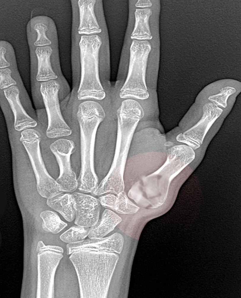
Bennett Fracture.
Contributed by S Bhimji, MD
(Click Image to Enlarge)
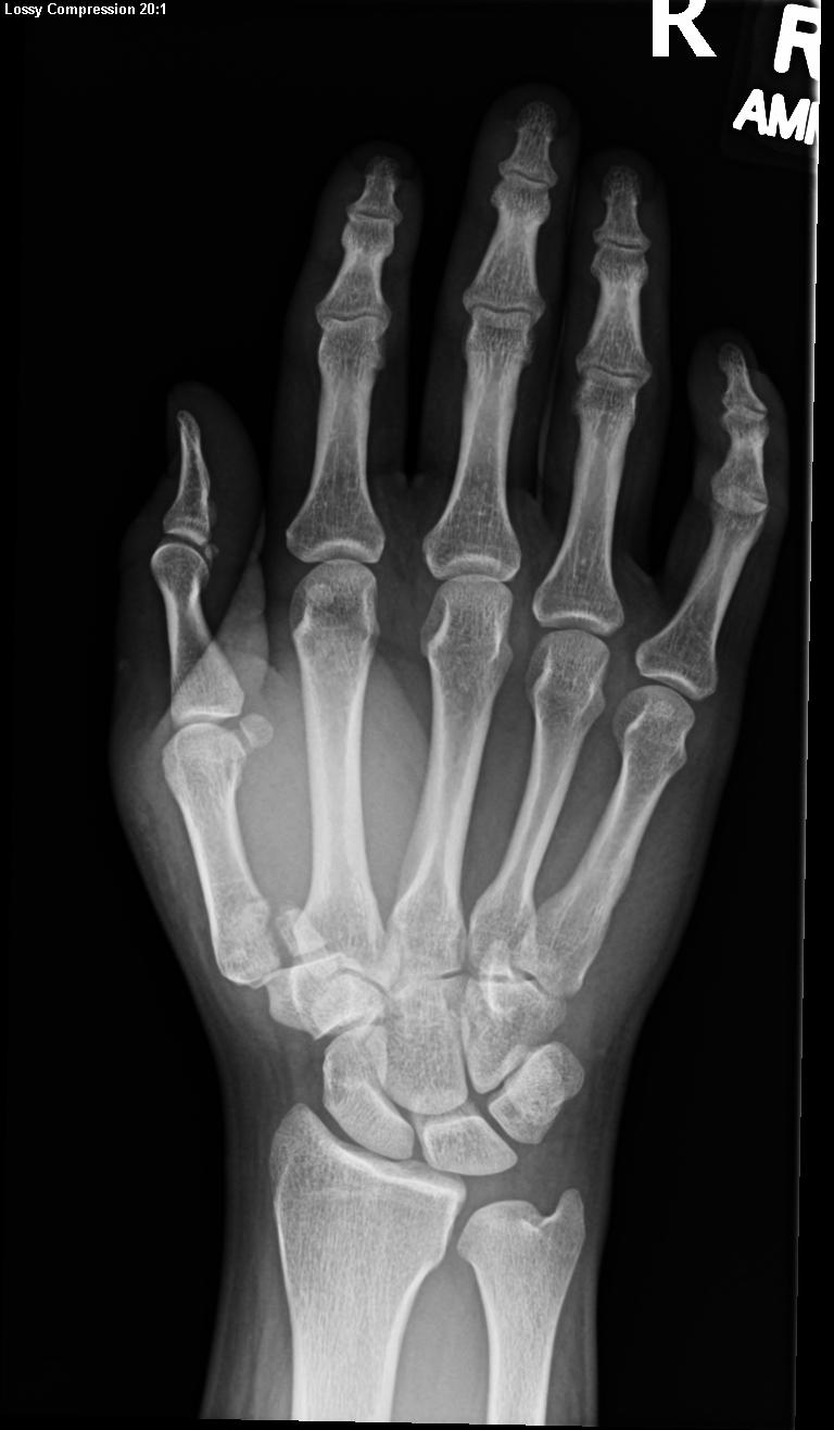
Frontal view of hand with a Bennett fracture Contributed by Kevin Carter, DO
(Click Image to Enlarge)
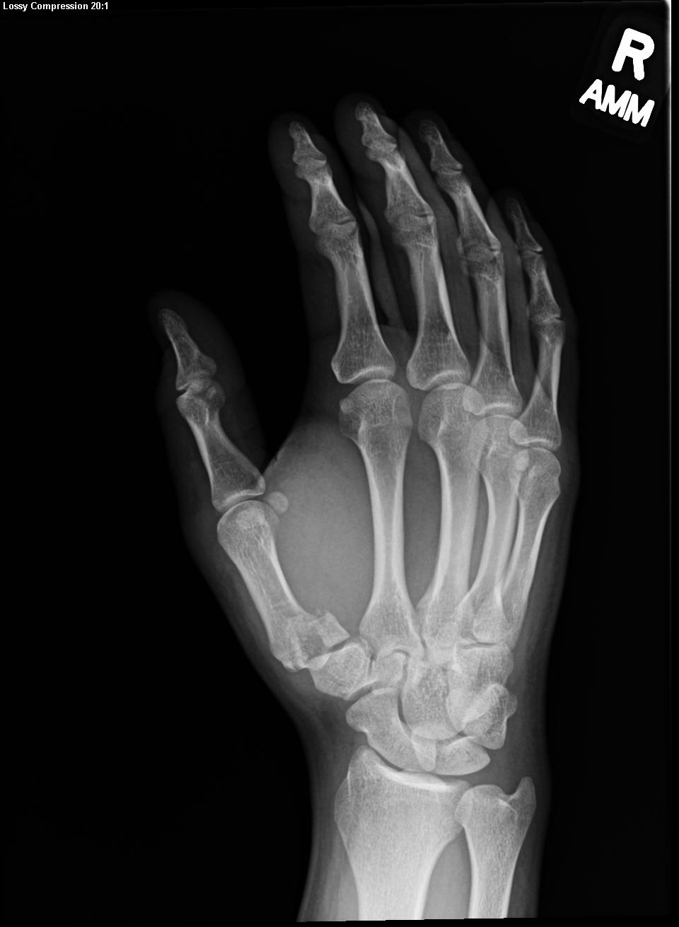
Oblique x-ray view of the hand with a Bennett fracture Contributed by Kevin Carter, DO
References
Edmunds JO. Traumatic dislocations and instability of the trapeziometacarpal joint of the thumb. Hand clinics. 2006 Aug:22(3):365-92 [PubMed PMID: 16843802]
McGuigan FX, Culp RW. Surgical treatment of intra-articular fractures of the trapezium. The Journal of hand surgery. 2002 Jul:27(4):697-703 [PubMed PMID: 12132098]
Level 2 (mid-level) evidenceWu WB, Du YF, Wang HX, Liang F. Bennett fracture combined with hamate fracture: carpometacarpal joint 'diagonal' fracture and dislocation: a case report. BMC musculoskeletal disorders. 2023 Jun 10:24(1):477. doi: 10.1186/s12891-023-06588-3. Epub 2023 Jun 10 [PubMed PMID: 37301961]
Level 3 (low-level) evidenceYounis Z, Hamid MA, Devasia T, Khan MM, Abdullah F, Singh R, Simons AW. Base of Thumb Fractures: A Review of Anatomy, Classification, and Management. Cureus. 2025 Jan:17(1):e76729. doi: 10.7759/cureus.76729. Epub 2025 Jan 1 [PubMed PMID: 39897322]
GEDDA KO. Studies on Bennett's fracture; anatomy, roentgenology, and therapy. Acta chirurgica Scandinavica. Supplementum. 1954:193():1-114 [PubMed PMID: 13188578]
Lambi AG, Rowland RJ, Brady NW, Rodriguez DE, Mercer DM. Metacarpal fractures. The Journal of hand surgery, European volume. 2023 Sep:48(2_suppl):42S-50S. doi: 10.1177/17531934231184119. Epub [PubMed PMID: 37704027]
Stanton JS, Dias JJ, Burke FD. Fractures of the tubular bones of the hand. The Journal of hand surgery, European volume. 2007 Dec:32(6):626-36 [PubMed PMID: 17993422]
Level 2 (mid-level) evidenceHove LM. Fractures of the hand. Distribution and relative incidence. Scandinavian journal of plastic and reconstructive surgery and hand surgery. 1993 Dec:27(4):317-9 [PubMed PMID: 8159947]
Carlsen BT, Moran SL. Thumb trauma: Bennett fractures, Rolando fractures, and ulnar collateral ligament injuries. The Journal of hand surgery. 2009 May-Jun:34(5):945-52. doi: 10.1016/j.jhsa.2009.03.017. Epub [PubMed PMID: 19411003]
Yin Y, Wang Y, Wang Z, Qu W, Tian W, Chen S. Accuracy of fluoroscopic examination in the treatment of Bennett's fracture. BMC musculoskeletal disorders. 2021 Jan 4:22(1):3. doi: 10.1186/s12891-020-03867-1. Epub 2021 Jan 4 [PubMed PMID: 33397336]
Cannon SR, Dowd GS, Williams DH, Scott JM. A long-term study following Bennett's fracture. Journal of hand surgery (Edinburgh, Scotland). 1986 Oct:11(3):426-31 [PubMed PMID: 3794490]
Demir E, Unglaub F, Wittemann M, Germann G, Sauerbier M. [Surgically treated intraarticular fractures of the trapeziometacarpal joint -- a clinical and radiological outcome study]. Der Unfallchirurg. 2006 Jan:109(1):13-21 [PubMed PMID: 16133289]
Cullen JP, Parentis MA, Chinchilli VM, Pellegrini VD Jr. Simulated Bennett fracture treated with closed reduction and percutaneous pinning. A biomechanical analysis of residual incongruity of the joint. The Journal of bone and joint surgery. American volume. 1997 Mar:79(3):413-20 [PubMed PMID: 9070532]
Oosterbos CJ, de Boer HH. Nonoperative treatment of Bennett's fracture: a 13-year follow-up. Journal of orthopaedic trauma. 1995 Feb:9(1):23-7 [PubMed PMID: 7714650]
Kjaer-Petersen K, Langhoff O, Andersen K. Bennett's fracture. Journal of hand surgery (Edinburgh, Scotland). 1990 Feb:15(1):58-61 [PubMed PMID: 2307882]
Timmenga EJ, Blokhuis TJ, Maas M, Raaijmakers EL. Long-term evaluation of Bennett's fracture. A comparison between open and closed reduction. Journal of hand surgery (Edinburgh, Scotland). 1994 Jun:19(3):373-7 [PubMed PMID: 8077832]
Ahlquist SA, Hookasian E, Gajewski CR, Lee B, Graham DJ, Sivakumar BS, Jones NF, Ebramzadeh E, Sangiorgio SN. A Biomechanical Cadaveric Comparison of Three Fixation Methods for Bennett Fractures. The Journal of hand surgery. 2025 Dec 13:():. pii: S0363-5023(25)00607-0. doi: 10.1016/j.jhsa.2025.10.021. Epub 2025 Dec 13 [PubMed PMID: 41389056]
Mercan N, Eravşar E, Safalı S, Uğur L, Özdemir A. Biomechanical insights through finite element analysis of Bennett fracture fixation: a comparative study of four surgical techniques. Journal of orthopaedic surgery and research. 2025 Jan 23:20(1):90. doi: 10.1186/s13018-025-05481-0. Epub 2025 Jan 23 [PubMed PMID: 39849521]
Level 2 (mid-level) evidenceDaher M, Roukoz S, Ghoul A, Tarchichi J, Aoun M, Sebaaly A. Management of Bennett's fracture: A systematic review and meta-analysis. JPRAS open. 2023 Dec:38():206-216. doi: 10.1016/j.jpra.2023.09.010. Epub 2023 Sep 30 [PubMed PMID: 37929064]
Level 1 (high-level) evidenceDuché T, Lambrey PJ, Chantelot C, Saab M. Functional and radiological outcomes of Bennett's fractures treated by Iselin's technique: About 29 cases with 6-year (2-14) follow-up. Orthopaedics & traumatology, surgery & research : OTSR. 2025 Sep:111(5):104023. doi: 10.1016/j.otsr.2024.104023. Epub 2024 Oct 15 [PubMed PMID: 39413871]
Level 3 (low-level) evidenceTorres Fuentes CE, Meneses Argalle JD, Romero León CA. Arthroscopic-Assisted Treatment of Bennett Fractures: A Potentially Advantageous Operative Procedure? Hand (New York, N.Y.). 2025 Mar:20(2):218-223. doi: 10.1177/15589447231211602. Epub 2023 Dec 4 [PubMed PMID: 38047463]
Vaz R, Branco PM, Rego P, Baptista C. Neglected Bennett Fracture: An Expanded Approach. Hand (New York, N.Y.). 2025 Nov 4:():15589447251387285. doi: 10.1177/15589447251387285. Epub 2025 Nov 4 [PubMed PMID: 41189437]
Hashiguchi H, Iwashita S, Yoneda M, Takai S. Factors influencing outcomes of nonsurgical treatment for baseball players with SLAP lesion. Asia-Pacific journal of sports medicine, arthroscopy, rehabilitation and technology. 2018 Oct:14():6-9. doi: 10.1016/j.asmart.2018.08.001. Epub 2018 Sep 5 [PubMed PMID: 30202738]
Fischborn T, Beckenbauer D, Held M, Daigeler A, Medved F. Analysis of Operative Techniques of Fractures of the First Metacarpal Base. Annals of plastic surgery. 2018 May:80(5):507-514. doi: 10.1097/SAP.0000000000001293. Epub [PubMed PMID: 29319570]
Velásquez-Girón E, Buitrago-Quiñonez ER, Hernández-Guevara J, Kafury ÁA. Outcomes of Patients with Bennett Fracture Treated with Three Different Surgical Techniques: A Systematic Review. The archives of bone and joint surgery. 2025:13(1):4-16. doi: 10.22038/ABJS.2024.81116.3702. Epub [PubMed PMID: 39886346]
Level 1 (high-level) evidenceKadow TR, Fowler JR. Thumb Injuries in Athletes. Hand clinics. 2017 Feb:33(1):161-173. doi: 10.1016/j.hcl.2016.08.008. Epub [PubMed PMID: 27886832]