 Tools
Tools
 Becker Melanosis
Introduction
Becker melanosis, also known as Becker nevus, is a benign acquired pigmentary disorder characterized by unilateral hyperpigmented patches that commonly emerge during adolescence and gradually enlarge over time. First described by S William Becker in 1949, as "concurrent melanosis and hypertrichosis in the distribution of nevus unius lateris" (see Image. Becker Melanosis).[1] Becker melanosis frequently affects the shoulder, scapular region, or upper trunk and may later develop associated hypertrichosis or acneiform changes.
Although the exact etiology remains incompletely understood, current evidence supports a significant role for androgen sensitivity, with studies demonstrating increased androgen receptor expression in lesional skin. Postzygotic beta-actin mutations have also been implicated in the pathogenesis of both Becker melanosis and Becker nevus syndrome. While most lesions remain stable and benign, atypical clinical variants, including follicular forms, bilateral involvement, checkerboard mosaic patterns, and midline lesions, can complicate diagnosis and management.
Recognition of Becker melanosis carries important clinical implications because some patients exhibit associated developmental abnormalities collectively termed Becker nevus syndrome. These abnormalities may include ipsilateral breast hypoplasia, musculoskeletal defects, smooth muscle hamartomas, scoliosis, neurologic manifestations, and pectoral muscle hypoplasia. Diagnosis remains primarily clinical, supported by characteristic histopathologic findings when biopsy is performed. Management focuses largely on cosmetic improvement and psychosocial support, with laser therapies representing the most commonly utilized treatment modalities for hyperpigmentation and hypertrichosis. Familiarity with the diverse clinical presentations, associated syndromic findings, and evolving therapeutic options remains essential for clinicians involved in dermatologic and interprofessional patient care.
Etiology
Register For Free And Read The Full Article
 Search engine and full access to all medical articles
- 10 free questions in your specialty
- Free CME/CE Activities
- Free daily question in your email
- Save favorite articles to your dashboard
- Emails offering discounts
Learn more about a Subscription to StatPearls Point-of-Care
Etiology
The exact etiology is not clear. Becker melanosis is considered a benign, late-onset type of epidermal nevus, and associated features like peripubertal development, male preponderance, hypertrichosis, and acneiform lesions suggest a role for androgens. An increase in the number of androgen receptors has been reported.[2]
Epidemiology
Men are more commonly affected (male-to-female ratio of about 5:1). Becker melanosis has an estimated prevalence of 0.5% among men. The lesion usually presents during puberty, but rarely cases of Becker melanosis presenting at birth or in early childhood have been reported; familial occurrence has also been reported. Some studies have shown a higher incidence among Black individuals.[3][4]
Pathophysiology
Androgen sensitivity and stimulation have been suggested to be among the main factors involved in the pathogenesis of Becker nevus. The onset of the lesions during adolescence, preponderance in men, and associated features like hypertrichosis and acneiform lesions have all been linked to the role of androgens in the pathophysiology. Some studies have reported an increased number of androgen receptors in lesional skin. Postzygotic mutations in beta-actin have been reported in association with both Becker nevus and Becker nevus syndrome.[5][6]
Histopathology
Typically, Becker melanosis shows primarily epidermal changes, including acanthosis and hyperkeratosis, with increased basal pigmentations. Dermal melanophages can be seen. Other histopathological features include elongation of the rete ridges, smooth muscle, and sebaceous gland hyperplasia.
History and Physical
Becker melanosis presents as a well-defined, unilateral, hyperpigmented tan or brown patch that increases in size, gradually often developing into a geographic pattern. Some studies have reported that the right side of the body is more commonly affected, with the most common sites being the shoulder, scapular region, and upper arms. Rarely, Becker melanosis may present with bilateral lesions at atypical sites, eg, the lower limbs. Significant hair growth over the lesions usually appears after hyperpigmentation is established and may take months to years to develop; acneiform lesions may also develop over the affected area.
Becker melanosis may be associated with other findings of ectodermal abnormalities, collectively known as Becker nevus syndrome. Reported associations include smooth muscle hamartomas, hypoplasia of the breast, pectoral muscle and fat, limb hypertrophy, adrenal gland hyperplasia, and accessory scrotum; a rare association with melanoma has been reported. Once developed, the natural course is for the lesions to persist indefinitely. The hypertrichosis typically develops after the hyperpigmentation, and the hairs become progressively coarse with time. Some studies suggest that hypertrichosis is not associated with most cases of Becker melanosis.[3][7][8][9][10]
Follicular Becker nevus represents a more recently described variant of Becker melanosis. Clinical presentation may include perifollicular macules with or without associated hypertrichosis.[11] Additional atypical clinical presentations include checkerboard mosaic patterns, wedge-shaped hyperpigmentation involving the forehead, and midline nevi.[12][13][14]
Evaluation
The diagnosis is mainly clinical. A skin biopsy shows mild acanthosis and hyperkeratosis. Increased melanin is seen in the basal layer, although the number of melanocytes tends to be normal. Dermal melanophages can be seen. Other histopathological features include elongation of the rete ridges, smooth muscle, and sebaceous gland hyperplasia.
The hyperproliferation of keratinocytes, melanocytes, arrector pili muscle, and dermal nerve fibers has been reported in recent studies. Some immunohistochemical studies have shown increased expression of certain markers, eg, epidermal Ki-67, melan-A, and keratin 15, in lesional and perilesional skin compared with normal skin. Dermal nerve fiber length and expression of smooth muscle actin have also been reported to be higher in the lesional skin of Becker melanosis.[15][10][16]
Treatment / Management
The hyperpigmentation of Becker melanosis usually remains stable, although some cases report spontaneous fading. Treatment is mainly indicated for cosmetic reasons, especially the rapid transformation of the lesion during adolescence. The hyperpigmentation and hypertrichosis respond to laser treatment. Various types of lasers are effective for Becker melanosis, including the Q-switched Ruby laser, Q-switched Nd:YAG, long-pulsed Alexandrite, and various fractional ablative lasers. The most commonly used lasers are the Q-switched ruby and the Q-switched Nd YAG lasers. However, both are associated with a high rate of recurrence. Hypertrichotic lesions have been reported to respond to combinations of fractional lasers (eg, the 1550 nm nonablative laser) with hair removal lasers. Multiple sessions are required for optimum results.
Combination lasers have been found to be effective in the treatment of Becker melanosis. Most frequently evaluated combinations include a combination of Nonablative Fractional Laser with Q-Switched Lasers. Combining long-pulsed 1064-nm Nd:YAG and 755-nm alexandrite lasers has been shown to successfully treat Becker melanosis. A recent case report described the use of a combination of low-fluence Q-switched Nd:YAG 1064 nm laser and long-pulsed Nd:YAG 1064 nm laser in the treatment of Becker melanosis. Other effective lasers include the nonablative 1927-nm fractional thulium fiber laser.[17][18][19]
Electrolysis has also been reported to treat the hypertrichosis associated with Becker melanosis effectively. Sun protection is advised, as sun exposure might make the lesions appear darker. Acneiform lesions have been found to respond to topical retinoids. In patients with associated breast hypoplasia, a novel yet effective treatment is breast lipofilling (eg, fat grafting to treat the cosmetic defect related to ipsilateral breast hypoplasia), which can be associated with Becker melanosis. Cosmetic camouflage can be useful in addressing the psychosocial issues and quality of life in patients who have lesions in relatively exposed areas.[20][21][22](B3)
Differential Diagnosis
The differential diagnoses for Becker melanosis include the following:
- Albright syndrome
- Congenital melanocytic nevus
- Congenital smooth muscle hamartoma
- Overdevelopment of a tissue such as the adrenal gland, limb, fingers, or toes
- Post-inflammatory hyperpigmentation
- Smooth muscle hamartoma
- Under the development of underlying structures such as the breast, fat, or limb
Prognosis
Becker melanosis is a benign nevus, and the primary concern in most cases is cosmetic. Cases with neurological associations need appropriate follow-up with specialists. The prognosis of surgical corrections for associations, eg, breast hypoplasia, depends on the extent of involvement in individual cases.[23]
Complications
Becker melanosis can be associated with neurological complications, including quadriparesis, attention-deficit hyperactivity disorder (ADHD), autistic spectrum disorder (ASD), and epilepsy.[24] In some patients, Becker’s melanosis is associated with underlying developmental anomalies, forming part of Becker nevus syndrome. These may include ipsilateral breast hypoplasia, skeletal abnormalities (eg, scoliosis), or muscular hypoplasia, especially involving the pectoralis major. Such associations, although uncommon, may contribute to functional and aesthetic impairment.
Cutaneous complications are otherwise minimal. The risk of malignant transformation has not been established. However, increased hair growth and pigmentation may be resistant to treatment, and therapeutic interventions, eg, laser therapy, can yield variable results and occasional adverse effects, including post-inflammatory dyspigmentation.
Postoperative and Rehabilitation Care
Standard postoperative care, as appropriate for individual patients, will be needed in cases that require interventions, such as breast reconstruction surgery.
Consultations
An interprofessional team approach is often required for optimum management of Becker melanosis. Dermatologists, pediatricians, neurologists, and plastic surgeons or laser specialists will often have to make combined decisions for individual cases.
Deterrence and Patient Education
Becker melanosis generally follows a benign and stable clinical course without a recognized risk of malignant transformation. Despite the absence of significant medical complications in most patients, visible hyperpigmentation and hypertrichosis can produce considerable psychosocial distress, particularly among adolescents and individuals with lesions involving exposed areas. Effective patient education should emphasize the benign nature of the disorder while addressing cosmetic concerns, body image issues, and the chronic persistence of lesions. Counseling should also include discussion regarding associated developmental abnormalities that may occur in Becker nevus syndrome, including ipsilateral breast hypoplasia, musculoskeletal defects, and neurologic manifestations, when clinically indicated. Sun protection measures should be encouraged because ultraviolet exposure may accentuate hyperpigmentation and increase cosmetic visibility of lesions.
Comprehensive counseling regarding treatment options remains essential for informed decision-making and realistic patient expectations. Patients should receive detailed explanations regarding available therapies, including laser treatment modalities, electrolysis for hypertrichosis, cosmetic camouflage, and management of associated acneiform lesions. Discussions should address expected clinical outcomes, the need for multiple treatment sessions, recurrence rates, possible adverse effects such as postinflammatory dyspigmentation, and the variable response associated with different laser systems. Shared decision-making that incorporates patient goals, psychosocial concerns, lesion severity, treatment tolerance, and cosmetic expectations supports improved satisfaction and long-term adherence to management plans.
Pearls and Other Issues
Although the typical presentation is a single, unilateral, hyperpigmented, or tan-colored macule over the shoulder or pectoral area, Becker melanosis has been reported to manifest in various atypical presentations. Associated abnormalities include unilateral hypoplasia of the breast, which can vary in magnitude, affecting the whole breast area or the nipple and areola alone. Supernumerary nipples can also occur as an association with aplasia of the ipsilateral pectoralis major muscle, ipsilateral limb shortening, localized lipoatrophy, spina bifida, scoliosis, pectus carinatum, quadriparesis, osteoma cutis, congenital adrenal hyperplasia, and accessory scrotum.
Becker melanosis has been found to occur in association with phakomatosis pigmentovascularis and neurofibromatosis. Becker melanosis associated with nevus depigmentosus has been described as a possible example of twin spotting. A case report has mentioned the occurrence of basal cell carcinoma occurring on the site of Becker melanosis in a sun-protected area. Hypohidrosis associated with Becker melanosis has been reported, and pityriasis versicolor localized to the area of Becker melanosis has been described.
Multiple and bilateral lesions have rarely been reported. A case of giant bilateral Becker melanosis, simulating the armor of a gladiator's arm, has been described. The same patient also had marginal osteophytes over the cervical vertebrae. Becker melanosis is associated with short stature, skeletal deformities, and intellectual disability. Bilateral, congenital Becker melanosis has been described. Becker melanosis occurs in siblings. Atypical sites of involvement include the lower limbs. Many cases may present with a later onset and absence of hypertrichosis.[8][25][26][27][28]
Enhancing Healthcare Team Outcomes
Becker melanosis, also known as Becker nevus, is a benign acquired pigmentary disorder characterized by unilateral hyperpigmented patches that frequently develop during adolescence and may later demonstrate hypertrichosis or acneiform changes. Proposed pathophysiologic mechanisms include androgen sensitivity with increased androgen receptor expression and postzygotic beta-actin mutations. Common sites include the shoulder, scapular region, and upper trunk, although atypical forms such as follicular Becker nevus, bilateral lesions, checkerboard mosaic patterns, and midline nevi have been reported. Diagnosis remains primarily clinical and may be supported by histopathologic findings, including acanthosis, basal layer hyperpigmentation, rete ridge elongation, and smooth muscle hyperplasia. Management primarily addresses cosmetic and psychosocial concerns through sun protection, laser therapies, electrolysis, acne treatment, and cosmetic camouflage. Evaluation for Becker nevus syndrome remains important because associated musculoskeletal, breast, and neurologic abnormalities may contribute to functional impairment and reduced quality of life.
Interprofessional collaboration improves diagnostic accuracy, patient counseling, treatment selection, and long-term follow-up in patients with Becker melanosis. Dermatologists, primary care clinicians, and advanced practitioners coordinate diagnostic evaluations, identify atypical presentations, and recognize associated syndromic abnormalities that require referral to specialists such as neurologists, orthopedic surgeons, plastic surgeons, or mental health professionals. Nurses reinforce patient education regarding treatment expectations, sun protection, procedural aftercare, and psychosocial support, while pharmacists provide guidance regarding topical therapies and medication safety. Laser specialists and cosmetic dermatology teams assist with individualized procedural planning and monitoring for complications such as dyspigmentation or recurrence. Shared decision-making remains essential because treatment goals frequently focus on cosmetic improvement and quality of life rather than medical necessity. Coordinated communication among healthcare professionals supports timely referral, individualized treatment planning, prevention of unnecessary interventions, and improved patient-centered outcomes.[22]
Media
(Click Image to Enlarge)
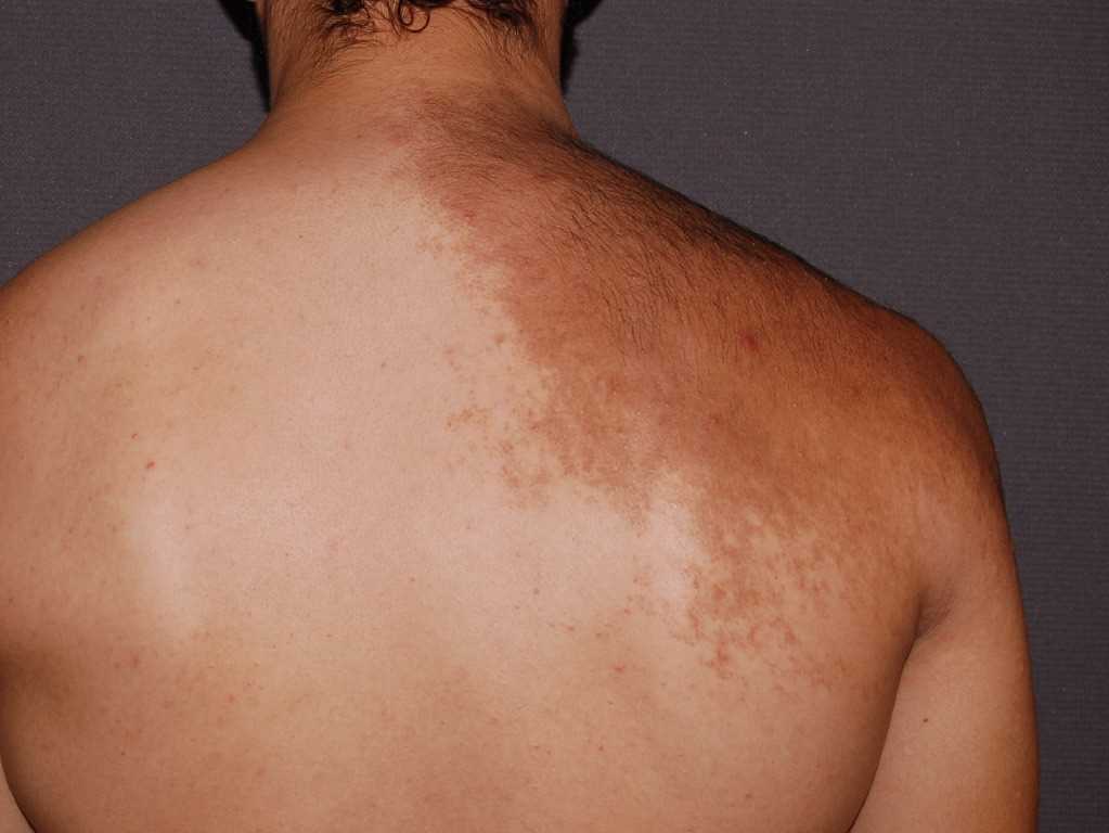
Becker Melanosis. Becker melanosis (Becker nevus) is a form of acquired hyperpigmentation.
References
BECKER SW. Concurrent melanosis and hypertrichosis in distribution of nevus unius lateris. Archives of dermatology and syphilology. 1949 Aug:60(2):155-60 [PubMed PMID: 18133444]
Patel P, Malik K, Khachemoune A. Sebaceus and Becker's Nevus: Overview of Their Presentation, Pathogenesis, Associations, and Treatment. American journal of clinical dermatology. 2015 Jun:16(3):197-204. doi: 10.1007/s40257-015-0123-y. Epub [PubMed PMID: 25782676]
Level 3 (low-level) evidencePatrizi A, Medri M, Raone B, Bianchi F, Aprile S, Neri I. Clinical characteristics of Becker's nevus in children: report of 118 cases from Italy. Pediatric dermatology. 2012 Sep-Oct:29(5):571-4. doi: 10.1111/j.1525-1470.2012.01734.x. Epub 2012 Apr 4 [PubMed PMID: 22471889]
Level 2 (mid-level) evidenceDanarti R, König A, Salhi A, Bittar M, Happle R. Becker's nevus syndrome revisited. Journal of the American Academy of Dermatology. 2004 Dec:51(6):965-9 [PubMed PMID: 15583590]
Kim YJ, Han JH, Kang HY, Lee ES, Kim YC. Androgen receptor overexpression in Becker nevus: histopathologic and immunohistochemical analysis. Journal of cutaneous pathology. 2008 Dec:35(12):1121-6. doi: 10.1111/j.1600-0560.2008.00988.x. Epub [PubMed PMID: 18616760]
Cai ED, Sun BK, Chiang A, Rogers A, Bernet L, Cheng B, Teng J, Rieger KE, Sarin KY. Postzygotic Mutations in Beta-Actin Are Associated with Becker's Nevus and Becker's Nevus Syndrome. The Journal of investigative dermatology. 2017 Aug:137(8):1795-1798. doi: 10.1016/j.jid.2017.03.017. Epub 2017 Mar 24 [PubMed PMID: 28347698]
Sheng P, Cheng YL, Cai CC, Guo WJ, Zhou Y, Shi G, Fan YM. Clinicopathological Features and Immunohistochemical Alterations of Keratinocyte Proliferation, Melanocyte Density, Smooth Muscle Hyperplasia and Nerve Fiber Distribution in Becker's Nevus. Annals of dermatology. 2016 Dec:28(6):697-703 [PubMed PMID: 27904268]
Dasegowda SB, Basavaraj G, Nischal K, Swaroop M, Umashankar N, Swamy SS. Becker's Nevus Syndrome. Indian journal of dermatology. 2014 Jul:59(4):421. doi: 10.4103/0019-5154.135530. Epub [PubMed PMID: 25071279]
Manoj J, Kaliyadan F, Hiran KR. Atypical presentation of Becker's melanosis. Indian dermatology online journal. 2011 Jan:2(1):42-3. doi: 10.4103/2229-5178.79856. Epub [PubMed PMID: 23130219]
AlGhamdi KM, AlKhalifah AI, AlSheikh AM, AlSaif FM. Clinicopathologic profile of Becker's melanosis with atypical features. Journal of drugs in dermatology : JDD. 2009 Aug:8(8):745-8 [PubMed PMID: 19663112]
Level 2 (mid-level) evidenceGaurav V, Ahuja R, Chandra AD, Ahuja S, Bhagwat A, Lal A, Danish M, Khan E, Taneja N. Follicular Becker's nevi: A case series and review of literature. Pediatric dermatology. 2024 Nov-Dec:41(6):1183-1187. doi: 10.1111/pde.15641. Epub 2024 Apr 30 [PubMed PMID: 38689523]
Level 2 (mid-level) evidenceSheehan CA, Agarwala SN, Hsu S. A midline Becker's nevus. JAAD case reports. 2025 Sep:63():59-60. doi: 10.1016/j.jdcr.2025.05.037. Epub 2025 Jun 25 [PubMed PMID: 40787057]
Level 3 (low-level) evidenceMakhija R, Gupta LK, Khare AK, Mittal A. Extensive Becker's Melanosis in a Checkerboard Mosaic Pattern. Indian dermatology online journal. 2020 Mar-Apr:11(2):294-295. doi: 10.4103/idoj.IDOJ_137_19. Epub 2020 Mar 9 [PubMed PMID: 32478009]
Yun SK, Lee IJ, Woo SH, Nam KH. Wedge-shaped hyperpigmentation of the forehead: a rare presentation of melanotic-type Becker's nevus. European journal of dermatology : EJD. 2023 Jun 1:33(3):296-297. doi: 10.1684/ejd.2023.4485. Epub [PubMed PMID: 37594338]
Bhawan J, Chang WH. Becker's melanosis: an ultrastructural study. Dermatologica. 1979:159(3):221-30 [PubMed PMID: 478060]
Level 3 (low-level) evidenceBoiron G, Surlève-Bazeille JE, Maleville J. [Becker's melanosis. Study of seven cases by photonic and electron microscopy. Contribution to the histogenesis of the cytoid bodies (author's transl)]. Annales de dermatologie et de venereologie. 1980 Aug-Sep:107(8-9):787-97 [PubMed PMID: 7447258]
Level 3 (low-level) evidenceWulkan AJ, McGraw T, Taylor M. Successful treatment of Becker's Nevus with long-pulsed 1064-nm Nd:YAG and 755-nm alexandrite laser and review of the literature. Journal of cosmetic and laser therapy : official publication of the European Society for Laser Dermatology. 2018 Aug:20(4):211-214. doi: 10.1080/14764172.2017.1326613. Epub 2018 Mar 26 [PubMed PMID: 28498021]
Chang R, Zhou L, Cen Q, Yu W, Han Y, Huang L, Zhu J, Lin X. Successful treatment of challenging Becker's Nevus with continuous low-fluence Q-switched Nd: YAG 1064 nm laser and long-pulsed Nd: YAG 1064 nm laser. Lasers in medical science. 2024 Oct 23:39(1):262. doi: 10.1007/s10103-024-04193-x. Epub 2024 Oct 23 [PubMed PMID: 39438308]
Alajlan A, Alsuwaidan S. Efficacy of non-ablative 1927-nm fractional thulium fiber laser for the treatment of Becker's nevus: Intra- and interobserver agreement study. Lasers in surgery and medicine. 2023 Aug:55(6):547-554. doi: 10.1002/lsm.23670. Epub 2023 Apr 23 [PubMed PMID: 37087675]
Kansal NK. Lightening Becker nevus: Role of topical therapies. Journal of the American Academy of Dermatology. 2019 Feb:80(2):e39. doi: 10.1016/j.jaad.2018.08.038. Epub 2018 Sep 6 [PubMed PMID: 30195570]
Al-Saif F, Al-Mekhadab E, Al-Saif H. Efficacy and safety of short-pulse erbium: Yttrium aluminum garnet laser treatment of Becker's nevus in Saudi patients: A pilot study. International journal of health sciences. 2017 Jul-Sep:11(3):14-17 [PubMed PMID: 28936145]
Level 3 (low-level) evidenceMomen S, Mallipeddi R, Al-Niaimi F. The use of lasers in Becker's naevus: An evidence-based review. Journal of cosmetic and laser therapy : official publication of the European Society for Laser Dermatology. 2016 Aug:18(4):188-92. doi: 10.3109/14764172.2015.1114647. Epub 2016 Mar 11 [PubMed PMID: 26735085]
Schlosser AK, Rais MS, Sanchez RJ. Breast Reconstruction in Becker Nevus Syndrome. Plastic and reconstructive surgery. Global open. 2025 Jul:13(7):e6957. doi: 10.1097/GOX.0000000000006957. Epub 2025 Jul 9 [PubMed PMID: 40642261]
Lopez-Balboa P, Knöpfel N, Ramwani M, Muwanga-Nanyonjo N, O'Neill C, Lomas D, Cullup T, Polubothu S, Kinsler VA. Becker naevi are associated with neurologic abnormalities. Journal of the American Academy of Dermatology. 2025 Apr:92(4):887-890. doi: 10.1016/j.jaad.2024.09.085. Epub 2024 Nov 21 [PubMed PMID: 39577695]
Bisht YS, Bhasin R, Manoj S, Sunita BS, Singhal E. Becker's nevus syndrome. Medical journal, Armed Forces India. 2015 Jul:71(Suppl 1):S89-91. doi: 10.1016/j.mjafi.2013.04.010. Epub 2013 Aug 30 [PubMed PMID: 26265883]
Cohen PR. Poland's Syndrome: Are Postzygotic Mutations in β-Actin Associated with its Pathogenesis? American journal of clinical dermatology. 2018 Feb:19(1):133-134. doi: 10.1007/s40257-017-0330-9. Epub [PubMed PMID: 29139054]
Ghosh SK, Majumder B, Agarwal M. Becker's nevus syndrome: a report of a rare disease with unusual associations. International journal of dermatology. 2017 Apr:56(4):458-460. doi: 10.1111/ijd.13378. Epub 2016 Sep 22 [PubMed PMID: 27655000]
Glinick SE, Alper JC, Bogaars H, Brown JA. Becker's melanosis: associated abnormalities. Journal of the American Academy of Dermatology. 1983 Oct:9(4):509-14 [PubMed PMID: 6355214]
Level 3 (low-level) evidence