 Tools
Tools
 Catheter Management Pulmonary Valvular Disorders
Introduction
Advances in pediatric cardiology, cardiac surgery, and perioperative care have dramatically improved long-term survival for children born with congenital heart disease. As a result, the prevalence of congenital heart disease in the adult population has risen to approximately 1 in 150, with roughly half of these patients having undergone at least 1 surgical repair in childhood. A significant proportion will require reintervention during adulthood.[1][2]
The right ventricular outflow tract (RVOT) is involved in about 20% of newborns with congenital heart disease, encompassing lesions such as tetralogy of Fallot, truncus arteriosus, pulmonary atresia with ventricular septal defect, and others. Surgical palliation of these defects commonly requires reconstruction with bioprosthetic valves, transannular patches, or extracardiac conduits between the right ventricle and the pulmonary artery.[3] These conduits are typically necessary when an anomalous left anterior descending coronary artery arising from the right coronary artery crosses the RVOT, precluding safe transannular patch repair. Although these conduits initially restore pulmonary valve competence, they undergo progressive calcification, intimal proliferation, and somatic growth, limiting long-term durability and often necessitating reintervention.[4]
Conduit dysfunction is multifactorial and depends on the patient’s age at implantation, the underlying defect, tissue type, surgical technique, and material used. Homograft valves, in particular, tend to degenerate, leading to significant pulmonary regurgitation, pulmonary stenosis, or mixed lesions, with roughly half of patients eventually requiring further intervention.[5][6] Surgical reoperation carries substantial morbidity and mortality risks related to dense adhesions, cardiac injury, ischemia, heart failure, and multi-organ dysfunction.[7][8][9][10][11][12]
Earlier repairs for tetralogy of Fallot that relied on generous transannular patches often resulted in free pulmonary insufficiency, progressive right ventricular dilation, tricuspid annular enlargement with regurgitation, and an arrhythmic substrate that can contribute to sudden cardiac death. Balloon angioplasty and bare-metal stenting were important early advances but often left significant residual pulmonary regurgitation. Transcatheter pulmonary valve implantation (TPVI) has emerged as a common, less invasive option in adult congenital heart disease. This procedure allows earlier restoration of pulmonary valve function before irreversible right ventricular remodeling sets in and may reduce the lifetime number of open surgical procedures.[13][14][15][16]
Anatomy and Physiology
Register For Free And Read The Full Article
 Search engine and full access to all medical articles
- 10 free questions in your specialty
- Free CME/CE Activities
- Free daily question in your email
- Save favorite articles to your dashboard
- Emails offering discounts
Learn more about a Subscription to StatPearls Point-of-Care
Anatomy and Physiology
The pulmonary valve is located between the right ventricle and the main pulmonary artery and normally consists of 3 thin semilunar cusps that permit unidirectional blood flow into the pulmonary circulation (see Image. Transverse Cross Section of the Heart). Congenital RVOT abnormalities, such as tetralogy of Fallot, pulmonary atresia, truncus arteriosus, or isolated pulmonary valve stenosis, often require surgical reconstruction with right ventricle-to-pulmonary artery conduits, transannular patches, or bioprosthetic valves. Over time, these repairs often fail due to calcification, stenosis, regurgitation, or conduit degeneration, leading to progressive right ventricular dilation, dysfunction, arrhythmias, and exercise intolerance. A thorough understanding of RVOT anatomy, conduit characteristics, coronary artery relationships, branch pulmonary artery anatomy, and prior surgical repairs is essential for appropriate patient selection and procedural planning for TPVI.
Optimal anatomy for TPVI depends on the transcatheter valve system used. Balloon-expandable valves are primarily indicated for dysfunctional RVOT conduits measuring at least 16 mm in diameter, where a stable landing zone can be achieved. In contrast, self-expanding valves and self-expanding stents followed by balloon-expandable valve implantation are approved and routinely used in patients with native or patched RVOTs following transannular patch repair, accommodating the larger, more variable geometry commonly encountered in these patients.[17][18][19] Off-label use in smaller conduits or complex RVOT anatomies may be considered at experienced centers when anatomic characteristics are otherwise favorable and surgical risk is prohibitive. Preprocedural imaging with echocardiography, cardiac computed tomography, and cardiac magnetic resonance imaging is critical for defining RVOT dimensions, assessing calcification, identifying conduit morphology, evaluating branch pulmonary arteries, and excluding coronary artery compression during valve deployment, thereby ensuring safe device selection and optimal procedural outcomes.
Indications
Optimizing the timing for RVOT intervention remains challenging. Standard TPVI criteria closely mirror those for surgical pulmonary valve replacement and focus on right ventricular volumetric data, systolic function, significant tricuspid regurgitation, RVOT obstruction, arrhythmias, QRS duration, and residual intracardiac defects. In asymptomatic patients with moderate to severe pulmonary regurgitation, TPVI is generally indicated when 1 or more of the following are present:
- Significant right ventricular dilation (right ventricular end-diastolic volume index >150–160 mL/m² or right ventricular end-systolic volume index >80 mL/m²)
- Right ventricular systolic dysfunction (ejection fraction <45%)
- Persistent atrial or ventricular arrhythmias
- RVOT obstruction with right ventricular systolic pressure greater than two-thirds systemic or >80 mm Hg
- Progressive tricuspid regurgitation
- Symptoms with moderate to severe pulmonary regurgitation/pulmonary stenosis and right ventricular systolic pressure >60 mm Hg [20][21]
The 2025 American College of Cardiology/American Heart Association/Heart Rhythm Society/International Society for Adult Congenital Heart Disease/Society for Cardiovascular Angiography and Interventions (ACC/AHA/HRS/ISACHD/SCAI) guideline assigns the following classes:
- Class 1 recommendation for pulmonary valve replacement (surgical or transcatheter) in symptomatic individuals with severe pulmonary regurgitation or pulmonary stenosis.
- Class 2a recommendation supports intervention in asymptomatic individuals with moderate or greater pulmonary valve dysfunction plus progressive right ventricular dilation, systolic dysfunction, or arrhythmias.
Guidelines from various societies continue to weigh risk factors such as age at initial repair, electrophysiologic markers (including QRS duration as a predictor of arrhythmic risk and sudden cardiac death), and the hemodynamic consequences of severe pulmonary regurgitation. Once clinical criteria are met, a detailed anatomic assessment of RVOT morphology is mandatory to determine the feasibility of TPVI.[22]
Contraindications
Active infection and occluded central venous access are absolute contraindications for TPVI. Unfavorable coronary artery anatomy that would result in coronary compression during balloon testing or device deployment is also an absolute contraindication and must be meticulously excluded. An RVOT diameter exceeding the largest available valve size prevents secure anchoring. Recurrent infective endocarditis and active intravenous drug use increase long-term infection risk and are often considered relative contraindications requiring careful shared decision-making.[20][22]
Equipment
Balloon-expandable valves were the first transcatheter systems approved for pulmonary use (see Image. Catheter Management of Pulmonary Valvular Disorders).[19] Experience has shown TPVI to be less invasive than surgery, with lower risks of bleeding, infection, and bypass-related morbidity. Two broad device categories are now used: balloon-expandable valves and self-expanding valves.
Balloon-expandable valves
Balloon-expandable valves remain the most commonly implanted. In patients with large, irregular, or heavily calcified homografts and right ventricular-to-pulmonary artery conduits at high risk of disruption during dilation, a self-expanding covered aortic endoprosthesis can first be deployed to create a stable, protected landing zone before balloon-expandable valve implantation. This strategy has proven feasible and effective for preparation of failed (stenotic or regurgitant) pulmonary homografts or right ventricular-to-pulmonary artery conduits.[23] A modified deployment technique for the self-expanding endoprosthesis, which adjusts the retention sleeve to improve anchoring, helps prevent graft displacement during delivery-system removal and has expanded its utility in congenital applications.[24]
Self-expanding valves
In select cases with very large native or patched RVOTs, dedicated self-expanding valves or self-expanding stents are used, followed by implantation of a balloon-expandable valve.[19] When the central RVOT is too wide, but the branch pulmonary arteries are suitable, bilateral implantation of branch pulmonary artery valves has been described, though it remains uncommon. Additional equipment includes extra-stiff guidewires, pigtail catheters for angiography, medium- and high-pressure balloons for sizing, predilation and postdilation, large sheaths, self-expanding endografts, and covered or bare-metal stents when further landing zone preparation is required.[19][23] Suture-mediated vascular closure devices are routinely used for hemostasis.[25]
Personnel
A cardiac team comprising interventional cardiologists, cardiac surgeons, noninvasive cardiologists, and cardiac anesthesiologists is crucial to achieving the best possible outcome for this complex procedure. This team should be experienced in treating RVOT interventions. In addition, consider extracorporeal membrane oxygenation (ECMO) facilities in the event of significant instability or deterioration, although the need for ECMO support with this procedure is quite rare.
Pediatric or interventional cardiologists with expertise in cardiac magnetic resonance imaging, cardiac computed tomography, and echocardiography are extremely helpful for preprocedure assessment and planning. Nurses must be available for pre- and post-procedure care, including assessing vital signs, performing baseline laboratory tests, confirming the patient’s ABO-Rh blood group, determining baseline renal function, and maintaining hydration. Nursing staff handles pre- and post-procedure care, including laboratory evaluation, hydration, blood readiness, and vigilant monitoring for complications.
Preparation
A detailed review of the patient’s history, with a thorough understanding of cardiac anatomy and specific attention to details of previous surgical interventions (ie, type of RVOT reconstruction, conduit size, conduit type, intraoperative concerns or challenges), is crucial. Review of other available diagnostic studies, such as previous hemodynamic evaluations, echocardiography, electrocardiograms, stress tests, and cross-sectional imaging with either cardiac magnetic resonance (CMR) imaging or computed tomography (CT), is crucial.[26][27] Specific attention to detail, including biventricular function, RVOT morphology, the degree of RVOT stenosis and/or regurgitation (spectral Doppler evaluation), and tricuspid regurgitation, should be assessed with echocardiography.[28][29][30] Considering potential routes for vascular access—such as the femoral or internal jugular veins or a hybrid approach—helps ensure vessel patency.
The preferred approach may depend on several factors, such as RVOT morphology, RVOT compliance and distensibility, and coronary artery anatomy. CT or MRI cross-sectional imaging with 3-dimensional reconstruction can help assess RVOT morphology, conduit type, and the degree of calcification.[18][31] The most challenging morphology for transcatheter intervention is a pyramidal shape that is narrow distally and wide proximally.
In contrast, patients with a homograft or conduit in place are conventionally more suitable.[32][33][34][35] Another key determinant of suitability is RVOT compliance and distensibility. CMR can identify pulsatility, which CT cannot, while balloon testing can better assess RVOT distensibility. Device selection is guided by RVOT anatomy: self-expanding valves or self-expanding stents (followed by a balloon-expandable valve) are preferred for native or patched RVOTs, whereas balloon-expandable valves are standard for conduits and surgical bioprostheses.[19]
Technique or Treatment
Moderate sedation is usually employed for the procedure with on-site surgical support. In select cases, general anesthesia is used.[36] Various vascular access sites can be implemented, including femoral, jugular, and subclavian veins. Femoral access is the most common, although the jugular approach may allow a more desirable anatomical curvature for valve delivery in certain anatomic arrangements and may provide access to a larger vessel in smaller patients in whom femoral access may be prohibitive.
Before valve implantation, administer heparin to maintain an activated clotting time greater than 250 seconds, and typically give prophylactic antibiotics before placing any stents or hardware. Perform a right heart catheterization in the standard fashion, with pressure measurements throughout the right heart. When performing a right heart assessment, use a balloon-tipped catheter that, with the balloon inflated, crosses the largest effective orifice of the tricuspid valve. This helps prevent the catheter from traversing small spaces within the tricuspid valve chordae or right ventricular trabeculations, which could make it difficult to pass larger sheaths and/or damage the tricuspid valve.
Angiograms are typically performed in the main pulmonary artery and right ventricle in 2 projections, although in patients with severe pulmonary regurgitation, a single pulmonary artery angiogram may be sufficient. Very often, the anteroposterior projection is optimized with cranial angulation (20 to 30 degrees) to best profile the RVOT/conduit to most accurately determine the conduit’s length, diameter, and morphology. Typically, a straight lateral projection with the lateral camera nicely defines the conduit and the proximity to the sternum, which may help predict how and where the conduit will dilate during balloon testing.
Aortic root injection and, often, selective coronary angiography are also performed to assess the proximity of the coronary arteries to the conduit, as discussed below. When performing angiograms, it helps to leave a catheter and/or wire in the RVOT to identify the conduit's exact location relative to the coronary arteries. After determining whether reasonable clinical and/or hemodynamic criteria support intervention, consider how best to prepare the conduit for valve placement.
In contemporary practice, a self-expanding covered endograft is deployed across the entire conduit length to line it from within.[23][24] The conduit is then dilated with the largest appropriate noncompliant balloon to fracture the conduit and achieve full expansion. Balloon size is selected based on the dimension of the main pulmonary artery just distal to the conduit anastomosis.
In patients with complex congenital heart disease, the reported incidence of coronary artery compression during conduit dilation is approximately 5%.[37] To mitigate this risk, selective coronary angiography is performed simultaneously with balloon inflation. If compression is observed, the inflation is repeated with the next smaller balloon, and the sequence is continued until a diameter is identified at which the coronary artery is no longer compressed. Then, an appropriately sized balloon-expandable pulmonary valve is implanted inside the rehabilitated right ventricle-to-pulmonary artery conduit.[23]
This contemporary approach has shortened procedural time and improved safety. Previously, the conduit was dilated stepwise, beginning at the narrowest diameter and advancing in 2-mm increments, with selective coronary angiography after each inflation. A super-stiff wire is essential throughout this portion of the procedure, and the tip should be positioned as distally as possible within the pulmonary branch arteries. Assess aortic root compression, with or without coronary compression, during balloon sizing, as it may cause significant aortic insufficiency in patients with a native RVOT or a transannular patch.[37]
Following valve implantation, right heart hemodynamics are reassessed, and a pulmonary artery angiogram is repeated to confirm valve position, valve competence, and unobstructed branch pulmonary artery flow. Additional balloon dilation is occasionally required to optimize valve expansion or to address a residual gradient or paravalvular leak. Hemostasis is achieved with a figure-of-8 suture, a vascular closure device, or manual pressure, and bleeding from the venous access site is rarely problematic despite the use of large-bore sheaths.
Most patients are observed overnight and discharged the following morning, although selected patients can be discharged on the same day. We prescribe daily aspirin at discharge, and patients continue it indefinitely while the valve remains in place. Despite these technical considerations, the procedure has been performed successfully in more than 96% of patients with RVOT dysfunction.[36]
In patients with severe pulmonary regurgitation arising from a native RVOT, preprocedural cardiac CT is routinely obtained to characterize the outflow geometry and to guide device selection. Three-dimensional reconstruction enables accurate assessment of the minimum and maximum RVOT diameters, the outflow's conical or cylindrical morphology, the length of the available landing zone, and the RVOT's proximity to the proximal coronary arteries. These measurements inform the choice between two principal transcatheter strategies.
When the RVOT is tapered and dynamic, a self-expanding transcatheter pulmonary valve can be deployed directly into the native outflow tract without further preparation.[38][39] When the RVOT is more cylindrical or oversized, a self-expanding prestent is first deployed to establish a defined cylindrical landing zone, and a balloon-expandable transcatheter pulmonary valve is then implanted within the prestent waist.[40][41] Coronary artery and aortic root proximity are reviewed in advance using simulated balloon and valve positions on the reconstructed dataset, and any concern for compression prompts adjustment of device size or overall strategy before the procedure.[42]
Complications
Although uncommon, several potential procedural complications can occur in patients with severe right ventricular dysfunction; a stiff wire traversing the tricuspid valve, combined with pulmonary regurgitation, can cause tricuspid regurgitation, hypotension, and hemodynamic instability. More significantly, severe hemodynamic compromise may result from valve dislodgment into the pulmonary artery, causing obstruction of the pulmonary blood flow, coronary compression causing coronary ischemia, and conduit rupture causing major hemorrhage. However, these are fortunately rare. Heavy calcification and the presence of homograft conduits have been identified as risk factors for rupture.[43]
In homografts and conduits, pre- and post-deployment balloon dilations can cause rupture or tearing. Most cases can be managed successfully with a covered stent or self-expanding endograft.[23] Surgical bailout is rarely required following conduit rupture.[17][44] Valve migration/embolization remains a potentially serious procedural complication that may require surgical intervention; however, with adequate conduit assessment and RVOT preparation, this remains rare.
If this complication occurs, valve deployment into a branch pulmonary artery has been proposed as a potential remedy if the valve embolizes distally. Retrieval with deployment of the embolized valve into the inferior vena cava (IVC), followed by stenting to open the valve leaflets, has also been proposed. Still, this procedure carries a significant risk of injury to the tricuspid valve, right ventricle, and IVC.
Longer-term complications include stent fracture, which remains the most common reason for reintervention with first-generation valves, even despite prestenting (5%–16%). Risk factors include younger age, higher pre- and procedural RVOT gradient, smaller angiographic conduit diameter, valve position directly under the sternum, stent recoil, or compression after deployment.[44][45] Type I fracture consists of 1 strut disruption without loss of stent integrity.
Type II includes stent integrity loss, and type III includes fractures with fragment separation. Type I occurs in up to 40% of patients; however, this type is not usually associated with adverse effects. Type II and III stent fractures are associated with early conduit restenosis and valve failure and may require surgical replacement or repeat TPVI. Clinically significant stent fracture with the second-generation valve in the pulmonary position has not yet been reported.
More recently, the development of infective endocarditis has emerged as a significant risk, with an incidence of approximately 2.4% per patient-year. Male sex, multiple stents, unprotected dental treatment, previous history of endocarditis, and noncompliance with aspirin constitute the risk factors.[46][47][48][49] Percutaneous pulmonary valve (PPV) endocarditis is characterized by vegetation visualized on the implant or as new evidence of PPV dysfunction associated with bloodstream infection.[50]
A wide spectrum of organisms, ranging from coagulase-negative staphylococci to Haemophilus, Aggregatibacter (previously Actinobacillus), Cardiobacterium, Eikenella, Kingella (HACEK) organisms, can cause TPVI-related endocarditis. Streptococcus viridans and Staphylococcus aureus are the most common causes. Occasionally, clinicians can treat and clear this medically. However, due to significant postinfection valve dysfunction, many patients require surgical valve replacement even if the bloodstream can be cleared of infection.
Clinical Significance
A decade after its approval in the United States, TPVI has emerged as a safe, effective nonsurgical therapy for RVOT dysfunction, often producing a peak cath gradient of less than 35 mm Hg and no more than mild insufficiency, with a complication rate of 6% to 13%.[51][52][53] Several studies have demonstrated the procedure's clinical efficacy, highlighting its potential to reduce right ventricular pressure and the RVOT gradient and eliminate pulmonary regurgitation.[30][44][54][55] Percutaneous restoration of pulmonary valve function has also been shown to significantly decrease right ventricular dimensions, improve stroke volumes, and improve New York Heart Association (NYHA) functional classes.[51][56][57][58]
Furthermore, patients experience improved exercise capacity and peak oxygen consumption.[56][59][60][61] At 1 year, 93.5 ± 2.4% of patients were free of valve dysfunction or need for reintervention.[44] The postapproval study's results showed freedom from PPV dysfunction to be 96.9%. Thus, results from multiple studies have demonstrated excellent success rates, hemodynamic outcomes, and low complication rates.
The long-term rate of reintervention with the first-generation valve remains relatively low, with 1- and 5-year freedom from reintervention greater than 90% and 76%, respectively, excluding patients with stent fracture.[53] Several risk factors for the need for reintervention have been identified: lack of prestenting, presence of a homograft, moderate-to-severe preprocedural tricuspid valve insufficiency, and postintervention RVOT gradient greater than 25 mm Hg.[47][53][62] Long-term survival for patients receiving a valve is excellent at 98% and 97% at 5 and 7 years, respectively.[47][53]
Likewise, second-generation valves decrease the RVOT gradient and the degree of pulmonary regurgitation, improve NYHA functional class, and decrease right ventricular systolic pressure, right ventricle-pulmonary artery gradient, and pulmonary artery systolic pressure.[63][64] Technical success rates for second-generation valve implantation are equivalent to those for first-generation valves, at about 96%, with similar potential complications (stent migration, ventricular arrhythmia, and conduit rupture). The technical advantage is its ability to be implanted in larger conduits.[65][66] Freedom from reintervention at 6 months is about 97%. Thus, both valve systems show promising results, particularly when care is taken to adequately prepare the existing conduit, especially when it presents with severe conduit stenosis.
Enhancing Healthcare Team Outcomes
Optimal catheter-based management of pulmonary valvular disorders requires coordinated collaboration among interventional cardiologists, congenital heart disease specialists, cardiothoracic surgeons, cardiac imaging specialists, anesthesiologists, advanced practice providers, nurses, pharmacists, and cardiac technologists. Successful patient selection depends on accurate assessment of right ventricular outflow tract anatomy, multimodality imaging, hemodynamic evaluation, and application of evidence-based criteria to determine the most appropriate transcatheter intervention. Advanced practice providers facilitate preprocedural evaluation, patient education, longitudinal follow-up, and surveillance coordination.
At the same time, nurses provide procedural preparation, intraoperative monitoring, patient education, and early recognition of vascular, bleeding, rhythm, or valve-related complications. Imaging specialists play a critical role in procedural planning by evaluating right ventricular outflow tract dimensions, conduit morphology, coronary artery relationships, and device suitability. A strategic, multidisciplinary approach enhances procedural safety and long-term outcomes through standardized protocols, shared decision-making, and clear communication before, during, and after intervention.
Pharmacists optimize antithrombotic therapy, antibiotic prophylaxis when indicated, medication reconciliation, and perioperative medication management while monitoring for drug interactions and adverse effects. Cardiac technologists and catheterization laboratory staff ensure appropriate equipment selection, device preparation, radiation safety, and procedural efficiency. Regular multidisciplinary heart team conferences facilitate collaborative treatment planning for complex anatomy and high-risk individuals, while coordinated postoperative surveillance enables early detection of valve dysfunction, endocarditis, arrhythmias, and right ventricular deterioration. Effective interprofessional communication and coordinated longitudinal care improve patient-centered outcomes, procedural success, patient safety, and overall team performance.
Media
(Click Image to Enlarge)
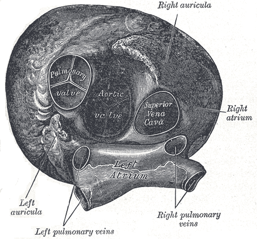
Transverse Cross Section of the Heart. This illustration highlights the anatomical features visible in a transverse cross section of the heart. This picture includes both atria and auriculae, the left and right pulmonary veins, the superior vena cava, and the aortic and pulmonary valves.
Henry Vandyke Carter, Public Domain, via Wikimedia Commons
(Click Image to Enlarge)
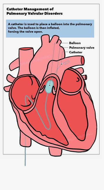
Catheter Management of Pulmonary Valvular Disorders. This illustration shows the balloon inflated within the pulmonary valve.
StatPearls Publishing Illustration
References
Marelli AJ, Mackie AS, Ionescu-Ittu R, Rahme E, Pilote L. Congenital heart disease in the general population: changing prevalence and age distribution. Circulation. 2007 Jan 16:115(2):163-72 [PubMed PMID: 17210844]
Level 2 (mid-level) evidenceWarnes CA, Liberthson R, Danielson GK, Dore A, Harris L, Hoffman JI, Somerville J, Williams RG, Webb GD. Task force 1: the changing profile of congenital heart disease in adult life. Journal of the American College of Cardiology. 2001 Apr:37(5):1170-5 [PubMed PMID: 11300418]
Level 2 (mid-level) evidenceVerheugt CL, Uiterwaal CS, van der Velde ET, Meijboom FJ, Pieper PG, van Dijk AP, Vliegen HW, Grobbee DE, Mulder BJ. Mortality in adult congenital heart disease. European heart journal. 2010 May:31(10):1220-9. doi: 10.1093/eurheartj/ehq032. Epub 2010 Mar 5 [PubMed PMID: 20207625]
Level 2 (mid-level) evidenceYuan SM, Mishaly D, Shinfeld A, Raanani E. Right ventricular outflow tract reconstruction: valved conduit of choice and clinical outcomes. Journal of cardiovascular medicine (Hagerstown, Md.). 2008 Apr:9(4):327-37. doi: 10.2459/JCM.0b013e32821626ce. Epub [PubMed PMID: 18334887]
Level 3 (low-level) evidenceOng K, Boone R, Gao M, Carere R, Webb J, Kiess M, Grewal J. Right ventricle to pulmonary artery conduit reoperations in patients with tetralogy of fallot or pulmonary atresia associated with ventricular septal defect. The American journal of cardiology. 2013 Jun 1:111(11):1638-43. doi: 10.1016/j.amjcard.2013.01.337. Epub 2013 Mar 4 [PubMed PMID: 23481618]
Level 2 (mid-level) evidenceBoethig D, Thies WR, Hecker H, Breymann T. Mid term course after pediatric right ventricular outflow tract reconstruction: a comparison of homografts, porcine xenografts and Contegras. European journal of cardio-thoracic surgery : official journal of the European Association for Cardio-thoracic Surgery. 2005 Jan:27(1):58-66 [PubMed PMID: 15621472]
Level 3 (low-level) evidenceElahi M, Dhannapuneni R, Firmin R, Hickey M. Direct complications of repeat median sternotomy in adults. Asian cardiovascular & thoracic annals. 2005 Jun:13(2):135-8 [PubMed PMID: 15905341]
Temeck BK, Katz NM, Wallace RB. An approach to reoperative median sternotomy. Journal of cardiac surgery. 1990 Mar:5(1):14-25 [PubMed PMID: 2133819]
van der Bom T, Zomer AC, Zwinderman AH, Meijboom FJ, Bouma BJ, Mulder BJ. The changing epidemiology of congenital heart disease. Nature reviews. Cardiology. 2011 Jan:8(1):50-60. doi: 10.1038/nrcardio.2010.166. Epub 2010 Nov 2 [PubMed PMID: 21045784]
Vida VL, Berggren H, Brawn WJ, Daenen W, Di Carlo D, Di Donato R, Lindberg HL, Corno AF, Fragata J, Elliott MJ, Hraska V, Kiraly L, Lacour-Gayet F, Maruszewski B, Rubay J, Sairanen H, Sarris G, Urban A, Van Doorn C, Ziemer G, Stellin G. Risk of surgery for congenital heart disease in the adult: a multicentered European study. The Annals of thoracic surgery. 2007 Jan:83(1):161-8 [PubMed PMID: 17184653]
Verheugt CL, Uiterwaal CS, Grobbee DE, Mulder BJ. Long-term prognosis of congenital heart defects: a systematic review. International journal of cardiology. 2008 Dec 17:131(1):25-32. doi: 10.1016/j.ijcard.2008.06.023. Epub 2008 Aug 6 [PubMed PMID: 18687485]
Level 1 (high-level) evidenceKhairy P, Aboulhosn J, Gurvitz MZ, Opotowsky AR, Mongeon FP, Kay J, Valente AM, Earing MG, Lui G, Gersony DR, Cook S, Ting JG, Nickolaus MJ, Webb G, Landzberg MJ, Broberg CS, Alliance for Adult Research in Congenital Cardiology (AARCC). Arrhythmia burden in adults with surgically repaired tetralogy of Fallot: a multi-institutional study. Circulation. 2010 Aug 31:122(9):868-75. doi: 10.1161/CIRCULATIONAHA.109.928481. Epub 2010 Aug 16 [PubMed PMID: 20713900]
Level 2 (mid-level) evidenceBonhoeffer P, Boudjemline Y, Saliba Z, Merckx J, Aggoun Y, Bonnet D, Acar P, Le Bidois J, Sidi D, Kachaner J. Percutaneous replacement of pulmonary valve in a right-ventricle to pulmonary-artery prosthetic conduit with valve dysfunction. Lancet (London, England). 2000 Oct 21:356(9239):1403-5 [PubMed PMID: 11052583]
Level 3 (low-level) evidencePeng LF, McElhinney DB, Nugent AW, Powell AJ, Marshall AC, Bacha EA, Lock JE. Endovascular stenting of obstructed right ventricle-to-pulmonary artery conduits: a 15-year experience. Circulation. 2006 Jun 6:113(22):2598-605 [PubMed PMID: 16735676]
Level 2 (mid-level) evidencePutman LM, van Gameren M, Meijboom FJ, de Jong PL, Roos-Hesselink JW, Witsenburg M, Takkenberg JJ, Bogers AJ. Seventeen years of adult congenital heart surgery: a single centre experience. European journal of cardio-thoracic surgery : official journal of the European Association for Cardio-thoracic Surgery. 2009 Jul:36(1):96-104; discussion 104. doi: 10.1016/j.ejcts.2009.01.046. Epub 2009 Mar 20 [PubMed PMID: 19303791]
Level 2 (mid-level) evidenceCoats L, Tsang V, Khambadkone S, van Doorn C, Cullen S, Deanfield J, de Leval MR, Bonhoeffer P. The potential impact of percutaneous pulmonary valve stent implantation on right ventricular outflow tract re-intervention. European journal of cardio-thoracic surgery : official journal of the European Association for Cardio-thoracic Surgery. 2005 Apr:27(4):536-43 [PubMed PMID: 15784347]
Level 2 (mid-level) evidenceBerman DP, McElhinney DB, Vincent JA, Hellenbrand WE, Zahn EM. Feasibility and short-term outcomes of percutaneous transcatheter pulmonary valve replacement in small ({30 kg) children with dysfunctional right ventricular outflow tract conduits. Circulation. Cardiovascular interventions. 2014 Apr:7(2):142-8. doi: 10.1161/CIRCINTERVENTIONS.113.000881. Epub 2014 Feb 25 [PubMed PMID: 24569596]
Level 2 (mid-level) evidenceSchievano S, Coats L, Migliavacca F, Norman W, Frigiola A, Deanfield J, Bonhoeffer P, Taylor AM. Variations in right ventricular outflow tract morphology following repair of congenital heart disease: implications for percutaneous pulmonary valve implantation. Journal of cardiovascular magnetic resonance : official journal of the Society for Cardiovascular Magnetic Resonance. 2007:9(4):687-95 [PubMed PMID: 17578725]
Level 2 (mid-level) evidencePatel ND, Levi DS, Cheatham JP, Qureshi SA, Shahanavaz S, Zahn EM. Transcatheter Pulmonary Valve Replacement: A Review of Current Valve Technologies. Journal of the Society for Cardiovascular Angiography & Interventions. 2022 Nov-Dec:1(6):100452. doi: 10.1016/j.jscai.2022.100452. Epub 2022 Sep 16 [PubMed PMID: 39132347]
Baumgartner H, De Backer J, Babu-Narayan SV, Budts W, Chessa M, Diller GP, Lung B, Kluin J, Lang IM, Meijboom F, Moons P, Mulder BJM, Oechslin E, Roos-Hesselink JW, Schwerzmann M, Sondergaard L, Zeppenfeld K, ESC Scientific Document Group. 2020 ESC Guidelines for the management of adult congenital heart disease. European heart journal. 2021 Feb 11:42(6):563-645. doi: 10.1093/eurheartj/ehaa554. Epub [PubMed PMID: 32860028]
Geva T. Repaired tetralogy of Fallot: the roles of cardiovascular magnetic resonance in evaluating pathophysiology and for pulmonary valve replacement decision support. Journal of cardiovascular magnetic resonance : official journal of the Society for Cardiovascular Magnetic Resonance. 2011 Jan 20:13(1):9. doi: 10.1186/1532-429X-13-9. Epub 2011 Jan 20 [PubMed PMID: 21251297]
Gurvitz M, Krieger EV, Fuller S, Davis LL, Kittleson MM, Aboulhosn JA, Bradley EA, Buber J, Daniels CJ, Dimopoulos K, Egbe A, Geoffrion TR, John A, Khairy P, Kim YY, Kreutzer J, Lewis MJ, Menachem JN, Moore JP, Osteen KA, Parikh PB, Saidi A, Salciccioli KB, Schunder RL, Valente AM, Wald RM. 2025 ACC/AHA/HRS/ISACHD/SCAI Guideline for the Management of Adults With Congenital Heart Disease: A Report of the American College of Cardiology/American Heart Association Joint Committee on Clinical Practice Guidelines. Journal of the American College of Cardiology. 2026 Feb 24:87(7):822-976. doi: 10.1016/j.jacc.2025.09.006. Epub 2025 Dec 18 [PubMed PMID: 41411480]
Level 1 (high-level) evidenceKrebushevski K, Cabalka AK, Pollak PM, Yu L, Anderson JH. Self-Expanding Aortic Endoprosthesis for Conduit Preparation Prior to Transcatheter Pulmonary Valve Replacement. Catheterization and cardiovascular interventions : official journal of the Society for Cardiac Angiography & Interventions. 2025 Oct:106(4):2244-2251. doi: 10.1002/ccd.70039. Epub 2025 Jul 29 [PubMed PMID: 40734327]
Anderson JH, Krebushevski K, Cabalka AK, Sutter AM, Taggart NW, Agasthi P. Modified deployment of a self-expanding aortic endoprosthesis for congenital aortic and conduit repair applications. The Journal of invasive cardiology. 2026 Jun:38(6):. doi: 10.25270/jic/25.00368. Epub [PubMed PMID: 41461099]
Hamid T, Rajagopal R, Pius C, Clarke B, Mahadevan VS. Preclosure of large-sized venous access sites in adults undergoing transcatheter structural interventions. Catheterization and cardiovascular interventions : official journal of the Society for Cardiac Angiography & Interventions. 2013 Mar:81(4):586-90. doi: 10.1002/ccd.24358. Epub 2012 Nov 8 [PubMed PMID: 22431302]
Chung R, Taylor AM. Imaging for preintervention planning: transcatheter pulmonary valve therapy. Circulation. Cardiovascular imaging. 2014 Jan:7(1):182-9. doi: 10.1161/CIRCIMAGING.113.000826. Epub [PubMed PMID: 24449547]
Hascoët S, Acar P, Boudjemline Y. Transcatheter pulmonary valvulation: current indications and available devices. Archives of cardiovascular diseases. 2014 Nov:107(11):625-34. doi: 10.1016/j.acvd.2014.07.048. Epub 2014 Oct 31 [PubMed PMID: 25444020]
Boudjemline Y, Sarquella-Brugada G, Kamache I, Patel M, Ladouceur M, Bonnet D, Boughenou FM, Fraisse A, Iserin L. Impact of right ventricular outflow tract size and substrate on outcomes of percutaneous pulmonary valve implantation. Archives of cardiovascular diseases. 2013 Jan:106(1):19-26. doi: 10.1016/j.acvd.2012.09.005. Epub 2012 Dec 25 [PubMed PMID: 23374968]
Alsulami G, Patel M, Malekzadeh-Milani S, Bonnet D, Boudjemline Y. Hyperacute flash pulmonary oedema after transcatheter pulmonary valve implantation: The melody of an overwhelmed left ventricle. Archives of cardiovascular diseases. 2014 Apr:107(4):219-24. doi: 10.1016/j.acvd.2014.03.007. Epub 2014 May 2 [PubMed PMID: 24793996]
Level 3 (low-level) evidenceKhambadkone S, Coats L, Taylor A, Boudjemline Y, Derrick G, Tsang V, Cooper J, Muthurangu V, Hegde SR, Razavi RS, Pellerin D, Deanfield J, Bonhoeffer P. Percutaneous pulmonary valve implantation in humans: results in 59 consecutive patients. Circulation. 2005 Aug 23:112(8):1189-97 [PubMed PMID: 16103239]
Schievano S, Petrini L, Migliavacca F, Coats L, Nordmeyer J, Lurz P, Khambadkone S, Taylor AM, Dubini G, Bonhoeffer P. Finite element analysis of stent deployment: understanding stent fracture in percutaneous pulmonary valve implantation. Journal of interventional cardiology. 2007 Dec:20(6):546-54 [PubMed PMID: 18042059]
Level 3 (low-level) evidenceGillespie MJ, Rome JJ, Levi DS, Williams RJ, Rhodes JF, Cheatham JP, Hellenbrand WE, Jones TK, Vincent JA, Zahn EM, McElhinney DB. Melody valve implant within failed bioprosthetic valves in the pulmonary position: a multicenter experience. Circulation. Cardiovascular interventions. 2012 Dec:5(6):862-70. doi: 10.1161/CIRCINTERVENTIONS.112.972216. Epub 2012 Dec 4 [PubMed PMID: 23212395]
Level 2 (mid-level) evidenceBoshoff DE, Cools BL, Heying R, Troost E, Kefer J, Budts W, Gewillig M. Off-label use of percutaneous pulmonary valved stents in the right ventricular outflow tract: time to rewrite the label? Catheterization and cardiovascular interventions : official journal of the Society for Cardiac Angiography & Interventions. 2013 May:81(6):987-95. doi: 10.1002/ccd.24594. Epub 2013 Feb 12 [PubMed PMID: 22887796]
Level 2 (mid-level) evidenceMeadows JJ, Moore PM, Berman DP, Cheatham JP, Cheatham SL, Porras D, Gillespie MJ, Rome JJ, Zahn EM, McElhinney DB. Use and performance of the Melody Transcatheter Pulmonary Valve in native and postsurgical, nonconduit right ventricular outflow tracts. Circulation. Cardiovascular interventions. 2014 Jun:7(3):374-80. doi: 10.1161/CIRCINTERVENTIONS.114.001225. Epub 2014 May 27 [PubMed PMID: 24867892]
Level 2 (mid-level) evidenceCao QL, Kenny D, Zhou D, Pan W, Guan L, Ge J, Hijazi ZM. Early clinical experience with a novel self-expanding percutaneous stent-valve in the native right ventricular outflow tract. Catheterization and cardiovascular interventions : official journal of the Society for Cardiac Angiography & Interventions. 2014 Dec 1:84(7):1131-7. doi: 10.1002/ccd.25544. Epub 2014 May 28 [PubMed PMID: 24824357]
Level 3 (low-level) evidenceAlkashkari W, Alsubei A, Hijazi ZM. Transcatheter Pulmonary Valve Replacement: Current State of Art. Current cardiology reports. 2018 Mar 15:20(4):27. doi: 10.1007/s11886-018-0966-y. Epub 2018 Mar 15 [PubMed PMID: 29546472]
Morray BH, McElhinney DB, Cheatham JP, Zahn EM, Berman DP, Sullivan PM, Lock JE, Jones TK. Risk of coronary artery compression among patients referred for transcatheter pulmonary valve implantation: a multicenter experience. Circulation. Cardiovascular interventions. 2013 Oct 1:6(5):535-42. doi: 10.1161/CIRCINTERVENTIONS.113.000202. Epub 2013 Sep 24 [PubMed PMID: 24065444]
Level 2 (mid-level) evidenceAgasthi P, Cabalka AK, Cetta F, Anderson JH. Harmony Transcatheter Pulmonary Valve Successfully Implanted via Transjugular Approach. Journal of the Society for Cardiovascular Angiography & Interventions. 2024 Feb:3(2):101207. doi: 10.1016/j.jscai.2023.101207. Epub 2023 Oct 21 [PubMed PMID: 39132210]
Morray BH, Gillespie MJ, Cheatham JP, Salavitabar A, Peng L, Jones TK, Levi DS, Gray RG, Asnes J, Cabalka AK, Fujimoto K, Qureshi AM, Bergersen L, Benson LN, Haugan D, McElhinney DB. Midterm Outcomes in a Pooled Cohort of Harmony Transcatheter Pulmonary Valve Recipients. Circulation. Cardiovascular interventions. 2025 Sep:18(9):e015196. doi: 10.1161/CIRCINTERVENTIONS.125.015196. Epub 2025 Jul 16 [PubMed PMID: 40665900]
Torres AJ, Dimas VV, Shahanavaz S, Balzer D, Morgan G, Lim DS, Armstrong AK, Berman D, Babaliaros V, Kim D, Gillespie MJ, Sommer R, Aboulhosn J, Jones TK, Mahadevan VS, Stapleton G, Ma Y, Shirali G, Parthiban A, Blanke P, Leipsic J, Zahn E. Transcatheter Pulmonary Valve Implantation With the Alterra Adaptive Prestent and SAPIEN 3 Transcatheter Heart Valve: 3-Year Pooled Outcomes of the ALTERRA Trials. Circulation. Cardiovascular interventions. 2026 Apr:19(4):e015873. doi: 10.1161/CIRCINTERVENTIONS.125.015873. Epub 2026 Feb 9 [PubMed PMID: 41657206]
Anderson JH, Cabalka AK. Transcatheter Pulmonary Valve Implantation: Valve-in-Frame Procedure. JACC. Cardiovascular interventions. 2022 Sep 26:15(18):1878-1879. doi: 10.1016/j.jcin.2022.05.021. Epub 2022 Jun 29 [PubMed PMID: 36137693]
Egbe AC, Cabalka AK, Taggart NW, Hagler DJ, Eleid MF, Rihal CS, Anderson JH. Transcatheter Valve Replacement in Adults With Congenital Heart Disease-The Mayo Clinic Experience. Circulation. Cardiovascular interventions. 2025 Dec:18(12):e015667. doi: 10.1161/CIRCINTERVENTIONS.125.015667. Epub 2025 Oct 27 [PubMed PMID: 41140155]
Boudjemline Y, Malekzadeh-Milani S, Patel M, Thambo JB, Bonnet D, Iserin L, Fraisse A. Predictors and outcomes of right ventricular outflow tract conduit rupture during percutaneous pulmonary valve implantation: a multicentre study. EuroIntervention : journal of EuroPCR in collaboration with the Working Group on Interventional Cardiology of the European Society of Cardiology. 2016 Jan 22:11(9):1053-62. doi: 10.4244/EIJY14M09_06. Epub [PubMed PMID: 25244126]
Level 2 (mid-level) evidenceMcElhinney DB, Cheatham JP, Jones TK, Lock JE, Vincent JA, Zahn EM, Hellenbrand WE. Stent fracture, valve dysfunction, and right ventricular outflow tract reintervention after transcatheter pulmonary valve implantation: patient-related and procedural risk factors in the US Melody Valve Trial. Circulation. Cardiovascular interventions. 2011 Dec 1:4(6):602-14. doi: 10.1161/CIRCINTERVENTIONS.111.965616. Epub 2011 Nov 9 [PubMed PMID: 22075927]
Level 2 (mid-level) evidenceMcElhinney DB, Bergersen L, Marshall AC. In situ fracture of stents implanted for relief of pulmonary arterial stenosis in patients with congenitally malformed hearts. Cardiology in the young. 2008 Aug:18(4):405-14. doi: 10.1017/S1047951108002424. Epub 2008 Jun 18 [PubMed PMID: 18559137]
Level 2 (mid-level) evidenceCheung G, Vejlstrup N, Ihlemann N, Arnous S, Franzen O, Bundgaard H, Søndergaard L. Infective endocarditis following percutaneous pulmonary valve replacement: diagnostic challenges and application of intra-cardiac echocardiography. International journal of cardiology. 2013 Nov 30:169(6):425-9. doi: 10.1016/j.ijcard.2013.10.016. Epub 2013 Oct 11 [PubMed PMID: 24182680]
Level 3 (low-level) evidenceLurz P, Coats L, Khambadkone S, Nordmeyer J, Boudjemline Y, Schievano S, Muthurangu V, Lee TY, Parenzan G, Derrick G, Cullen S, Walker F, Tsang V, Deanfield J, Taylor AM, Bonhoeffer P. Percutaneous pulmonary valve implantation: impact of evolving technology and learning curve on clinical outcome. Circulation. 2008 Apr 15:117(15):1964-72. doi: 10.1161/CIRCULATIONAHA.107.735779. Epub 2008 Apr 7 [PubMed PMID: 18391109]
Level 2 (mid-level) evidenceChen XJ, Smith PB, Jaggers J, Lodge AJ. Bioprosthetic pulmonary valve replacement: contemporary analysis of a large, single-center series of 170 cases. The Journal of thoracic and cardiovascular surgery. 2013 Dec:146(6):1461-6. doi: 10.1016/j.jtcvs.2012.09.081. Epub 2012 Nov 2 [PubMed PMID: 23122698]
Level 2 (mid-level) evidenceBuber J, Bergersen L, Lock JE, Gauvreau K, Esch JJ, Landzberg MJ, Valente AM, Sandora TJ, Marshall AC. Bloodstream infections occurring in patients with percutaneously implanted bioprosthetic pulmonary valve: a single-center experience. Circulation. Cardiovascular interventions. 2013 Jun:6(3):301-10. doi: 10.1161/CIRCINTERVENTIONS.112.000348. Epub 2013 Jun 11 [PubMed PMID: 23756696]
Level 2 (mid-level) evidencePatel M, Malekzadeh-Milani S, Ladouceur M, Iserin L, Boudjemline Y. Percutaneous pulmonary valve endocarditis: incidence, prevention and management. Archives of cardiovascular diseases. 2014 Nov:107(11):615-24. doi: 10.1016/j.acvd.2014.07.052. Epub 2014 Oct 16 [PubMed PMID: 25445753]
Biernacka EK, Rużyłło W, Demkow M, Kowalski M, Śpiewak M, Piotrowski W, Kuśmierczyk M, Banaś S, Różanski J, Hoffman P. Transcatheter pulmonary valve implantation in patients with right ventricular outflow tract dysfunction: early and mid-term results. The Journal of invasive cardiology. 2015 Jun:27(6):E82-9 [PubMed PMID: 26028663]
Cheatham JP, Hellenbrand WE, Zahn EM, Jones TK, Berman DP, Vincent JA, McElhinney DB. Clinical and hemodynamic outcomes up to 7 years after transcatheter pulmonary valve replacement in the US melody valve investigational device exemption trial. Circulation. 2015 Jun 2:131(22):1960-70. doi: 10.1161/CIRCULATIONAHA.114.013588. Epub 2015 May 5 [PubMed PMID: 25944758]
Vezmar M, Chaturvedi R, Lee KJ, Almeida C, Manlhiot C, McCrindle BW, Horlick EM, Benson LN. Percutaneous pulmonary valve implantation in the young 2-year follow-up. JACC. Cardiovascular interventions. 2010 Apr:3(4):439-48. doi: 10.1016/j.jcin.2010.02.003. Epub [PubMed PMID: 20398873]
Asoh K, Walsh M, Hickey E, Nagiub M, Chaturvedi R, Lee KJ, Benson LN. Percutaneous pulmonary valve implantation within bioprosthetic valves. European heart journal. 2010 Jun:31(11):1404-9. doi: 10.1093/eurheartj/ehq056. Epub 2010 Mar 15 [PubMed PMID: 20231157]
Level 2 (mid-level) evidenceDemkow M, Biernacka EK, Spiewak M, Kowalski M, Siudalska H, Wolski P, Sondergaard L, Miśko J, Hoffman P, Rużyłło W. Percutaneous pulmonary valve implantation preceded by routine prestenting with a bare metal stent. Catheterization and cardiovascular interventions : official journal of the Society for Cardiac Angiography & Interventions. 2011 Feb 15:77(3):381-9. doi: 10.1002/ccd.22700. Epub [PubMed PMID: 20602475]
Coats L, Khambadkone S, Derrick G, Hughes M, Jones R, Mist B, Pellerin D, Marek J, Deanfield JE, Bonhoeffer P, Taylor AM. Physiological consequences of percutaneous pulmonary valve implantation: the different behaviour of volume- and pressure-overloaded ventricles. European heart journal. 2007 Aug:28(15):1886-93 [PubMed PMID: 17595193]
Zahn EM, Hellenbrand WE, Lock JE, McElhinney DB. Implantation of the melody transcatheter pulmonary valve in patients with a dysfunctional right ventricular outflow tract conduit early results from the u.s. Clinical trial. Journal of the American College of Cardiology. 2009 Oct 27:54(18):1722-9. doi: 10.1016/j.jacc.2009.06.034. Epub [PubMed PMID: 19850214]
Martins JD, Ewert P, Sousa L, Freitas I, Trigo C, Jalles N, Matos P, Agapito A, Ferreira R, Pinto FF. Percutaneous pulmonary valve implantation: initial experience. Revista portuguesa de cardiologia : orgao oficial da Sociedade Portuguesa de Cardiologia = Portuguese journal of cardiology : an official journal of the Portuguese Society of Cardiology. 2010 Dec:29(12):1839-46 [PubMed PMID: 21428139]
Batra AS, McElhinney DB, Wang W, Zakheim R, Garofano RP, Daniels C, Yung D, Cooper DM, Rhodes J. Cardiopulmonary exercise function among patients undergoing transcatheter pulmonary valve implantation in the US Melody valve investigational trial. American heart journal. 2012 Feb:163(2):280-7. doi: 10.1016/j.ahj.2011.10.017. Epub [PubMed PMID: 22305848]
Borik S, Crean A, Horlick E, Osten M, Lee KJ, Chaturvedi R, Friedberg MK, McCrindle BW, Manlhiot C, Benson L. Percutaneous pulmonary valve implantation: 5 years of follow-up: does age influence outcomes? Circulation. Cardiovascular interventions. 2015 Feb:8(2):e001745. doi: 10.1161/CIRCINTERVENTIONS.114.001745. Epub [PubMed PMID: 25652317]
Level 2 (mid-level) evidenceHolzer RJ, Hijazi ZM. Transcatheter pulmonary valve replacement: State of the art. Catheterization and cardiovascular interventions : official journal of the Society for Cardiac Angiography & Interventions. 2016 Jan 1:87(1):117-28. doi: 10.1002/ccd.26263. Epub 2015 Oct 1 [PubMed PMID: 26423185]
Armstrong AK, Balzer DT, Cabalka AK, Gray RG, Javois AJ, Moore JW, Rome JJ, Turner DR, Zellers TM, Kreutzer J. One-year follow-up of the Melody transcatheter pulmonary valve multicenter post-approval study. JACC. Cardiovascular interventions. 2014 Nov:7(11):1254-62. doi: 10.1016/j.jcin.2014.08.002. Epub 2014 Nov 17 [PubMed PMID: 25459038]
Kenny D, Hijazi ZM, Kar S, Rhodes J, Mullen M, Makkar R, Shirali G, Fogel M, Fahey J, Heitschmidt MG, Cain C. Percutaneous implantation of the Edwards SAPIEN transcatheter heart valve for conduit failure in the pulmonary position: early phase 1 results from an international multicenter clinical trial. Journal of the American College of Cardiology. 2011 Nov 15:58(21):2248-56. doi: 10.1016/j.jacc.2011.07.040. Epub [PubMed PMID: 22078433]
Level 1 (high-level) evidenceFaza N, Kenny D, Kavinsky C, Amin Z, Heitschmidt M, Hijazi ZM. Single-center comparative outcomes of the Edwards SAPIEN and Medtronic Melody transcatheter heart valves in the pulmonary position. Catheterization and cardiovascular interventions : official journal of the Society for Cardiac Angiography & Interventions. 2013 Oct 1:82(4):E535-41. doi: 10.1002/ccd.24680. Epub 2013 Mar 18 [PubMed PMID: 23008193]
Level 2 (mid-level) evidenceKheiwa A, Divanji P, Mahadevan VS. Transcatheter pulmonary valve implantation: will it replace surgical pulmonary valve replacement? Expert review of cardiovascular therapy. 2018 Mar:16(3):197-207. doi: 10.1080/14779072.2018.1435273. Epub 2018 Feb 12 [PubMed PMID: 29433351]
Suradi HS, Hijazi ZM. Percutaneous pulmonary valve implantation. Global cardiology science & practice. 2015:2015(2):23. doi: 10.5339/gcsp.2015.23. Epub 2015 Sep 8 [PubMed PMID: 26535223]