 Tools
Tools
 Auditory Pathway
Introduction
The auditory system governs the detection and interpretation of environmental sound. Peripheral and central components constitute this organ system. The outer, middle, and inner ear serve as the peripheral auditory structures, while the cochlear nuclei, superior olivary nuclei, lateral lemniscus, inferior colliculus, medial geniculate nuclei, and auditory cortex form the central auditory structures (see Image. Peripheral and Central Structures of the Auditory System).
Central auditory pathways encode sound frequency, intensity, and spatial location. Integration of these acoustic features supports accurate interpretation of environmental input. Environmental context, attentional state, and perceived relevance of sound stimuli vary over time. Therefore, perception undergoes continuous modulation within the central auditory pathways. Understanding the fundamental principles of audition and auditory processing enhances clinical evaluation of hearing disorders and guides accurate localization of lesions within the auditory pathway.
Structure and Function
Register For Free And Read The Full Article
 Search engine and full access to all medical articles
- 10 free questions in your specialty
- Free CME/CE Activities
- Free daily question in your email
- Save favorite articles to your dashboard
- Emails offering discounts
Learn more about a Subscription to StatPearls Point-of-Care
Structure and Function
Peripheral Auditory System: How Sound Reaches the Brain
Sounds are produced by energy waves traveling through a medium, generating particle motion within that medium. Particle displacement produces local air-pressure fluctuations, consisting of alternating compression and rarefaction. "Sound frequency" refers to the number of compression–rarefaction cycle repetitions within a defined time interval and is measured in Hertz (Hz) or cycles per second. Humans typically perceive frequencies spanning 20 to 20,000 Hz.[1]
Sound waves enter the outer ear and pass through the external acoustic meatus to the tympanic membrane. Contact with incoming waves produces tympanic membrane displacement, initiating vibration of the middle ear ossicles—the malleus, incus, and stapes. These 3 ossicles transmit tympanic membrane motion to the oval (vestibular) window and function as amplifiers and impedance-matching structures during transmission of sound from the air-filled middle ear to the perilymphatic fluid contacting the oval window (see Image. Right Auditory Ossicles). The oval window forms the entry point to the vestibule of the inner ear.
The vestibule connects to both the cochlea and the semicircular canals (see Image. Inner Ear Anatomy). The cochlea completes approximately 2½ turns around the modiolus, and perilymph fills its fluid spaces.
The cochlea contains 3 chambers: the scala tympani, scala vestibuli, and scala media (see Image. Diagrammatic Longitudinal Section of the Cochlea). The scala tympani occupies the outermost portion of the cochlea, whereas the scala vestibuli lies in the innermost region. The scala media, or cochlear duct, separates these chambers along their course, except at the helicotrema, where the scala tympani communicates with the scala vestibuli. Perilymph fills the scala tympani and scala vestibuli. The scala media contains endolymph, the basilar membrane, and the organ of Corti. Endolymph within the scala media has a uniquely high potassium concentration compared with other body fluids.
Oscillations of the oval window generate pressure waves in the perilymph, which propagate through the scala vestibuli and scala tympani. These waves displace the basilar membrane, forming the floor of the scala media.
The organ of Corti rests on the basilar membrane within the scala media (see Image. Organ of Corti). This structure contains the mechanoreceptor population: 3 rows of outer hair cells and 1 row of inner hair cells. The bases of the hair cells anchor to the basilar membrane. Stereocilia project from the apical surface of each cell and attach to the tectorial membrane. Potassium channels reside on the surfaces of the stereocilia.
The basilar membrane shifts in response to oscillations within the scala vestibuli and scala tympani. Displacement of the basilar membrane bends the stereocilia against the tectorial membrane, leading to the opening or closing of potassium channels, depending on the direction of deflection. Hair cells lack axons. Depolarization produces direct synaptic transmission from the cell body to the auditory nerve. Therefore, sound energy is converted to electrical activity within the inner ear.
The positions of the tectorial and basilar membranes within the cochlea determine their frequency-specific responses. The cochlear base, which lies near the oval window, contains stiffer membranes and shorter stereocilia, yielding maximal sensitivity to high-frequency stimuli. Membrane compliance increases, and stereocilia lengthen progressively toward the cochlear apex. Stereocilia at the apex exceed twice the length of those at the base, producing greater responsiveness to low-frequency sound.[2] This spatial organization constitutes the tonotopic gradient, which enables the brain to map distinct sound frequencies.
Spiral ganglion peripheral axons, which contribute to the auditory nerve, synapse at the bases of the hair cell bodies. Approximately 90% of auditory nerve fibers receive input from inner hair cells.[3] Therefore, inner hair cells process most auditory stimuli, with the remaining input arising from outer hair cells.
The auditory cortex uses spiral ganglion neurons to protect the inner ear from excessive sound exposure. Activation of these neurons increases basilar membrane stiffness, thereby reducing hair cell excitation and decreasing hair cell sensitivity to sound.[4] Exposure to loud stimuli triggers this descending modulation to limit the hair cell response. Transient hearing reduction can occur afterward as the descending auditory pathways and peripheral auditory structures readjust to a quieter environment.
Central Auditory System
Peripheral auditory impulses reach the auditory cortex via a series of ascending auditory pathways. Sensory information from the auditory nerve ascends along the following route:
- Ipsilateral auditory nerve
- Ipsilateral cochlear nucleus
- Superior olivary complex*
- Contralateral and ipsilateral lateral lemniscus nuclei
- Contralateral and ipsilateral inferior colliculus
- Contralateral and ipsilateral medial geniculate nuclei
- Contralateral and ipsilateral transverse temporal gyrus (auditory region)
*The majority of fibers decussate to the contralateral superior olivary complex, while a smaller subset remains on the ipsilateral side.
Most of the auditory pathway is binaural, receiving input from both right and left auditory systems. This bilateral organization facilitates precise localization of auditory stimuli (see Image. Auditory Pathways).[5]
Types of Processing
The central auditory pathways process several critical sound attributes, including attenuation, spatial location, frequency, and combination sensitivity. Each auditory nucleus exhibits a tonotopic organization, as demonstrated in the cochlea, which enables frequency-specific information to ascend to the auditory cortex, where it is perceived. Perception of a specific frequency corresponds to the pitch of the sound.
Attenuation is encoded by neurons firing at variable rates. Specialized neurons at multiple levels of the auditory pathway respond optimally to sounds within defined intensity ranges, permitting discrimination between louder and softer environmental stimuli.
Spatial location is encoded within the superior olivary complex through comparison of auditory inputs from the right and left sides of the head. A sound originating from the midline reaches both ears simultaneously, producing synchronous input to the superior olivary complex. In contrast, a sound originating from the right side generates a temporal delay, with the right-ear input arriving before the left-ear input. This interaural timing difference enables the superior olivary complex to determine the spatial location of a sound.[6]
Combination-sensitive units constitute a subset of auditory neurons that respond selectively to specific acoustic elements. These neurons, which may exhibit facilitatory or inhibitory responses, are present in the cochlear nucleus, lateral lemniscus, inferior colliculus, medial geniculate, and auditory cortex.[7][8][9][10][11][12] These neurons contribute to the detection of salient auditory cues required for communication.[13]
Descending Circuits
Ascending auditory information is modulated by an extensive network of descending auditory pathways. Projections from the auditory cortex reach each level of the auditory system through both direct and indirect routes.[14][15][16][17][18] The auditory cortex maintains direct and indirect connections with numerous higher-order brain regions, including the limbic system, hippocampus, nucleus basalis of Meynert, prefrontal cortex, and multiple other sensory cortical areas. These regions influence auditory processing via descending pathways, adjusting perception based on stimulus relevance, attentional state, or learned behavioral responses.[19][20][21][22][23][24][25][26]
Embryology
The cochlea begins development from the surface ectoderm on gestational day 4, initially forming the otic vesicle, which subsequently differentiates into the membranous labyrinth of the inner ear. The dorsal portion of the labyrinth gives rise to the utricle and semicircular ducts, whereas the ventral portion develops into the cochlea and saccule.
Auditory pathway nuclei originate from distinct regions of the brain. The auditory cortex derives from the telencephalon, the medial geniculate nucleus from the diencephalon, the inferior colliculus from the mesencephalon, and the cochlear and superior olivary nuclei from the rhombencephalon. These regions are fully operational at birth despite their continued structural and functional plasticity throughout life.[27]
Blood Supply and Lymphatics
The external ear receives arterial supply primarily from the posterior auricular branch of the external carotid artery. The middle ear is vascularized by mastoid branches of the posterior auricular and occipital arteries, as well as the deep auricular arteries. The inner ear receives blood from multiple sources, including the anterior tympanic branch of the maxillary artery, the stylomastoid branch of the posterior auricular artery, the petrosal branch of the middle meningeal artery, and the labyrinthine artery, a branch of either the basilar or anterior inferior cerebellar artery.[28]
Lymphatic drainage of the external ear occurs via the preauricular lymph nodes.[29] The middle ear drains into the retroauricular and junctional lymph nodes.[30] Lymphatic drainage of the inner ear remains uncertain. Salt and Hirose proposed that perilymph and surrounding bone facilitate diffuse drainage rather than through conventional lymphatic channels (see Image. Lymph Nodes of the Head and Neck).[31]
Muscles
Three muscles influence audition. The levator veli palatini and tensor veli palatini assist in opening the auditory tube (also known as the eustachian or pharyngotympanic tube), thereby helping to regulate air pressure within the middle ear (see Image. Muscles Around the Pharyngotympanic Tube). The stapedius muscle originates from the pyramidal eminence of the temporal bone and inserts on the neck of the stapes. Contraction of the stapedius limits stapes movement, reducing excessive vibration on the oval window. Descending neural circuits trigger stapedius contraction in response to loud sounds, dampening auditory input and protecting the cochlea from potential damage.
Physiologic Variants
Morphological and physiological variations within the auditory system are typically congenital. Most cases (approximately 80%) are caused by membranous malformations involving inner ear hair cells, resulting in sensorineural hearing loss. The remaining congenital malformations involve abnormalities in the ossicles or other middle ear structures, leading to bone conduction deficits and decreased hearing in the affected ear. These variants often occur bilaterally, producing substantial hearing loss in many individuals.[32]
Surgical Considerations
The auditory system presents multiple surgical considerations, determined by the type and mechanism of pathology. Recurrent otitis media may be managed with drainage tube insertion. Tympanoplasty can repair damaged ossicles, as well as tympanic membrane perforations and scars.[33][34]
Vestibular schwannomas may occur sporadically or form in association with neurofibromatosis type 2. Vestibular schwannomas related to neurofibromatosis type 2 exhibit faster proliferation, greater lobularity, and reduced vascularity compared with sporadic vestibular schwannomas. Surgical removal of these tumors carries a high risk of deafness on the affected side. Some microsurgical techniques have been developed that offer partial preservation of audition. The success of auditory preservation depends strongly on tumor size and surgeon experience.
Due to the faster growth of neurofibromatosis type 2–associated vestibular schwannomas, the prognosis for hearing preservation is less favorable than in sporadic cases. In both types, decompression of the internal acoustic meatus and early tumor resection prior to systemic treatment achieve higher rates of auditory preservation than noncompression approaches.[35]
Surgeons should consider that patients with vestibular schwannomas may have auditory implants that can shift during magnetic resonance imaging (MRI) due to the strong magnetic field. Before MRI, the implant magnet must be removed or the head secured to prevent movement during imaging.[36]
Auditory brainstem or cochlear implants can restore audition following surgical resection of a vestibular schwannoma. Implantation may be performed via a transmastoid or intradural approach. Complication rates are low when experienced surgical teams perform the procedure.[37]
Clinical Significance
Acoustic Neuroma
The auditory nerve, located at the junction of the pons, medulla, and cerebellum, is the most common site of a slow-growing tumor termed an "acoustic neuroma."[38] Tumor enlargement can compress surrounding structures, notably cranial nerves VIII, VII, and IX, as well as the cerebellum and brainstem. Hearing loss is usually the initial symptom. Additional deficits develop ipsilateral to the lesion as the tumor progresses, including tinnitus, vertigo, nystagmus, facial drooping, decreased corneal reflex, hoarseness, dysphagia, ataxia, and dysarthria.[39][40]
Tinnitus
Tinnitus is the perception of a sound not originating from the environment, typically described as ringing or buzzing in the ear. The sensation results from hyperexcitation within a specific tonotopic region of the auditory cortex.[41] Patients with tinnitus may report difficulty distinguishing sounds or conversations in noisy environments due to elevated background neural activity. Severe tinnitus can disrupt sleep and negatively affect social and emotional functioning.[42][43]
Tinnitus usually develops gradually after several years of repeated exposure to loud noise. Cochlear damage from such exposure is thought to induce changes in auditory processing, resulting in hyperexcitation of the auditory cortex. This cortical excitation produces the perception of a sound absent from the environment.[44]
Tinnitus has no definitive cure. Biofeedback therapy may reduce, but not eliminate, the symptom. This therapy employs 1 or more sounds at or near the patient’s perceived tinnitus frequency, engaging inhibitory limbic circuits to suppress aberrant cortical activity.[45][46] Background white noise at bedtime can improve sleep in individuals experiencing tinnitus-related sleep disturbances.[47][48]
Hearing Loss
The most common forms of hearing loss result from damage to either the bony conduction system of the middle ear or the neural conduction pathway within the cochlea. Bone conduction hearing aids can bypass middle ear deficits, transmitting vibrations directly to the cochlea to restore auditory perception.[49] For individuals with neural conduction deficits in the cochlea, cochlear implants can directly stimulate cochlear regions to enable hearing.[50]
Clinical tests to differentiate between bone and neural conduction deficits were developed in 1855 and are known as the Weber and Rinne tests. The Weber test places a tuning fork at the center of the skull and asks the patient which ear perceives the sound more clearly. Perception in the ear with the deficit indicates a bone conduction problem, whereas perception in the unaffected ear suggests a sensory (neural) conduction issue. The Rinne test positions a tuning fork in the air near the ear and then on the mastoid process. If the patient hears the tone better via the mastoid process in the affected ear, a bone conduction problem is present. A neural conduction deficit is detected if neither air nor bone conduction elicits a response.[51]
Cauliflower Ear
Repeated trauma can produce outer ear deformities. Trauma may induce an auricular hematoma, in which blood accumulates between the perichondrium and auricular cartilage. Over time, compromised blood flow to the auricular cartilage can lead to fibrosis and distortion of the outer ear.[52] This distortion and swelling can lead to narrowing of the external auditory canal, resulting in conductive hearing loss due to impaired sound transmission.
Otitis Media
Otitis media is inflammation of the middle ear lining. The condition is more common in children due to horizontally oriented auditory tubes, which limit fluid drainage. The incidence of this condition declines in adulthood as the auditory tubes assume a more lateral and caudal orientation. Middle ear inflammation causes swelling and increased pressure against the tympanic membrane. In severe cases, the tympanic membrane can rupture and reduce auditory acuity.[53]
Other Issues
Most individuals respond well to conventional hearing tests. However, factors such as undeveloped speech, disease, or trauma can limit a patient’s ability to provide reliable feedback. The auditory brainstem response (ABR) offers a more universal method for auditory assessment, as it does not require active patient participation. ABR measures electrical activity in the brainstem auditory pathways in response to sound stimuli. Deviations from normative values indicate auditory dysfunction. The test evaluates multiple auditory nuclei, enabling identification of trauma or disease within the auditory system.[54] ABR is a standard assessment for infants prior to the development of verbal communication.[55]
Media
(Click Image to Enlarge)
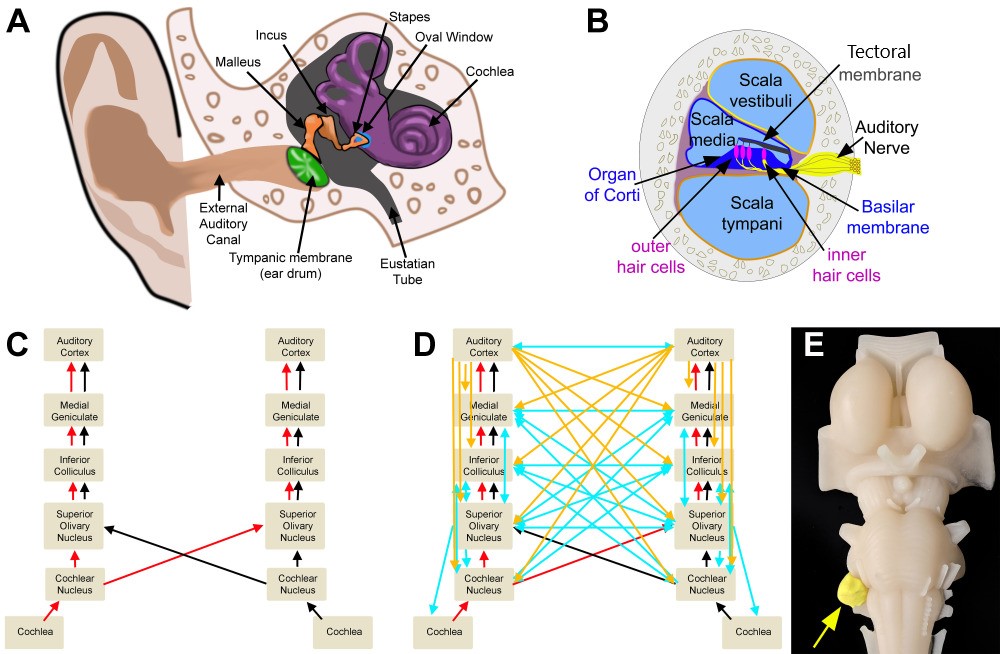
Peripheral and Central Structures of the Auditory System. Panel A shows the outer, middle, and inner ear. Panel B depicts a cross-section of the cochlea. Panel C illustrates the ascending auditory pathway. Panel D demonstrates the ascending pathways (red/black), descending pathways from the cortex (orange) and brainstem (blue), and crossed auditory pathways (blue). Panel E presents a model representation of an acoustic neuroma.
All imaging was created by Diana Peterson for the current manuscript.
(Click Image to Enlarge)
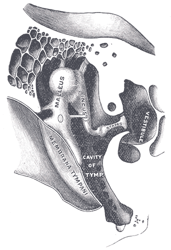
Right Auditory Ossicles. This image shows the anterior aspect of the right chain of ossicles (malleus, incus, and stapes) and their ligaments. Other structures in this illustration include the tympanic membrane, tympanic cavity, and vestibule.
Henry Vandyke Carter, Public Domain, via Wikimedia Commons
(Click Image to Enlarge)
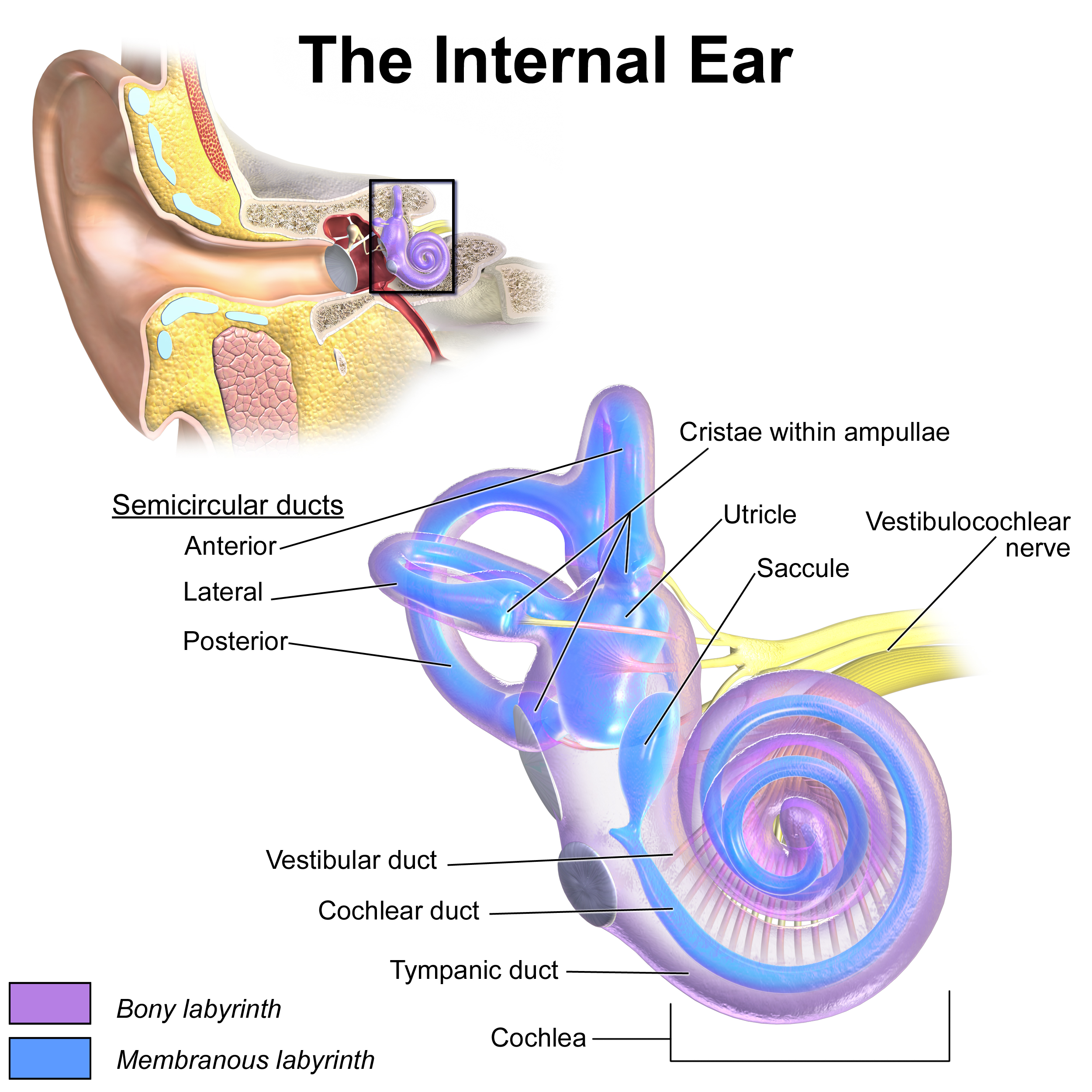
Inner Ear Anatomy. This illustration shows the semicircular ducts and parts of the cochlea.
Blausen.com staff. Medical Gallery of Blausen Medical 2014. WikiJournal of Medicine. doi: 10.15347/wjm/2014.010.
(Click Image to Enlarge)
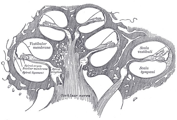
Diagrammatic Longitudinal Section of the Cochlea. This illustration shows the scala vestibuli, scala media, scala tympani and cochlear nerve.
Henry Vandyke Carter, Public Domain, via Wikimedia Commons
(Click Image to Enlarge)
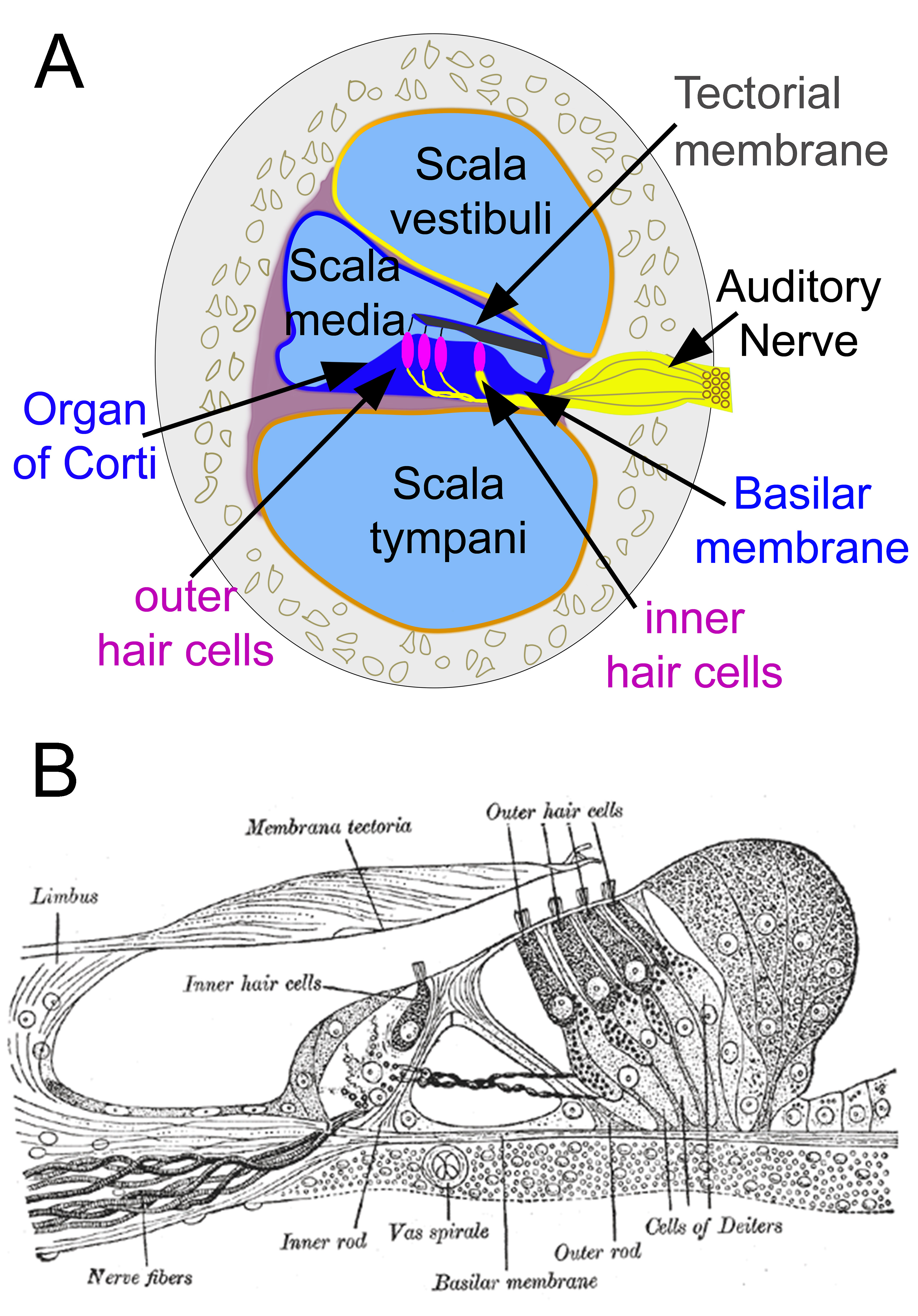
Cochlear Cross-Section and Organ of Corti. Panel A illustrates a cross-sectional view of the cochlea, showing the scala vestibuli, scala media, and scala tympani. The organ of Corti lies within the scala media, rests on the basilar membrane, and is situated beneath the tectorial membrane. Panel B demonstrates the cellular architecture of the organ of Corti, including inner and outer hair cells, supporting cells, the Corti tunnel, and associated nerve fibers.
(A) Contributed by D Peterson, MD
(B) Henry Vandyke Carter, Public Domain, via Wikimedia Commons
(Click Image to Enlarge)
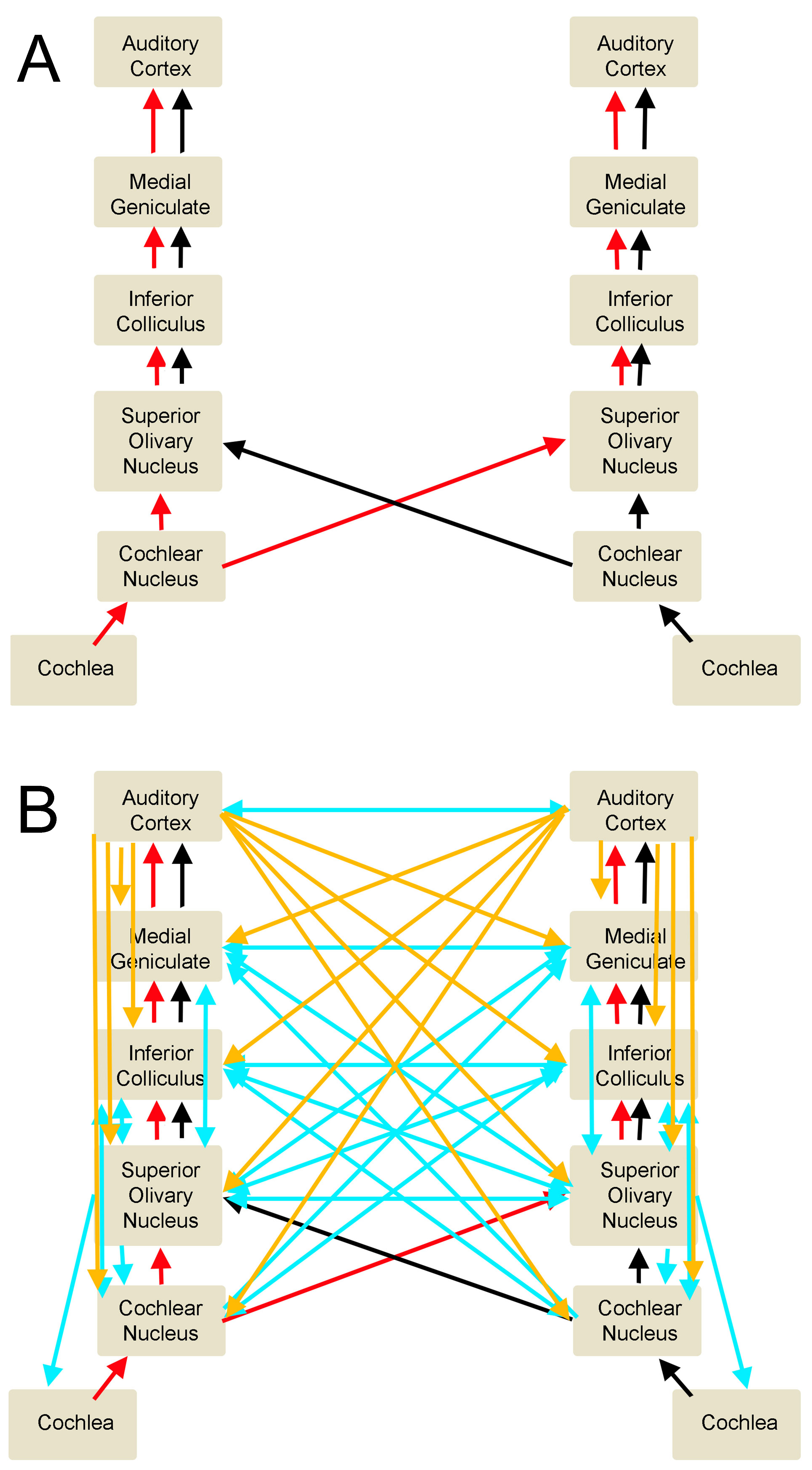
Auditory Pathways. A. This illustration represents the ascending auditory circuits. B. In this combined illustration, the ascending auditory pathway is represented by the red and black arrows, which stand for binaural ascending signals. The descending pathway is represented by the orange arrows (for cortical impulses) and blue arrows (for brainstem signals). Crossing arrows represent the interconnections between the various auditory centers.
Contributed and created by Diana Peterson.
(Click Image to Enlarge)
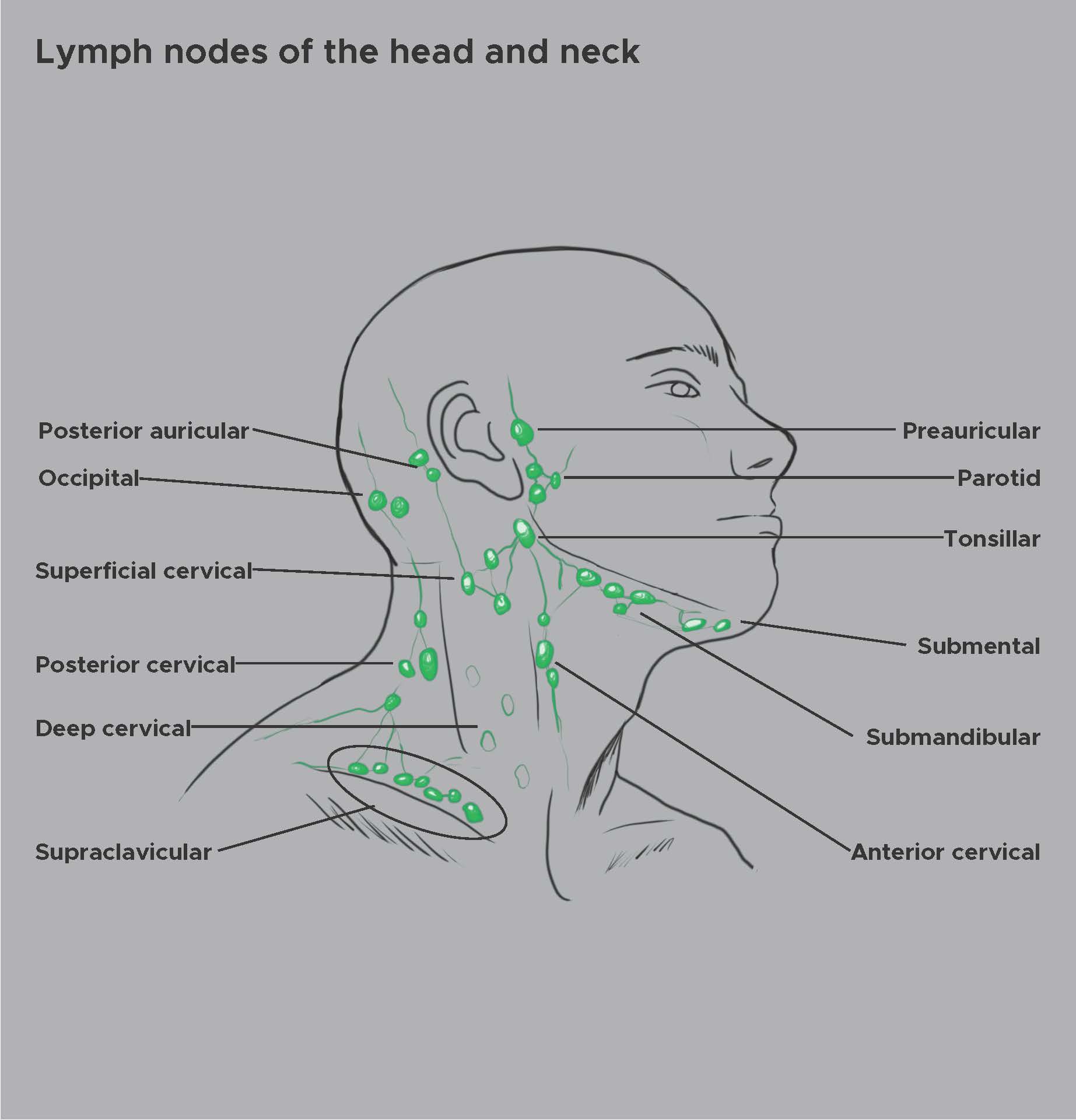
Lymph Nodes of the Head and Neck. The illustration shows major lymph node groups of the head and neck, labeled in anatomical context. Nodes are indicated as green ovals, with regional names identifying their location.
Contributed by Chelsea Rowe
(Click Image to Enlarge)
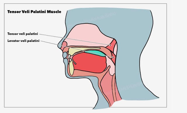
Muscles Around the Pharyngotympanic Tube. This illustration shows the tensor veli palatini and levator veli palatini, which assist in opening the pharyngotympanic tube, also known as the eustachian or auditory tube.
Illustration by E Gregory
References
Luengrungrus K, Thanawirattananit P, Teeramatwanich W. Normative Data of Extended High Frequency Audiometry in Normal Hearing Subjects with Different Aged Groups. Audiology research. 2024 Dec 9:14(6):1084-1092. doi: 10.3390/audiolres14060089. Epub 2024 Dec 9 [PubMed PMID: 39727612]
Zhao B, Müller U. The elusive mechanotransduction machinery of hair cells. Current opinion in neurobiology. 2015 Oct:34():172-9. doi: 10.1016/j.conb.2015.08.006. Epub 2015 Sep 3 [PubMed PMID: 26342686]
Level 3 (low-level) evidenceDelacroix L, Malgrange B. Cochlear afferent innervation development. Hearing research. 2015 Dec:330(Pt B):157-69. doi: 10.1016/j.heares.2015.07.015. Epub 2015 Jul 29 [PubMed PMID: 26231304]
Mulders WH, Robertson D. Evidence for direct cortical innervation of medial olivocochlear neurones in rats. Hearing research. 2000 Jun:144(1-2):65-72 [PubMed PMID: 10831866]
Level 3 (low-level) evidenceLynch KM, Bodison SC, Cabeen RP, Toga AW, Voelker CCJ. The Spatial Organization of Ascending Auditory Pathway Microstructural Maturation From Infancy Through Adolescence Using a Novel Fiber Tracking Approach. Human brain mapping. 2024 Dec 15:45(18):e70091. doi: 10.1002/hbm.70091. Epub [PubMed PMID: 39676439]
Felix RA 2nd, Gourévitch B, Portfors CV. Subcortical pathways: Towards a better understanding of auditory disorders. Hearing research. 2018 May:362():48-60. doi: 10.1016/j.heares.2018.01.008. Epub 2018 Jan 31 [PubMed PMID: 29395615]
Level 3 (low-level) evidenceWenstrup JJ. Frequency organization and responses to complex sounds in the medial geniculate body of the mustached bat. Journal of neurophysiology. 1999 Nov:82(5):2528-44 [PubMed PMID: 10561424]
Level 3 (low-level) evidencePeterson DC, Voytenko S, Gans D, Galazyuk A, Wenstrup J. Intracellular recordings from combination-sensitive neurons in the inferior colliculus. Journal of neurophysiology. 2008 Aug:100(2):629-45. doi: 10.1152/jn.90390.2008. Epub 2008 May 21 [PubMed PMID: 18497365]
Level 3 (low-level) evidencePeterson DC, Nataraj K, Wenstrup J. Glycinergic inhibition creates a form of auditory spectral integration in nuclei of the lateral lemniscus. Journal of neurophysiology. 2009 Aug:102(2):1004-16. doi: 10.1152/jn.00040.2009. Epub 2009 Jun 10 [PubMed PMID: 19515958]
Level 3 (low-level) evidenceGans D, Sheykholeslami K, Peterson DC, Wenstrup J. Temporal features of spectral integration in the inferior colliculus: effects of stimulus duration and rise time. Journal of neurophysiology. 2009 Jul:102(1):167-80. doi: 10.1152/jn.91300.2008. Epub 2009 Apr 29 [PubMed PMID: 19403742]
Level 3 (low-level) evidenceYavuzoglu A, Schofield BR, Wenstrup JJ. Circuitry underlying spectrotemporal integration in the auditory midbrain. The Journal of neuroscience : the official journal of the Society for Neuroscience. 2011 Oct 5:31(40):14424-35. doi: 10.1523/JNEUROSCI.3529-11.2011. Epub [PubMed PMID: 21976527]
Level 3 (low-level) evidenceMarsh RA, Nataraj K, Gans D, Portfors CV, Wenstrup JJ. Auditory responses in the cochlear nucleus of awake mustached bats: precursors to spectral integration in the auditory midbrain. Journal of neurophysiology. 2006 Jan:95(1):88-105 [PubMed PMID: 16148270]
Peterson DC, Wenstrup JJ. Selectivity and persistent firing responses to social vocalizations in the basolateral amygdala. Neuroscience. 2012 Aug 16:217():154-71. doi: 10.1016/j.neuroscience.2012.04.069. Epub 2012 May 6 [PubMed PMID: 22569154]
Level 3 (low-level) evidenceCoomes DL, Schofield BR. Projections from the auditory cortex to the superior olivary complex in guinea pigs. The European journal of neuroscience. 2004 Apr:19(8):2188-200 [PubMed PMID: 15090045]
Level 3 (low-level) evidenceSchofield BR, Coomes DL. Projections from auditory cortex contact cells in the cochlear nucleus that project to the inferior colliculus. Hearing research. 2005 Aug:206(1-2):3-11 [PubMed PMID: 16080994]
Level 3 (low-level) evidenceSchofield BR, Coomes DL. Pathways from auditory cortex to the cochlear nucleus in guinea pigs. Hearing research. 2006 Jun-Jul:216-217():81-9 [PubMed PMID: 16874906]
Level 3 (low-level) evidenceSchofield BR, Coomes DL, Schofield RM. Cells in auditory cortex that project to the cochlear nucleus in guinea pigs. Journal of the Association for Research in Otolaryngology : JARO. 2006 Jun:7(2):95-109 [PubMed PMID: 16557424]
Level 3 (low-level) evidenceCoomes Peterson D, Schofield BR. Projections from auditory cortex contact ascending pathways that originate in the superior olive and inferior colliculus. Hearing research. 2007 Oct:232(1-2):67-77 [PubMed PMID: 17643879]
Level 3 (low-level) evidenceWeinberger NM. Associative representational plasticity in the auditory cortex: a synthesis of two disciplines. Learning & memory (Cold Spring Harbor, N.Y.). 2007 Jan-Feb:14(1-2):1-16 [PubMed PMID: 17202426]
Level 3 (low-level) evidenceSuga N. Role of corticofugal feedback in hearing. Journal of comparative physiology. A, Neuroethology, sensory, neural, and behavioral physiology. 2008 Feb:194(2):169-83. doi: 10.1007/s00359-007-0274-2. Epub 2008 Jan 29 [PubMed PMID: 18228080]
Level 3 (low-level) evidenceForbes CE, Grafman J. The role of the human prefrontal cortex in social cognition and moral judgment. Annual review of neuroscience. 2010:33():299-324. doi: 10.1146/annurev-neuro-060909-153230. Epub [PubMed PMID: 20350167]
Level 3 (low-level) evidenceWu C, Stefanescu RA, Martel DT, Shore SE. Listening to another sense: somatosensory integration in the auditory system. Cell and tissue research. 2015 Jul:361(1):233-50. doi: 10.1007/s00441-014-2074-7. Epub 2014 Dec 21 [PubMed PMID: 25526698]
Opoku-Baah C, Schoenhaut AM, Vassall SG, Tovar DA, Ramachandran R, Wallace MT. Visual Influences on Auditory Behavioral, Neural, and Perceptual Processes: A Review. Journal of the Association for Research in Otolaryngology : JARO. 2021 Jul:22(4):365-386. doi: 10.1007/s10162-021-00789-0. Epub 2021 May 20 [PubMed PMID: 34014416]
Price CN, Moncrieff D. Defining the Role of Attention in Hierarchical Auditory Processing. Audiology research. 2021 Mar 13:11(1):112-128. doi: 10.3390/audiolres11010012. Epub 2021 Mar 13 [PubMed PMID: 33805600]
Zhang L, Wang J, Sun H, Feng G, Gao Z. Interactions between the hippocampus and the auditory pathway. Neurobiology of learning and memory. 2022 Mar:189():107589. doi: 10.1016/j.nlm.2022.107589. Epub 2022 Feb 3 [PubMed PMID: 35124220]
De Franceschi G, Barkat TR. Task-induced modulations of neuronal activity along the auditory pathway. Cell reports. 2021 Dec 14:37(11):110115. doi: 10.1016/j.celrep.2021.110115. Epub [PubMed PMID: 34910908]
Litovsky R. Development of the auditory system. Handbook of clinical neurology. 2015:129():55-72. doi: 10.1016/B978-0-444-62630-1.00003-2. Epub [PubMed PMID: 25726262]
Level 3 (low-level) evidenceMei X, Atturo F, Wadin K, Larsson S, Agrawal S, Ladak HM, Li H, Rask-Andersen H. Human inner ear blood supply revisited: the Uppsala collection of temporal bone-an international resource of education and collaboration. Upsala journal of medical sciences. 2018 Sep:123(3):131-142. doi: 10.1080/03009734.2018.1492654. Epub 2018 Sep 11 [PubMed PMID: 30204028]
Pan WR, le Roux CM, Levy SM, Briggs CA. Lymphatic drainage of the external ear. Head & neck. 2011 Jan:33(1):60-4. doi: 10.1002/hed.21395. Epub [PubMed PMID: 20848416]
Lim DJ, Hussl B. Macromolecular transport by the middle ear and its lymphatic system. Acta oto-laryngologica. 1975 Jul-Aug:80(1-2):19-31 [PubMed PMID: 1166775]
Level 3 (low-level) evidenceSalt AN, Hirose K. Communication pathways to and from the inner ear and their contributions to drug delivery. Hearing research. 2018 May:362():25-37. doi: 10.1016/j.heares.2017.12.010. Epub 2017 Dec 19 [PubMed PMID: 29277248]
Sennaroğlu L, Bajin MD. Classification and Current Management of Inner Ear Malformations. Balkan medical journal. 2017 Sep 29:34(5):397-411. doi: 10.4274/balkanmedj.2017.0367. Epub 2017 Aug 25 [PubMed PMID: 28840850]
Hoberman A, Preciado D, Paradise JL, Chi DH, Haralam M, Block SL, Kearney DH, Bhatnagar S, Muñiz Pujalt GB, Shope TR, Martin JM, Felten DE, Kurs-Lasky M, Liu H, Yahner K, Jeong JH, Cohen NL, Czervionke B, Nagg JP, Dohar JE, Shaikh N. Tympanostomy Tubes or Medical Management for Recurrent Acute Otitis Media. The New England journal of medicine. 2021 May 13:384(19):1789-1799. doi: 10.1056/NEJMoa2027278. Epub [PubMed PMID: 33979487]
Noorbakhsh KA, Liu H, Kurs-Lasky M, Smith KJ, Hoberman A, Shaikh N. Cost-Effectiveness of Management Strategies in Recurrent Acute Otitis Media. The Journal of pediatrics. 2023 May:256():11-17.e2. doi: 10.1016/j.jpeds.2022.11.032. Epub 2022 Dec 5 [PubMed PMID: 36470464]
Gugel I, Grimm F, Teuber C, Kluwe L, Mautner VF, Tatagiba M, Schuhmann MU. Management of NF2-associated vestibular schwannomas in children and young adults: influence of surgery and clinical factors on tumor volume and growth rate. Journal of neurosurgery. Pediatrics. 2019 Nov 1:24(5):584-592. doi: 10.3171/2019.6.PEDS1947. Epub 2019 Aug 23 [PubMed PMID: 31443078]
Obholzer R. Clinical evaluation and investigation of vestibular schwannoma in NF2-related schwannomatosis. Handbook of clinical neurology. 2025:212():93-96. doi: 10.1016/B978-0-12-824534-7.00029-9. Epub [PubMed PMID: 41052869]
Colletti V, Shannon RV, Carner M, Veronese S, Colletti L. Complications in auditory brainstem implant surgery in adults and children. Otology & neurotology : official publication of the American Otological Society, American Neurotology Society [and] European Academy of Otology and Neurotology. 2010 Jun:31(4):558-64. doi: 10.1097/MAO.0b013e3181db7055. Epub [PubMed PMID: 20393378]
Briggs RJ, Fabinyi G, Kaye AH. Current management of acoustic neuromas: review of surgical approaches and outcomes. Journal of clinical neuroscience : official journal of the Neurosurgical Society of Australasia. 2000 Nov:7(6):521-6 [PubMed PMID: 11029233]
Level 2 (mid-level) evidenceGreene J, Al-Dhahir MA. Acoustic Neuroma(Archived). StatPearls. 2026 Jan:(): [PubMed PMID: 29262098]
Ruiz-García C, Lassaletta L, López-Larrubia P, Varela-Nieto I, Murillo-Cuesta S. Tumors of the nervous system and hearing loss: Beyond vestibular schwannomas. Hearing research. 2024 Jun:447():109012. doi: 10.1016/j.heares.2024.109012. Epub 2024 Apr 21 [PubMed PMID: 38703433]
Møller AR. The role of neural plasticity in tinnitus. Progress in brain research. 2007:166():37-45 [PubMed PMID: 17956769]
Level 3 (low-level) evidenceCrocetti A, Forti S, Del Bo L. Neurofeedback for subjective tinnitus patients. Auris, nasus, larynx. 2011 Dec:38(6):735-8. doi: 10.1016/j.anl.2011.02.003. Epub 2011 May 17 [PubMed PMID: 21592701]
Weise C, Heinecke K, Rief W. Biofeedback-based behavioral treatment for chronic tinnitus: results of a randomized controlled trial. Journal of consulting and clinical psychology. 2008 Dec:76(6):1046-57. doi: 10.1037/a0013811. Epub [PubMed PMID: 19045972]
Level 1 (high-level) evidenceLauer AM, Jimenez SV, Delano PH. Olivocochlear efferent effects on perception and behavior. Hearing research. 2022 Jun:419():108207. doi: 10.1016/j.heares.2021.108207. Epub 2021 Feb 18 [PubMed PMID: 33674070]
Buizza C, Franco E, Ghilardi A, Cela H. A Brief Biofeedback Training, Integrated with Breathing and Relaxation Exercises, in Treating Tinnitus Disorders within Routine Medical Care. Applied psychophysiology and biofeedback. 2025 Sep:50(3):453-464. doi: 10.1007/s10484-025-09694-1. Epub 2025 Feb 6 [PubMed PMID: 39913019]
Ma X, Wang F, Zhang C, Shen W, Yang S. Self-Directed Neurofeedback Treatment for Subjective Tinnitus Patients Evaluated by Multimodal Functional Imaging. Neural plasticity. 2022:2022():5114721. doi: 10.1155/2022/5114721. Epub 2022 Oct 18 [PubMed PMID: 36304208]
Handscomb L. Use of bedside sound generators by patients with tinnitus-related sleeping difficulty: which sounds are preferred and why? Acta oto-laryngologica. Supplementum. 2006 Dec:(556):59-63 [PubMed PMID: 17114145]
Kubota Y, Takahashi K, Nonomura Y, Yamagishi T, Ohshima S, Izumi S, Morita Y, Aizawa N, Horii A. Effects of sound source localization of masking sound on perception level of simulated tinnitus. Scientific reports. 2022 Jan 27:12(1):1452. doi: 10.1038/s41598-022-05535-x. Epub 2022 Jan 27 [PubMed PMID: 35087148]
Manuelli M, Migliorelli A, Moretti C, Borin M, Malagutti N, Bianchini C, Pelucchi S, Stomeo F, Ciorba A. Bone-conduction Hearing Aids: A Scoping Review. Indian journal of otolaryngology and head and neck surgery : official publication of the Association of Otolaryngologists of India. 2024 Dec:76(6):5071-5079. doi: 10.1007/s12070-024-05042-7. Epub 2024 Sep 11 [PubMed PMID: 39559027]
Level 2 (mid-level) evidenceHamed N, Alajmi N, Alkoblan FI, Alghtani YA, Abdelsamad Y, Alhussien A, Alhajress RI, Alhabib SF. The Chronological Evolution of Cochlear Implant Contraindications: A Comprehensive Review. Journal of clinical medicine. 2024 Apr 18:13(8):. doi: 10.3390/jcm13082337. Epub 2024 Apr 18 [PubMed PMID: 38673610]
Huizing EH. The early descriptions of the so-called tuning-fork tests of Weber, Rinne, Schwabach, and Bing. II. The "Rhine Test" and its first description by Polansky. ORL; journal for oto-rhino-laryngology and its related specialties. 1975:37(2):88-91 [PubMed PMID: 1093081]
Greywoode JD, Pribitkin EA, Krein H. Management of auricular hematoma and the cauliflower ear. Facial plastic surgery : FPS. 2010 Dec:26(6):451-5. doi: 10.1055/s-0030-1267719. Epub 2010 Nov 17 [PubMed PMID: 21086231]
Fireman P. Otitis media and eustachian tube dysfunction: connection to allergic rhinitis. The Journal of allergy and clinical immunology. 1997 Feb:99(2):S787-97 [PubMed PMID: 9042072]
Norrix LW, Velenovsky D. Clinicians' Guide to Obtaining a Valid Auditory Brainstem Response to Determine Hearing Status: Signal, Noise, and Cross-Checks. American journal of audiology. 2018 Mar 8:27(1):25-36. doi: 10.1044/2017_AJA-17-0074. Epub [PubMed PMID: 29392291]
Kumar S, Dutta A, Natraj R, Kumar MS, Gupta M. A Comparative Analysis of Click ABR and Multi-ASSR in Assessing Infant Hearing: A Cross-Sectional Study. Indian journal of otolaryngology and head and neck surgery : official publication of the Association of Otolaryngologists of India. 2024 Aug:76(4):3176-3182. doi: 10.1007/s12070-024-04639-2. Epub 2024 Mar 16 [PubMed PMID: 39130223]
Level 2 (mid-level) evidence