 Tools
Tools
 Atrophoderma of Pasini and Pierini
Introduction
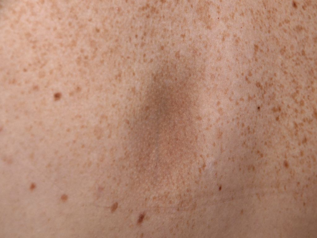
Atrophoderma of Pasini and Pierini is a rare cutaneous disorder characterized by dermal atrophy. Clinically, APP presents as single or multiple sharply demarcated, hyperpigmented, nonindurated patches with no evident signs of inflammation (see Image. Atrophoderma Pierini Pasini). Pasini first described the condition in 1923 as progressive idiopathic atrophoderma. In 1936, Pierini and Vivoli further characterized the condition and suggested a possible association with morphea.
Etiology
Register For Free And Read The Full Article
 Search engine and full access to all medical articles
- 10 free questions in your specialty
- Free CME/CE Activities
- Free daily question in your email
- Save favorite articles to your dashboard
- Emails offering discounts
Learn more about a Subscription to StatPearls Point-of-Care
Etiology
The cause of APPi remains unknown. Some authors have linked it to infection with Borrelia burgdorferi.[1] Buechner and Rufi studied sera from 26 patients with typical lesions; 53% had positive IgG antibodies against B burgdorferi compared with 14% of control subjects. No IgM antibodies were detected. Familial cases of APP have been described, but a genetic association has not been confirmed.[1] Although definitive evidence is lacking, a neurogenic cause has been suggested because of the apparent zosteriform distribution of lesions in some cases.[2] There is growing evidence that APP represents a manifestation within the spectrum of localized scleroderma.[3][4]
Several systems have been proposed to classify the clinical types of morphea. The first, suggested in 1995 by Paterson, classifies morphea into 5 subtypes: plaque, generalized, bullous, linear, and deep. Atrophoderma of Pasini and Pierini was included within the plaque-type spectrum, along with morphea en plaque, guttate morphea, keloidal morphea, and lichen sclerosus et atrophicus. Generalized morphea is defined as involving 2 or more body areas. Bullous and linear morphea include linear morphea of the extremities, morphea en coup de sabre, and progressive hemiatrophy. Deep morphea encompasses morphea profunda, subcutaneous morphea, eosinophilic fasciitis, and pansclerotic morphea.
Cases of morphea with a burnt appearance may be atrophic and histologically similar to APP. Plaques of morphea and atrophoderma lesions may occur in the same patient, with atrophic patches appearing in previously normal skin. One case report described a patient with typical APP who developed progressive systemic sclerosis.[5]
Epidemiology
Atrophoderma of Pasini and Pierini is a rare condition, with fewer than 100 cases reported in the literature. It most commonly presents in the second or third decades of life, although onset has been described in both younger and older patients. Congenital cases have also been reported.[2] Atrophoderma of Pasini and Pierini shows a marked female predominance, with a female-to-male ratio of approximately 6:1. It is most commonly reported in White individuals of European descent.
Histopathology
Microscopic findings in APP are generally subtle. In many cases, the epidermis appears normal, although mild atrophy with flattened rete ridges may be observed. The most consistent finding is a reduction in dermal thickness. Collagen changes can include atrophy, sclerosis, fragmentation, and hyalinization.[6] Elastic fiber alterations are variable, and reductions and fragmentation resembling those seen in anetoderma have been reported.[5] Dilatation of superficial blood vessels may be present, often accompanied by a sparse perivascular round-cell inflammatory infiltrate. Sweat glands, pilosebaceous units, and other appendages typically appear normal.
History and Physical
From a clinical perspective, APP is typically asymptomatic and progresses insidiously. Lesions most often first appear on the trunk, particularly the back, and may extend to the chest, arms, and abdomen. Rarely, they may be associated with pain, pruritus, or paresthesia.
The condition is characterized by single or multiple irregular round patches that vary in size from a few millimeters to several centimeters.[3] Lesions are slightly depressed relative to the surrounding skin, with sharply demarcated, cliff-drop–like borders. Unlike morphea, an erythematous or lilac-ring border is absent. Lesions are usually hyperpigmented; however, Saleh et al reported in a retrospective study of 16 Lebanese patients that 56% of lesions were hypopigmented and 25% were flesh-colored.[5]
A lack of inflammatory features is typical. Lesions do not exhibit sclerosis or induration on palpation, and the surrounding skin appears normal. Lesions are often bilateral, though unilateral zosteriform or Blaschkoid patterns have been documented.[3]
Evaluation
Evaluation of APP may include serologic testing, imaging, dermatoscopy, and histopathology in select cases. An enzyme-linked immunosorbent assay can be performed to detect anti–B burgdorferi antibodies. Thickness of the lesions and the surrounding skin can be assessed with magnetic resonance imaging or 13-MHz B-mode ultrasonography to document dermal atrophy, although such imaging is often unnecessary. Dermoscopy is not routinely performed; however, a recent case report of a 50-year-old patient demonstrated a violaceous pigmented network sparing the perifollicular region, predominant perifollicular brownish pigmentation, and white dots.[7][8]
Histopathologic changes are generally minimal, with reduced dermal volume relative to normal skin. Additional findings may include varying degrees of collagen homogenization and clumping in the mid and reticular dermis, interstitial edema, and a mild perivascular infiltrate of lymphocytes and histiocytes. Sweat glands, pilosebaceous units, and other appendages are typically normal. The overlying epidermis is usually unremarkable but may exhibit mild atrophy with flattened rete ridges.
Treatment / Management
No treatment has been consistently effective for APP. Patients with a positive B burgdorferi antibody titer sometimes respond to tetracyclines. Doxycycline (200 mg/d) has been reported to prevent the appearance of new lesions; however, the overall efficacy of antibiotics in seropositive patients remains inconclusive. In a retrospective study of 25 patients treated with either oral penicillin (2 million IU/d) or oral tetracycline (500 mg three times daily) for 2 to 3 weeks, 20 patients showed clinical improvement. Notably, 4 of 6 untreated patients in the same study did not develop progressive disease.[9] (B3)
Other therapeutic approaches include topical corticosteroids and antimalarials such as hydroxychloroquine, which may be beneficial in cases associated with lupus. Topical calcineurin inhibitors have also been used, though responses are variable.[10] Laser therapy with a Q-switched alexandrite laser has been reported to improve hyperpigmented lesions.[11] A recent case report described a patient with long-standing APP who responded positively to treatment with methylprednisolone acetate, representing a novel therapeutic approach in this condition.[12](B3)
Differential Diagnosis
Atrophoderma of Pasini and Pierini closely resembles several other conditions and must be distinguished from atrophoderma of Moulin, anetoderma, lichen sclerosus, morphea, and postinflammatory hyperpigmentation.
Prognosis
Atrophoderma of Pasini and Pierini typically progresses over 10 to 20 years. Eventually, disease activity stabilizes, but existing lesions generally do not resolve. Progression to systemic sclerosis is extremely rare. One reported case by Bisaccia et al in 1982 described a 42-year-old woman with typical lesions on the back who subsequently developed progressive systemic sclerosis, including Raynaud phenomenon, sclerodactyly, and pulmonary fibrosis.[13]
Complications
Complications of APP are primarily cosmetic and psychological, as the condition is benign and nonprogressive. Persistent depressed, hyperpigmented patches can cause noticeable disfigurement, leading to reduced self-esteem and psychosocial distress, particularly in young patients.[14] Residual dyspigmentation and textural changes often persist long-term. Rarely, overlap with morphea may occur, suggesting potential progression to localized sclerosis. There is no risk of malignancy or systemic involvement; however, misdiagnosis may result in unnecessary interventions.[15]
Deterrence and Patient Education
Patient education and deterrence in APP focus on providing reassurance, avoiding unnecessary interventions, and promoting long-term skin care. Patients should be counseled that the condition is benign, noncontagious, and typically nonprogressive, which can help alleviate anxiety and prevent overtreatment. Emphasizing the chronic, yet stable, nature of lesions is essential for setting realistic expectations, as complete resolution is uncommon.
Patients should avoid trauma, excessive friction, and unproven topical agents that may worsen dyspigmentation. Photoprotection is recommended to minimize contrast between affected and normal skin. Although no definitive preventive measures exist, early recognition may help distinguish the condition from morphea and prevent aggressive therapies.
Psychological support may be beneficial for individuals with cosmetic concerns. Discussion of limited treatment efficacy and emphasis on cosmetic camouflage strategies can improve patient satisfaction. Routine follow-up is generally not required but may be considered in atypical cases to monitor for rare overlap with localized scleroderma.
Enhancing Healthcare Team Outcomes
Atrophoderma of Pasini and Pierini is a rare disorder that primary care providers and nurse practitioners are unlikely to encounter. When patients present with unknown skin lesions, referral to a dermatologist for definitive diagnosis is recommended, followed by coordinated follow-up within the interprofessional team. The condition shows a marked female predominance, with a female-to-male ratio of 6:1, and is more frequently reported in White individuals of European descent. While the disorder may progress in some individuals, little is known about its long-term course.
Management of APP benefits from an interprofessional approach. Pharmacists can support medication reconciliation and treatment planning, particularly if topical corticosteroids, antimalarials, or antibiotics are prescribed. Pharmacists also play a role in patient education and monitoring adherence. Clinicians should work with dermatology specialty nurses to provide patient and family education, ensure regular monitoring, and coordinate follow-up visits. Any concerning findings should be communicated promptly to the interprofessional team. Optimal outcomes are achieved through a coordinated strategy involving treatment, education, and ongoing monitoring.
Media
(Click Image to Enlarge)
References
González-Morán A, Martín-López R, Ramos ML, Román C, González-Asensio MP. [Idiopathic atrophoderma of Pasini and Pierini. Study of 4 cases]. Actas dermo-sifiliograficas. 2005 Jun:96(5):303-6 [PubMed PMID: 16476392]
Level 3 (low-level) evidenceBassi A, Remaschi G, Difonzo EM, Greco A, Buccoliero AM, Giani T, Facchini F, Cimaz R, de Martino M. Idiopathic congenital atrophoderma of Pasini and Pierini. Archives of disease in childhood. 2015 Dec:100(12):1184. doi: 10.1136/archdischild-2015-309498. Epub 2015 Sep 15 [PubMed PMID: 26374755]
Arif T, Adil M, Amin SS, Ahmed M. Atrophoderma of Pasini and Pierini in a blaschkoid pattern. Journal der Deutschen Dermatologischen Gesellschaft = Journal of the German Society of Dermatology : JDDG. 2017 Jun:15(6):663-664. doi: 10.1111/ddg.13198. Epub 2017 Mar 27 [PubMed PMID: 28346752]
Ling X, Shi X. Idiopathic atrophoderma of Pasini-Pierini associated with morphea: the same disease spectrum? Giornale italiano di dermatologia e venereologia : organo ufficiale, Societa italiana di dermatologia e sifilografia. 2016 Feb:151(1):127-8 [PubMed PMID: 26373865]
Saleh Z, Abbas O, Dahdah MJ, Kibbi AG, Zaynoun S, Ghosn S. Atrophoderma of Pasini and Pierini: a clinical and histopathological study. Journal of cutaneous pathology. 2008 Dec:35(12):1108-14. doi: 10.1111/j.1600-0560.2008.00986.x. Epub 2008 Jun 18 [PubMed PMID: 18616761]
Level 2 (mid-level) evidenceVieira-Damiani G, Lage D, Christofoletti Daldon PÉ, Tibúrcio Alves CR, Cintra ML, Metze K, Adur J, Pelegati VB, Carvalho HF, Cesar CL. Idiopathic atrophoderma of Pasini and Pierini: A case study of collagen and elastin texture by multiphoton microscopy. Journal of the American Academy of Dermatology. 2017 Nov:77(5):930-937. doi: 10.1016/j.jaad.2017.02.044. Epub 2017 Apr 25 [PubMed PMID: 28389037]
Level 3 (low-level) evidenceJaiswal S, Uprety S, Thapa P, Lamichhane P. Dermatoscopy of atrophoderma of Pasini and Pierini: A case-based insight. JAAD case reports. 2025 Nov:65():89-91. doi: 10.1016/j.jdcr.2025.08.021. Epub 2025 Sep 2 [PubMed PMID: 41078424]
Level 3 (low-level) evidenceSong CX, Zhang YT, Tan C. Dermoscopic and Reflectance Confocal Microscopy Features of Superficial Morphea on Preexisting Atrophoderma of Pasini and Pierini. Dermatology practical & conceptual. 2022 May:12(2):e2022048. doi: 10.5826/dpc.1202a48. Epub 2022 Apr 1 [PubMed PMID: 35646456]
Buechner SA, Rufli T. Atrophoderma of Pasini and Pierini. Clinical and histopathologic findings and antibodies to Borrelia burgdorferi in thirty-four patients. Journal of the American Academy of Dermatology. 1994 Mar:30(3):441-6 [PubMed PMID: 8113457]
Level 3 (low-level) evidencePope E, Laxer RM. Diagnosis and management of morphea and lichen sclerosus and atrophicus in children. Pediatric clinics of North America. 2014 Apr:61(2):309-19. doi: 10.1016/j.pcl.2013.11.006. Epub 2013 Dec 27 [PubMed PMID: 24636648]
Arpey CJ, Patel DS, Stone MS, Qiang-Shao J, Moore KC. Treatment of atrophoderma of Pasini and Pierini-associated hyperpigmentation with the Q-switched alexandrite laser: a clinical, histologic, and ultrastructural appraisal. Lasers in surgery and medicine. 2000:27(3):206-12 [PubMed PMID: 11013382]
Level 3 (low-level) evidenceCeballos-Pérez M, Gutierrez Villarreal IM, Cano Lizarraga IE, Gómez López DE, Solís Ramírez MY, Cantu G, Ancona Castro C. Idiopathic Atrophoderma of Pasini and Pierini: Response to Mycophenolic Acid Treatment. Cureus. 2025 Jan:17(1):e77439. doi: 10.7759/cureus.77439. Epub 2025 Jan 14 [PubMed PMID: 39949423]
Jablonska S, Blaszczyk M. Scleroderma overlap syndromes. Advances in experimental medicine and biology. 1999:455():85-92 [PubMed PMID: 10599327]
Level 3 (low-level) evidenceHubail R, Karaidi N, Al Awadhi A. Idiopathic Atrophoderma of Pasini and Pierini: A Case Report and Literature Review. Cureus. 2022 Jul:14(7):e26571. doi: 10.7759/cureus.26571. Epub 2022 Jul 5 [PubMed PMID: 35936167]
Level 3 (low-level) evidencePathak R, Neupane P, Shrestha S. A Clinical Case of Idiopathic Atrophoderma of Pasini and Pierini With Literature Review. Case reports in dermatological medicine. 2025:2025():8886954. doi: 10.1155/crdm/8886954. Epub 2025 Mar 14 [PubMed PMID: 40123740]
Level 3 (low-level) evidence