 Tools
Tools
 Ascites
Introduction
Ascites is the pathological accumulation of fluid within the peritoneal cavity, most frequently associated with liver disease (especially cirrhosis), heart disease, kidney disease, pancreatic disease, or chylous ascites, in which lymph fluid collects in the peritoneal space. Conversely, in most healthy individuals, very little free intraperitoneal fluid is present. Some women may have approximately 20 mL of free intraperitoneal fluid during the menstrual cycle; however, healthy men have negligible amounts.
Etiology
Register For Free And Read The Full Article
 Search engine and full access to all medical articles
- 10 free questions in your specialty
- Free CME/CE Activities
- Free daily question in your email
- Save favorite articles to your dashboard
- Emails offering discounts
Learn more about a Subscription to StatPearls Point-of-Care
Etiology
Ascites represents the pathological accumulation of fluid within the peritoneal cavity and reflects a complex interplay of hepatic, cardiac, renal, infectious, and malignant processes. The condition often signifies advanced disease and contributes to substantial morbidity and mortality. Understanding its diverse etiologies is essential for accurate diagnosis, effective management, and prevention of complications.
Cirrhosis
Ascites develops as a consequence of multiple underlying conditions, with cirrhosis representing the predominant cause in the United States and accounting for approximately 80% of cases. Among patients with cirrhosis, ascites represents the most frequent complication and affects nearly 50% of those who have experienced decompensated cirrhosis for more than 10 years.[1][2][3] The onset of ascites often marks the transition from compensated to decompensated cirrhosis. Mortality associated with cirrhotic ascites rises due to complications, eg, spontaneous bacterial peritonitis and hepatorenal syndrome, reaching 15% at 1 year and 44% at 5 years.
Alcohol-related liver disease, metabolic dysfunction-associated steatotic liver disease (MASLD), and chronic viral hepatitis contribute most often to cirrhosis complicated by ascites. Cirrhotic ascites occurs more frequently in individuals younger than 65 years, in those with alcohol-related cirrhosis, or with hepatic encephalopathy and varices.[4] The risk increases among patients taking nonselective beta-blockers but decreases with atorvastatin use or antiviral therapy for hepatitis C.[5] Up to 19% of cirrhotic patients develop hemorrhagic ascites, most often spontaneously, with 72% attributed to bloody lymph and 13% to hepatocellular carcinoma, though paracentesis may also trigger its development.[6][5]
Chylous Ascites
Chylous ascites, a rare variant characterized by its milky appearance and high triglyceride content, results from lymphatic fluid leakage into the peritoneal cavity. In children, lymphatic malformations represent the most frequent cause, while in adults, the condition is more commonly associated with cirrhosis and intra-abdominal malignancy in developed nations.[7]
Infections, eg, tuberculosis and filariasis, predominate as causes in developing and Eastern countries. Chylous ascites often arises from trauma, rupture of lymphatic channels secondary to obstruction, or elevated peritoneal lymphatic pressure from inflammation. Postoperative cases, as well as congenital or acquired forms, frequently accompany portal hypertension or traumatic injury.
Pancreatic Ascites
Pancreatic ascites may develop through iatrogenic mechanisms following procedures, eg, endoscopic retrograde cholangiopancreatography, renal or adrenal surgery, colectomy with splenic flexure mobilization, partial pancreatic resection, or splenectomy. External pancreatic fistulas, typically after surgery, may arise from anterior ductal disruptions leading to peripancreatic fluid collections in the lesser sac or from posterior ductal disruptions resulting in pancreatic-pleural fistula formation and pleural effusions. Noniatrogenic causes include chronic pancreatitis, persistent pancreatic fistulas to the peritoneum, pseudocysts, or walled-off necrosis.[8][9]
Additional Etiologies
Peritoneal cancer accounts for approximately 10% of ascites cases, most often resulting from breast, ovarian, bronchial, gastric, colon, or pancreatic malignancies, while 20% originate from cancers of unknown primary sites.[10] Congestive heart failure, usually linked to pulmonary hypertension, and constrictive pericarditis cause about 3% of cases. Tuberculosis accounts for 2%, nephrotic syndrome or dialysis for 1%, and pancreatic ascites for another 1%.
Other etiologies, accounting for approximately 2% of cases, include noncirrhotic alcohol-related liver disease, portal vein thrombosis, Budd-Chiari syndrome, lymphoma, other peritoneal malignancies, intravenous drug use, obesity, inferior vena cava obstruction or web formation, hypercholesterolemia, type 2 diabetes, severe malnutrition, infections, and ovarian lesions.
Epidemiology
Ascites affects approximately 60,000 per 100,000 individuals worldwide and occurs across all ages, sexes, and races. However, cirrhotic ascites demonstrates distinct epidemiologic trends. The male-to-female ratio reaches 2.5:1, reflecting a higher prevalence among men.
Mexican-Americans and Black populations experience a greater incidence, primarily due to the underlying distribution of cirrhosis. Individuals with lower education levels and those living below the poverty line also face a higher risk, highlighting socioeconomic influences on disease development and access to preventive care.[11]
Pathophysiology
The first stage in the development of cirrhotic ascites is portal hypertension. Portal pressure increases above a critical threshold, and circulating nitric oxide levels increase, leading to splanchnic arterial vasodilatation. As vasodilatation progresses, reduced effective arterial blood volume, increased permeability, and elevated pressure in splanchnic capillaries cause sodium retention through the induction of the renin-angiotensin-aldosterone system, arginine vasopressin secretion, and the sympathetic nervous system.[12] Increased lymph production, exceeding lymph reabsorption, also contributes to the accumulation of ascitic fluid. Portal hypertension also permits bacterial translocation, leading to increased pathogen-associated molecular patterns (PAMPs) and a consequent increased risk of infection, particularly spontaneous bacterial peritonitis.[13]
Additionally, by producing proteinaceous fluid, tumor cells lining the peritoneum can also cause ascites. In high-output or low-output heart failure or nephrotic syndrome, effective arterial blood volume is decreased, and the vasopressin, renin-angiotensin-aldosterone, and sympathetic nervous systems are activated, leading to renal vasoconstriction and, ultimately, sodium and water retention.[14]
Pancreatic ascites results from leakage from a pseudocyst or walled-off necrosis into the peritoneum through pancreatic duct disruption.[14] Alternatively, pancreatic duct disruption without a pseudocyst leads to the formation of a fistulous tract. Fistulas from an anterior pancreatic duct disruption cause secretions to empty directly into the peritoneum. Posterior pancreatic duct ruptures cause fistula formation through the aortic or esophageal hiatus and, possibly, through the diaphragmatic dome, leading to pleural effusions.[15]
In cases of chylous ascites, traumatic rupture of lymphatic vessels or excessive peritoneal lymphatic pressure due to obstruction can cause lymph to collect in the peritoneal space. In portal hypertension, endothelial compromise can occur due to elevated lymphatic pressure, leading to dilation of lymphatic channels, which can rupture and cause chylous ascites. In nonportal instances, lymph passes through a retroperitoneal fistula due to congenital or acquired megalymphatics. When the amount of fluid exceeds the intestinal mucosa's capacity to reabsorb it, chylous ascites and a protein-losing enteropathy can develop, manifested by malabsorption, malnutrition, and steatorrhea.[15]
History and Physical
Clinical History
Patients typically report progressive abdominal distension, which may be painless or associated with abdominal discomfort, weight gain, early satiety, shortness of breath, and dyspnea, resulting from fluid accumulation and increased abdominal pressure. Symptoms, eg, fever, abdominal tenderness, and confusion, can be seen in spontaneous bacterial peritonitis.
Patients with malignant ascites can have symptoms related to malignancy, which may include weight loss. On the other hand, patients with ascites due to heart failure may report dyspnea, orthopnea, and peripheral edema, and those with chylous ascites report diarrhea, steatorrhea, malnutrition, edema, nausea, enlarged lymph nodes, early satiety, fevers, and night sweats.
Physical Examination Findings
Typical physical findings include abdominal distention, flank dullness to percussion, shifting dullness, a fluid wave, and evidence of pleural effusions. Other findings are related to the underlying cause of the ascites, eg, stigmata of cirrhosis, including spider angiomas, palmar erythema, abdominal wall collaterals, muscle wasting, gynecomastia, leukonychia, and jaundice. In heart failure, physical examination findings may include jugular venous distension, pulmonary congestion, or peripheral edema. A palpable umbilical nodule, eg, a Sister Mary Joseph nodule, is a metastatic lesion most often from a gastrointestinal, pancreatic, or genitourinary cancer as the cause of ascites. Other symptoms and physical findings are related to the underlying cause of ascites formation.
Evaluation
Initial Evaluation
Diagnostic abdominal paracentesis with the appropriate ascitic fluid analysis is the most rapid and cost-effective method of diagnosing the underlying cause of ascites in affected patients.[16][17] The initial tests that should be performed on the ascitic fluid include:
- Complete blood cell count: A complete blood cell count with total nucleated cell and polymorphonuclear neutrophil (PMN) count. A PMN count ≥250/mm³ indicates spontaneous bacterial peritonitis, although similar findings may occur in pancreatic ascites.
- Bacterial culture: Bacterial culture may be performed through bedside inoculation of blood culture bottles.
- Cytology: This study is performed to identify evidence of malignancy.
- Serum-ascites albumin gradient (SAAG): SAAG equals the serum albumin level minus the ascitic albumin level (g/dL) and predicts the presence or absence of portal hypertension with 97% accuracy. The traditional method of classifying ascitic fluid as transudative or exudative based on protein concentration has been largely abandoned due to its limited accuracy of 56%.[18]
- SAAG ≥1.1 g/dL: Indicative of portal hypertension and is seen in cirrhosis, alcoholic hepatitis, diffuse hepatic cancer obstructing portal vessels, Budd-Chiari syndrome (hepatic vein thrombosis), portal vein thrombosis, idiopathic portal fibrosis, congestive heart failure, and pericarditis.
- SAAG <1.1 g/dL: Indicates the patient does not have portal hypertension and occurs in peritoneal carcinomatosis, pancreatitis, serositis, nephrotic syndrome, and peritoneal tuberculosis.
Additional Evaluation Studies
Additional tests should target specific clinical suspicions. A serum amylase level greater than 1000 U/L or more than 3 times the upper limit of normal supports a diagnosis of pancreatic ascites. Mycobacterial culture is appropriate when tuberculosis is strongly suspected. Ascitic fluid lactate dehydrogenase (LDH) and glucose levels assist in identifying secondary peritonitis.
Milky-appearing ascitic fluid suggests chylous ascites. Lipoprotein electrophoresis serves as the gold standard for detecting chylomicrons, but limited laboratory availability often necessitates triglyceride testing instead. A triglyceride level greater than 200 mg/dL typically confirms chylous ascites, while recent data support a diagnostic threshold of 187 mg/dL, yielding up to 95% sensitivity and specificity.[19] SAAG values in chylous ascites vary depending on etiology. The tumor marker CA-125 in ascitic fluid is frequently elevated in cirrhotic and chylous ascites. Increased adenosine deaminase is associated with tuberculous ascites. Other indices, including lactate and pH, add minimal diagnostic value.
Imaging Studies
Ultrasound offers the highest sensitivity for detecting ascites, revealing homogenous, freely mobile anechoic fluid within the peritoneal cavity, most prominently in the Morrison pouch (see Image. Abdominal Ascites). Computed tomography (CT) can detect fluid accumulation and identify mass lesions. In pancreatic ascites, CT demonstrates intraperitoneal fluid with a density greater than 15 Hounsfield units, reflecting high protein content, and often reveals collapsed pseudocysts, parenchymal atrophy, dilated main pancreatic duct, or pancreatic calcifications. Magnetic resonance or endoscopic cholangiopancreatography can further delineate ductal disruptions.[20]
Lymphangiography and nuclear lymphoscintigraphy using 99Tc antimony sulfide colloid and dextran or albumin assist in diagnosing chylous ascites and locating lymphatic leaks or fistulas.[7] Single-photon emission computed tomography (SPECT) provides additional localization information, particularly when surgical intervention is required.[21]
Treatment / Management
Targeted Medical Therapies
Appropriate management of ascites depends on identifying and addressing the underlying cause of fluid retention. The primary therapeutic goals include reducing ascitic fluid accumulation and peripheral edema while maintaining adequate intravascular volume.[22][23][24] Sodium restriction and diuretics represent the first-line approach. In cases of high-SAAG ascites, treatment involves alcohol abstinence, dietary sodium restriction to 88 mEq (2000 mg) daily, and diuretic therapy with once-daily spironolactone and furosemide in a 100:40 mg ratio.
Patients with treatable liver diseases, eg, autoimmune hepatitis, chronic hepatitis B with reactivation, hemochromatosis, or Wilson disease, should receive targeted therapies. Although certain nonalcoholic or nonhepatitis B causes of cirrhosis may show partial reversibility, most remain progressive. By the time ascites develops, patients often benefit more from liver transplantation than from prolonged medical therapy. Low-SAAG ascites commonly results from nonovarian peritoneal carcinomatosis, where diuretics provide minimal benefit and repeated therapeutic paracentesis becomes necessary. Ovarian malignancy-associated ascites may improve with surgical debulking and chemotherapy, while lupus serositis-related ascites often responds to glucocorticoids.
Tuberculous peritonitis requires anti-tuberculous treatment, and infections caused by chlamydia or other bacteria demand appropriate antibiotic therapy. Postoperative lymphatic leaks following distal splenorenal shunt or radical lymphadenectomy may resolve spontaneously. Pancreatic ascites can often be managed medically; however, endoscopic pancreatic duct stenting may be needed to close ductal disruptions. Supportive measures, eg, nil per os status and parenteral nutrition, may suffice.
However, persistent ascites may require intermittent therapeutic paracentesis combined with diuretics and somatostatin or octreotide to reduce pancreatic exocrine secretion and promote ductal healing.[25] Please see StatPearls' companion resource, "Octreotide," for further information.
Invasive Therapies
When conservative measures fail, endoscopic retrograde cholangiopancreatography (ERCP) with transpapillary pancreatic stent placement can be performed.[25] Surgical options depend on ductal caliber and leak location and may include pseudocyst-jejunal anastomosis, distal pancreatectomy with ductal ligation, or Roux-en-Y pancreaticojejunostomy.[26]
Patients with chylous ascites unresponsive to underlying etiologic therapy may undergo therapeutic paracentesis or transjugular intrahepatic portosystemic shunt (TIPS) placement. When these approaches fail, a peritoneovenous shunt may provide symptomatic relief by diverting lymph into the venous circulation. However, such shunts carry significant risks, including electrolyte imbalance, septic shock, small-bowel obstruction, disseminated intravascular coagulation, air embolism, and shunt occlusion due to lymph viscosity.[27](B3)
Therapeutic paracentesis offers symptomatic relief for tense ascites refractory to diuretics (see Images. Paracentesis and Ascites Pocket). Albumin administration following large-volume paracentesis helps prevent hypotension and postparacentesis circulatory dysfunction (PPCD), defined as a more than 50% increase in renin activity above baseline. PPCD may lead to hyponatremia, renal impairment, or hepatorenal syndrome.[28] The American Association for the Study of Liver Diseases (AASLD) recommends albumin replacement after removal of 5 or more liters of fluid; typically, 6 to 8 grams of albumin should be replaced per liter of fluid removed.[29] However, data to confirm whether albumin should be given after removing 3 to 5 L are lacking.[30] Updated studies suggest dosing albumin based on total volume removed: 25 g for 5 to 6 L, 50 g for 7 to 10 L, and 75 g for more than 10 L.[30]
TIPS, a side-to-side portacaval shunt inserted via the internal jugular vein, is an effective treatment for carefully selected patients with refractory ascites who are intolerant of frequent paracenteses. This procedure reduces portal pressure directly. Patients with Child C liver disease, a MELD score of 18 or greater, sarcopenia, congestive heart failure, or unexplained hepatic encephalopathy face higher risks of post-TIPS complications and generally do not qualify for the procedure.[31] Individuals with Child A or B liver disease and those receiving a covered stent less than 10 mm have better outcomes.[32] Earlier TIPS placement during ascites development improves long-term results, and patients unresponsive to TIPS should be evaluated for liver transplantation.
Differential Diagnosis
Other diagnoses that should be considered when evaluating patients with ascites include:
- Acute liver failure
- Chronic liver failure/cirrhosis
- Alcoholic hepatitis
- Portal hypertension
- Hepatitis B, C, D, and E
- Advanced metabolic dysfunction-associated steathepatitis (MASH)
- Liver cancer
- Hepatic adenomas
- Biliary tract disease
- Budd-Chiari syndrome, ie, hepatic vein thrombosis (Please see StatPearls' companion resource, "Budd-Chiari Syndrome," for further information.)
- Dilated cardiomyopathy
- Nephrotic syndrome
- Hepatorenal syndrome (Please see StatPearls' companion resource, "Hepatorenal Syndrome," for further information.)
- Dilated or restrictive cardiomyopathy
- Pancreatitis (pancreatic ascites)
- Protein-losing enteropathy
- Familial Mediterranean Fever
- Tuberculosis
- Pregnancy (associated with preeclampsia and HELLP syndrome of hemolysis, elevated liver enzymes, and low platelets)
- Ovarian cysts
- Hypoalbuminemia
- Chylous ascites
Prognosis
The prognosis of patients with ascites depends on the cause and chronicity. Acute underlying disorders that respond to treatment have a more favorable prognosis than those that do not. Overall, the prognosis is much worse for patients who have decompensated cirrhosis compared to those with compensated cirrhosis. Patients with cirrhotic ascites have a 1-year mortality rate of 40% that increases to 50% at 2 years.[33] Mortality is higher in patients with hepatic encephalopathy, sarcopenia, renal dysfunction, low urinary sodium levels, and hyponatremia.[34] Patients with refractory ascites have a 1-year survival of less than 50%.
Mortality was 27.6 times greater in otherwise similar individuals, and survival in people with people was 1.08 years (interquartile range [IQR] of 0.26 to 1.75 years) and 0.38 years (IQR of 0.1 to 1.3 years) in those who required therapeutic paracentesis.[35]
Complications
Complications of Ascites
Complications of ascites include:
- Spontaneous bacterial peritonitis
- Hepatic hydrothorax
- Pleural effusion
- Acute kidney injury due to hepatorenal syndrome
- Kidney failure
- Hyponatremia
- Sarcopenia
- Cellulitis
- Abdominal wall hernias
- Refractory/tense ascites
Complications related to paracentesis treatment include:
- Infection
- Electrolyte imbalance
- Bowel perforation
- Bleeding
- Leakage of fluid through the abdominal wall
- Injury to the kidneys
Postoperative and Rehabilitation Care
Patients with ascites require long-term monitoring to assess treatment effectiveness. Body weight, electrolyte levels, BUN, creatinine, and urinary sodium levels should be monitored in all patients treated with diuretics. The goal of treatment is to minimize edema and ensure the patient's ability to perform activities of daily living. A low-sodium diet is often recommended.
Consultations
Intraprofessional team members who may be involved in the management of ascites include:
- Gastroenterologists
- Hepatologists if available
- Liver transplant hepatologists and surgeons for patients whose ascites remains refractory to all other conservative measures
- Cardiologists, nephrologists, or other specialists and subspecialists, depending on the cause of ascites
- Neurologists to assess mental status
- Radiologists to perform TIPS in patients resistant to diuretics
- A dietitian to promote and provide information about nutrition and dietary restrictions
Deterrence and Patient Education
Deterrence and patient education for ascites focus on preventing disease progression, reducing recurrence, and minimizing complications through early intervention and lifestyle modification. Hepatitis B vaccination should be advised to reduce the risk of this etiology. Additionally, patients should receive clear education on the importance of alcohol cessation, adherence to sodium restriction (≤2000 mg daily), and compliance with prescribed diuretics to maintain optimal fluid balance. Regular monitoring of weight, abdominal girth, and signs of infection or encephalopathy helps detect worsening disease early.
Clinicians should counsel patients on avoiding hepatotoxic medications and over-the-counter drugs that may exacerbate liver injury. Nutrition counseling from a dietitian supports adherence to dietary restrictions and prevents malnutrition, while pharmacists reinforce medication safety and dosing. Patients with cirrhosis or other chronic liver diseases require education on vaccination, follow-up imaging, and laboratory monitoring to identify complications such as spontaneous bacterial peritonitis or hepatorenal syndrome. Comprehensive education fosters self-management, enhances adherence, and improves long-term outcomes through active patient engagement and interprofessional coordination.
Pearls and Other Issues
Transfusion of blood products (fresh frozen plasma or platelets) routinely before paracentesis in patients with cirrhosis and coagulopathy, presumably to prevent hemorrhagic complications, is not supported by data. Contraindications to paracentesis include coagulopathy in the presence of DIC, massive ileus with bowel distension unless the procedure is image-guided to guarantee that the bowel is not entered.
Enhancing Healthcare Team Outcomes
Ascites, the pathological accumulation of fluid within the peritoneal cavity, most often results from cirrhosis but may also stem from heart, kidney, pancreatic, or other etiologies, including malignant disease. Though frequently perceived as a gastrointestinal disorder, ascites can arise from multiple organ systems, and the underlying illnesses carry significant morbidity and mortality. The development of ascites can be a harbinger of the progression from compensated to decompensated liver disease and, as such, may signal worsening hepatic function and an increased risk of complications, such as spontaneous bacterial peritonitis, hepatorenal syndrome, and porto-systemic encephalopathy. Accurate diagnosis through ascitic fluid analysis, interpretation of the serum-ascites albumin gradient, and imaging is essential to guide targeted treatment and improve outcomes. Early identification and evidence-based management reduce hospitalizations and enhance quality of life.
A streamlined, interprofessional care model is critical for effective management.[3][36] Gastroenterologists and hepatologists, along with related advanced practitioners, oversee diagnostic evaluation and therapy, while primary care clinicians can monitor systemic stability and coagulation. Nephrologists contribute by tracking renal function and providing valuable assistance in the monitoring of the effectiveness and adverse effects of diuretics. Neurologists are essential in assessing and managing neurologic changes, eg, hepatic encephalopathy, other neurologic conditions that may arise, and adverse effects of medications. Radiologists can provide guidance in the diagnostic evaluation of ascites and perform TIPS in refractory cases. Surgeons may be required for decompressive procedures or liver transplantation. Pharmacists ensure safe medication use, and nurses are essential for monitoring fluid status and weight, as well as providing patient education. Physical therapists promote safe ambulation to prevent deconditioning, while dietitians provide essential nutritional counseling. Strong interprofessional communication, coordinated care planning, and shared decision-making enhance patient-centered outcomes, reduce complications, and strengthen team performance in the management of ascites.
Media
(Click Image to Enlarge)
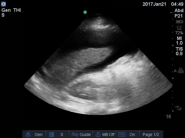
Abdominal Ascites. Ultrasound image showing abdominal ascites.
Contributed by EM Aponte, MD
(Click Image to Enlarge)
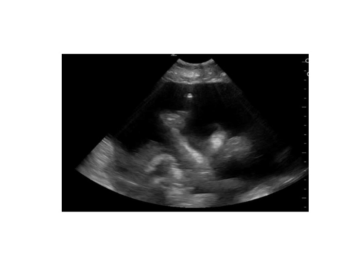
Paracentesis. Image demonstrating ascites surrounding the bowel; the bright spot indicates the position of the paracentesis needle tip.
Contributed by M Mitarai, MD
(Click Image to Enlarge)
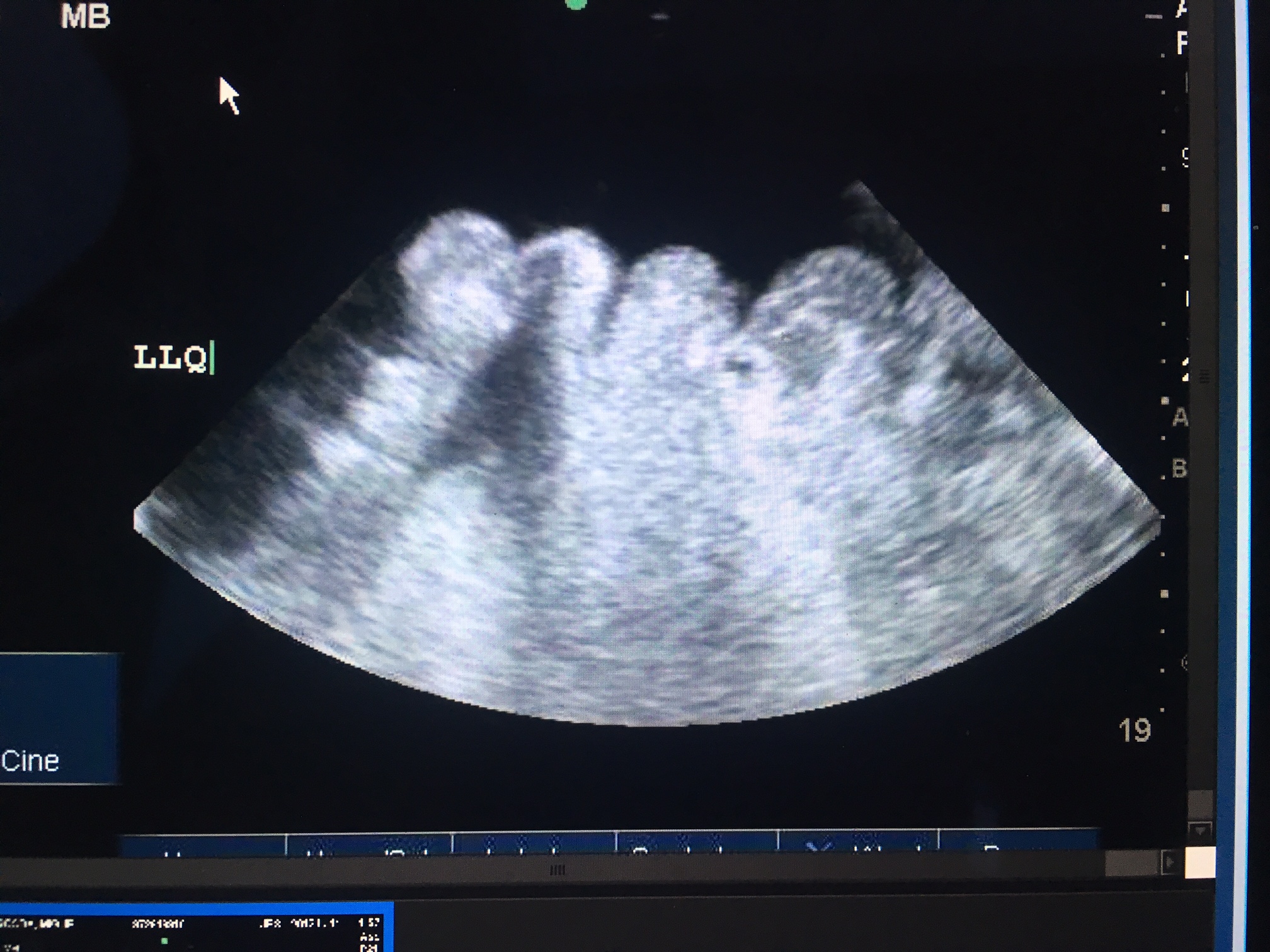
Ascites Pocket. Image demonstrating a pocket of ascites ideal for paracentesis.
Contributed by M O'Rourke, MD
References
Lee JC, Kim JS, Kim HW, Cho IK, Lee J, Jang ES, Lee SH, Hwang JH, Kim JW, Jeong SH, Kim J. Outcome of endoscopic retrograde cholangiopancreatography in patients with clinically defined decompensated liver cirrhosis. Journal of digestive diseases. 2018 Oct:19(10):605-613. doi: 10.1111/1751-2980.12661. Epub 2018 Sep 23 [PubMed PMID: 30126061]
Privitera G, Figorilli F, Jalan R, Mehta G. Portosystemic Shunt Embolization and Recurrent Ascites: A Single-Center Case Series. Gastroenterology. 2018 Nov:155(5):1649-1650. doi: 10.1053/j.gastro.2018.06.092. Epub 2018 Aug 15 [PubMed PMID: 30118744]
Level 2 (mid-level) evidenceSarin SK, Choudhury A. Management of acute-on-chronic liver failure: an algorithmic approach. Hepatology international. 2018 Sep:12(5):402-416. doi: 10.1007/s12072-018-9887-5. Epub 2018 Aug 16 [PubMed PMID: 30116993]
Tapper EB, Zhao Z, Mazumder N, Parikh ND. Incidence of, Risk Factors for, and Outcomes After Ascites in a Population-Based Cohort of Older Americans. Digestive diseases and sciences. 2022 Nov:67(11):5327-5335. doi: 10.1007/s10620-022-07454-3. Epub 2022 Mar 9 [PubMed PMID: 35262903]
Kibrit J, Khan R, Jung BH, Koppe S. Clinical Assessment and Management of Portal Hypertension. Seminars in interventional radiology. 2018 Aug:35(3):153-159. doi: 10.1055/s-0038-1660793. Epub 2018 Aug 6 [PubMed PMID: 30087517]
Conangla-Planes M, Serres X, Persiva O, Augustín S. Imaging diagnosis of portal hypertension. Radiologia. 2018 Jul-Aug:60(4):290-300. doi: 10.1016/j.rx.2017.12.010. Epub 2018 Feb 19 [PubMed PMID: 29472014]
Bhardwaj R, Vaziri H, Gautam A, Ballesteros E, Karimeddini D, Wu GY. Chylous Ascites: A Review of Pathogenesis, Diagnosis and Treatment. Journal of clinical and translational hepatology. 2018 Mar 28:6(1):105-113. doi: 10.14218/JCTH.2017.00035. Epub 2017 Dec 4 [PubMed PMID: 29577037]
Sharbidre KG, Galgano SJ, Morgan DE. Traumatic pancreatitis. Abdominal radiology (New York). 2020 May:45(5):1265-1276. doi: 10.1007/s00261-019-02241-7. Epub [PubMed PMID: 31576413]
Mehdorn AS, Schwieters AK, Mardin WA, Senninger N, Strücker B, Pascher A, Vowinkel T, Becker F. Pancreatic Fistula and Biochemical Leak after Splenectomy: Incidence and Risk Factors-A Retrospective Single-Center Analysis. Langenbeck's archives of surgery. 2022 Sep:407(6):2517-2525. doi: 10.1007/s00423-022-02531-7. Epub 2022 May 4 [PubMed PMID: 35508768]
Level 2 (mid-level) evidenceMoore CM, Van Thiel DH. Cirrhotic ascites review: Pathophysiology, diagnosis and management. World journal of hepatology. 2013 May 27:5(5):251-63. doi: 10.4254/wjh.v5.i5.251. Epub [PubMed PMID: 23717736]
Scaglione S, Kliethermes S, Cao G, Shoham D, Durazo R, Luke A, Volk ML. The Epidemiology of Cirrhosis in the United States: A Population-based Study. Journal of clinical gastroenterology. 2015 Sep:49(8):690-6. doi: 10.1097/MCG.0000000000000208. Epub [PubMed PMID: 25291348]
Bernardi M, Moreau R, Angeli P, Schnabl B, Arroyo V. Mechanisms of decompensation and organ failure in cirrhosis: From peripheral arterial vasodilation to systemic inflammation hypothesis. Journal of hepatology. 2015 Nov:63(5):1272-84. doi: 10.1016/j.jhep.2015.07.004. Epub 2015 Jul 17 [PubMed PMID: 26192220]
Jalan R, D'Amico G, Trebicka J, Moreau R, Angeli P, Arroyo V. New clinical and pathophysiological perspectives defining the trajectory of cirrhosis. Journal of hepatology. 2021 Jul:75 Suppl 1():S14-S26. doi: 10.1016/j.jhep.2021.01.018. Epub [PubMed PMID: 34039485]
Level 3 (low-level) evidenceFukui H, Kawaratani H, Kaji K, Takaya H, Yoshiji H. Management of refractory cirrhotic ascites: challenges and solutions. Hepatic medicine : evidence and research. 2018:10():55-71. doi: 10.2147/HMER.S136578. Epub 2018 Jul 3 [PubMed PMID: 30013405]
Agin M, Tumgor G, İcil S, İskit S, Alabaz D, Ballı HT. A rare cause of acute abdominal distention: opening of the pancreatic duct into hydatic cyst. Archivos argentinos de pediatria. 2016 Oct 1:114(5):e346-8. doi: 10.5546/aap.2016.eng.e346. Epub 2016 Oct 1 [PubMed PMID: 27606660]
Szkodziak PR, Czuczwar P, Wrona W, Paszkowski T, Szkodziak F, Woźniak S. Ascites Index - a novel technique to evaluate ascites in ovarian hyperstimulation syndrome: a concept-proof study. Ginekologia polska. 2018:89(4):182-8. doi: 10.5603/GP.a2018.0031. Epub [PubMed PMID: 29781072]
Long B, Koyfman A. The emergency medicine evaluation and management of the patient with cirrhosis. The American journal of emergency medicine. 2018 Apr:36(4):689-698. doi: 10.1016/j.ajem.2017.12.047. Epub 2017 Dec 23 [PubMed PMID: 29290508]
Runyon BA, Montano AA, Akriviadis EA, Antillon MR, Irving MA, McHutchison JG. The serum-ascites albumin gradient is superior to the exudate-transudate concept in the differential diagnosis of ascites. Annals of internal medicine. 1992 Aug 1:117(3):215-20 [PubMed PMID: 1616215]
Thaler MA, Bietenbeck A, Schulz C, Luppa PB. Establishment of triglyceride cut-off values to detect chylous ascites and pleural effusions. Clinical biochemistry. 2017 Feb:50(3):134-138. doi: 10.1016/j.clinbiochem.2016.10.008. Epub 2016 Oct 15 [PubMed PMID: 27750038]
Drake LM, Anis M, Lawrence C. Accuracy of magnetic resonance cholangiopancreatography in identifying pancreatic duct disruption. Journal of clinical gastroenterology. 2012 Sep:46(8):696-9. doi: 10.1097/MCG.0b013e31825003b3. Epub [PubMed PMID: 22565603]
Weiss M, Schwarz F, Wallmichrath J, Baumeister R, Frick A, Bartenstein P, Rominger A. Chylothorax and chylous ascites. Clinical utility of planar scintigraphy and tomographic imaging with SPECT/CT. Nuklearmedizin. Nuclear medicine. 2015:54(5):231-40. doi: 10.3413/Nukmed-0723-15-02. Epub 2015 Jul 17 [PubMed PMID: 26183818]
Burgos AC, Thornburg B. Transjugular Intrahepatic Portosystemic Shunt Placement for Refractory Ascites: Review and Update of the Literature. Seminars in interventional radiology. 2018 Aug:35(3):165-168. doi: 10.1055/s-0038-1661347. Epub 2018 Aug 6 [PubMed PMID: 30087519]
Adebayo D, Neong SF, Wong F. Refractory Ascites in Liver Cirrhosis. The American journal of gastroenterology. 2019 Jan:114(1):40-47. doi: 10.1038/s41395-018-0185-6. Epub [PubMed PMID: 29973706]
Storni F, Stirnimann G, Banz V, De Gottardi A. Treatment of Malignant Ascites Using an Automated Pump Device. The American journal of gastroenterology. 2018 Jul:113(7):1060-1061. doi: 10.1038/s41395-018-0100-1. Epub 2018 Jun 5 [PubMed PMID: 29867179]
Segal I, Parekh D, Lipschitz J, Gecelter G, Myburgh JA. Treatment of pancreatic ascites and external pancreatic fistulas with a long-acting somatostatin analogue (Sandostatin). Digestion. 1993:54 Suppl 1():53-8 [PubMed PMID: 8359569]
da Cunha JE, Machado M, Bacchella T, Penteado S, Mott CB, Jukemura J, Pinotti HW. Surgical treatment of pancreatic ascites and pancreatic pleural effusions. Hepato-gastroenterology. 1995 Sep-Oct:42(5):748-51 [PubMed PMID: 8751245]
Makino Y, Shimanuki Y, Fujiwara N, Morio Y, Sato K, Yoshimoto J, Gunji Y, Suzuki T, Sasaki S, Iwase A, Kawasaki S, Takahashi K, Seyama K. Peritoneovenous shunting for intractable chylous ascites complicated with lymphangioleiomyomatosis. Internal medicine (Tokyo, Japan). 2008:47(4):281-5 [PubMed PMID: 18277030]
Level 3 (low-level) evidenceKulkarni AV, Kumar P, Sharma M, Sowmya TR, Talukdar R, Rao PN, Reddy DN. Pathophysiology and Prevention of Paracentesis-induced Circulatory Dysfunction: A Concise Review. Journal of clinical and translational hepatology. 2020 Mar 28:8(1):42-48. doi: 10.14218/JCTH.2019.00048. Epub 2020 Mar 26 [PubMed PMID: 32274344]
Biggins SW, Angeli P, Garcia-Tsao G, Ginès P, Ling SC, Nadim MK, Wong F, Kim WR. Diagnosis, Evaluation, and Management of Ascites, Spontaneous Bacterial Peritonitis and Hepatorenal Syndrome: 2021 Practice Guidance by the American Association for the Study of Liver Diseases. Hepatology (Baltimore, Md.). 2021 Aug:74(2):1014-1048. doi: 10.1002/hep.31884. Epub [PubMed PMID: 33942342]
Tanaka T, Vander Weg M, Jones MP, Wehby G. Assessment of the 2021 AASLD Practice Guidance for Albumin Infusion in Elective Therapeutic Paracentesis: A Regression Discontinuity Design. The American journal of gastroenterology. 2024 Oct 1:119(10):2045-2051. doi: 10.14309/ajg.0000000000002767. Epub 2024 Mar 19 [PubMed PMID: 38501671]
Riggio O, Nardelli S, Moscucci F, Pasquale C, Ridola L, Merli M. Hepatic encephalopathy after transjugular intrahepatic portosystemic shunt. Clinics in liver disease. 2012 Feb:16(1):133-46. doi: 10.1016/j.cld.2011.12.008. Epub 2012 Jan 4 [PubMed PMID: 22321469]
Trebicka J, Bastgen D, Byrtus J, Praktiknjo M, Terstiegen S, Meyer C, Thomas D, Fimmers R, Treitl M, Euringer W, Sauerbruch T, Rössle M. Smaller-Diameter Covered Transjugular Intrahepatic Portosystemic Shunt Stents Are Associated With Increased Survival. Clinical gastroenterology and hepatology : the official clinical practice journal of the American Gastroenterological Association. 2019 Dec:17(13):2793-2799.e1. doi: 10.1016/j.cgh.2019.03.042. Epub 2019 Mar 30 [PubMed PMID: 30940552]
D'Amico G, Morabito A, D'Amico M, Pasta L, Malizia G, Rebora P, Valsecchi MG. Clinical states of cirrhosis and competing risks. Journal of hepatology. 2018 Mar:68(3):563-576. doi: 10.1016/j.jhep.2017.10.020. Epub 2017 Oct 27 [PubMed PMID: 29111320]
Llach J, Ginès P, Arroyo V, Rimola A, Titó L, Badalamenti S, Jiménez W, Gaya J, Rivera F, Rodés J. Prognostic value of arterial pressure, endogenous vasoactive systems, and renal function in cirrhotic patients admitted to the hospital for the treatment of ascites. Gastroenterology. 1988 Feb:94(2):482-7 [PubMed PMID: 3335320]
Ede CJ, Nikolova D, Brand M. Surgical portosystemic shunts versus devascularisation procedures for prevention of variceal rebleeding in people with hepatosplenic schistosomiasis. The Cochrane database of systematic reviews. 2018 Aug 3:8(8):CD011717. doi: 10.1002/14651858.CD011717.pub2. Epub 2018 Aug 3 [PubMed PMID: 30073663]
Level 1 (high-level) evidenceSiddique SM, Lane-Fall M, McConnell MJ, Jakhete N, Crismale J, Porges S, Khungar V, Mehta SJ, Goldberg D, Li Z, Schiano T, Regan L, Orloski C, Shea JA. Exploring opportunities to prevent cirrhosis admissions in the emergency department: A multicenter multidisciplinary survey. Hepatology communications. 2018 Mar:2(3):237-244. doi: 10.1002/hep4.1141. Epub 2018 Jan 26 [PubMed PMID: 29507899]
Level 3 (low-level) evidence