 Tools
Tools
 Arachnoiditis
Introduction
The meninges are intracranial and intraspinal membranes that protect the central nervous system (CNS) and provide a supportive framework for the brain and spinal vascular structures (see Images. Relationship of the Meninges to the Skull and Brain; Spinal Cord and Meninges). The meninges consist of 3 layers: dura mater (dura), arachnoid mater (arachnoid), and pia mater (pia). The cerebrospinal fluid (CSF) circulates in the subarachnoid space between the arachnoid and pia, delivering nutrients to neural tissues and maintaining electrolyte and extracellular fluid balance within the CNS. The choroid plexuses, located in the ventricular system, produce the CSF, which fills the ventricles and subarachnoid space, providing cushioning and mechanical protection against brain displacement.
The dura is adherent to the skull and the internal surface of the vertebrae and consists of 2 layers: the periosteal layer externally and the meningeal layer internally. The periosteal layer lines the inner surface of the skull, while the fibrous meningeal layer forms a strong membrane continuous at the foramen magnum, where the cranial and spinal dural layers meet. The epidural space is a potential space between the bone and dura that expands when fluid or blood accumulates.
The dural venous sinuses are endothelium-lined cavities separating the periosteal and meningeal layers of the dura. Collections of arachnoid villi, termed "arachnoid granulations," project through the meningeal layer into these sinuses, facilitating CSF absorption into the venous system. The subdural space is a potential space between the dura and arachnoid.
The arachnoid and pia are closely apposed membranes derived from a single mesenchymal layer enveloping the embryonic brain. These layers are collectively referred to as the "pia-arachnoid," "leptomeninx," or "leptomeninges." Fluid-filled intervals within the leptomeningeal layers form the subarachnoid space. Arachnoid trabeculae extending across this space create a web-like appearance, which gives the arachnoid mater its name.
The arachnoid contains collagen, elastic fibers, and fibroblasts. CSF pressure maintains contact between this avascular membrane and the meningeal layer of the dura. The pia is a thin, highly vascularized membrane that adheres closely to the surface of the brain and spinal cord.
The subarachnoid space lies between the arachnoid and pia and, unlike the epidural and subdural spaces, constitutes a true anatomic space containing trabecular cells and blood vessels. Freely circulating CSF within this space supports and cushions the CNS.
Arachnoiditis is a chronic inflammatory disorder involving the arachnoid mater and subarachnoid space, most commonly within the spinal canal. The condition is poorly understood but is frequently associated with prior spinal surgery, infection, subarachnoid hemorrhage (SAH), lumbar epidural injections, exposure to oil-based myelographic contrast agents, or chemical irritation. Idiopathic forms have also been described.
Inflammation produces leptomeningeal thickening, dural adhesions, fibrosis, and nerve root clumping. Fibrotic tissue may envelop the nerve roots, obstruct CSF flow, and progress to chronic adhesive arachnoiditis. Arachnoiditis ossificans, characterized by ossification of the arachnoid following prolonged inflammation, may cause progressive neurological impairment (see Image. Arachnoiditis Ossificans). Advanced disease can manifest with spinal cord swelling or syringomyelia.[1][2]
Etiology
Register For Free And Read The Full Article
 Search engine and full access to all medical articles
- 10 free questions in your specialty
- Free CME/CE Activities
- Free daily question in your email
- Save favorite articles to your dashboard
- Emails offering discounts
Learn more about a Subscription to StatPearls Point-of-Care
Etiology
The etiology of arachnoiditis is unknown. The interval between exposure to a potential insult and the onset of clinical manifestations is variable. Inconsistent imaging findings further complicate evaluation. Risk factors associated with the development of arachnoiditis are classified as chemical, mechanical, inflammatory, or infectious in origin, as follows:
- Chemical
- Exposure to neurotoxic substances, such as sulfite-containing preservatives
- Blood, such as from SAH [3]
- Direct local anesthetic toxicity
- Contrast myelography
- Spinal anesthetic agents
- Accidental chemical injection
- Mechanical
- Trauma
- Surgical meningeal manipulation
- Spinal surgery
- Disk herniation
- Inflammatory
- Ankylosing spondylitis
- Guillain-Barré syndrome
- Autoimmune vasculitis
- Infectious
- Syphilis
- Tuberculosis
- HIV
- Viral infection
- Candida and other fungi [4]
Meningeal scar tissue develops following exposure to an insult, encasing the nerve roots, impairing blood flow, reducing oxygen and nutrient delivery, and promoting the accumulation of metabolic waste. These processes result in pain and neurological dysfunction.
Previous trauma, spinal surgery, and SAH are among the most frequently reported causes of arachnoiditis. Chemical exposure may be intentional or unintentional.[5][6][7][8] Arachnoiditis may also arise without a preceding inciting event.
Epidemiology
The condition has been more extensively studied and recognized since its first description by Horsley in 1909. The incidence of lumbar arachnoiditis has increased in recent years, paralleling the growing number of lumbar spine surgeries and improved awareness and diagnostic techniques. Arachnoiditis may present subclinically in some patients and is, therefore, likely underdiagnosed and underreported.
Recent estimates indicate that about 25,000 new cases of arachnoiditis occur globally each year, mainly concentrated in North and South America, Europe, and Asia, corresponding to higher rates of spinal operative procedures in these areas. (Source: Orphanet, 2010) The true incidence is difficult to ascertain because of the condition’s rarity, diagnostic complexity, and the inconsistent terminology used in reports, including "chronic spinal meningitis" and "meningitis serosa circumscripta spinalis."
Pathophysiology
Exposure to an insult induces inflammation within the subarachnoid or subdural spaces, leading to collagen deposition and subsequent fibrosis. Scar formation reduces CSF flow and disrupts circulation, clearance of metabolic byproducts, oxygenation, and nutrient delivery to neural tissues. Progressive fibrosis results in nerve root encasement, compression, injury, and atrophy. Arachnoiditis frequently manifests with pain and may be accompanied by neurological deficits.[9]
History and Physical
The clinical presentation of arachnoiditis is highly variable. The clinical history often reveals a gradual progression from back and leg pain to multifocal sensory, motor, or reflex deficits. Symptom severity ranges from mild to disabling, and the disease course may remain static or progress over time. Identifying the precipitating cause is often difficult because the interval between exposure and symptom onset is inconsistent. For example, spinal adhesive arachnoiditis has been reported up to 10 months after aneurysmal rupture with SAH.
Clinical manifestations depend on the spinal levels affected and the extent of inflammation. Lumbar and thoracic levels are most frequently involved, followed by lumbosacral disease. Panspinal involvement is uncommon. Severe arachnoiditis produces disabling back pain, numbness, paresthesias, myeloradiculopathy of the lower extremities, bowel or bladder dysfunction, sexual dysfunction, difficulty sitting for extended periods, and motor weakness.[10]
The most frequent symptoms include back pain, radicular pain, and sensory disturbances. Motor weakness, gait imbalance, and neurogenic bowel or bladder occur less commonly. Urinary symptoms develop late in approximately 23% of patients and often include urgency, frequency, or, less commonly, incontinence. Paraplegia and isolated low back pain have also been described.
Neurological manifestations vary with disease severity and site of involvement. A complete neurological examination should be performed in all patients with suspected arachnoiditis to guide diagnostic evaluation and management.
Evaluation
Arachnoiditis is a clinical diagnosis. Laboratory testing and neurophysiologic studies, including electromyography and nerve conduction studies, provide inconsistent results and are seldom used to establish the diagnosis.[11][12]
Magnetic resonance imaging (MRI) and computed tomography (CT) myelography demonstrate high sensitivity, specificity, and accuracy for identifying chronic adhesive arachnoiditis and are valuable in supporting clinical findings.[13] However, radiologic abnormalities do not consistently correlate with clinical presentation or disease severity. Direct visualization by thecaloscopy may reveal loculated arachnoid cysts when MRI findings are inconclusive.[14] Spinal cord biopsy is occasionally indicated to exclude neoplasia when MRI demonstrates cord swelling or an intramedullary increased signal.[15]
Typical MRI findings include loculated arachnoid cysts involving multiple vertebral levels or, in severe cases, the entire spinal axis. Additional features include spinal cord swelling with or without T2 hyperintensity, cord displacement or atrophy, nerve root clumping, syrinx formation, and arachnoid septations. CT myelography commonly demonstrates interrupted intrathecal contrast flow, thickened or tethered nerve roots, intrathecal soft tissue masses, and calcification within the arachnoid space.
Between the 2 modalities, MRI is preferred for evaluating adhesive arachnoiditis because it allows distinction between benign meningeal calcification and true arachnoid ossification. However, noncontrast-enhanced CT is more sensitive than MRI for the detection of arachnoiditis ossificans.
Treatment / Management
Arachnoiditis has no definitive cure and is difficult to manage. Treatment is primarily supportive, directed toward symptom control and improvement of quality of life. Pain often leads to functional impairment. Therefore, multimodal and interprofessional pain management is recommended. Pharmacologic management may include nonsteroidal anti-inflammatory drugs and opioids. Adjunct medications such as duloxetine, gabapentin, pregabalin, and the muscle relaxant baclofen may be used in combination with analgesics.[16]
Physical therapy and psychological interventions, including cognitive behavioral therapy, biofeedback, and guided imagery, can help modulate pain perception and improve overall function. Patients who experience severe sitting intolerance may benefit from motorized assistive devices such as standing wheelchairs, which can enhance mobility and quality of life.
Thecaloscopy with dissection of arachnoid cysts and adhesions, as well as pain-modulating neurostimulation, may provide symptom relief, though recurrence is common. Intrathecal therapy has also been attempted but may, in some cases, exacerbate the condition.
The role of surgery in arachnoiditis is unclear. Surgical intervention can release a tethered cord and restore CSF flow. Reported procedures include shunting, cyst fenestration, myelotomy, duraplasty, adhesiolysis, and laminectomy.
The long-term prognosis after surgery is poor. Surgery may produce immediate improvement but is frequently followed by recurrence and progressive worsening of symptoms. Decompressive resection of isolated ossified plaques in arachnoiditis ossificans can provide temporary relief, but long-term outcomes are unfavorable. Several procedures can restore CSF flow in advanced chronic adhesive arachnoiditis with obstruction, though these interventions do not prevent recurrence.
Preventing arachnoiditis through early treatment of an epidural infection may be the only situation in which early surgical intervention is beneficial. Timely local resolution of infection can prevent inflammation from spreading within the thecal sac and developing into severe adhesive arachnoiditis.
Symptomatic improvement after epidural steroid injections has been reported, but this approach is generally discouraged because chemical spinal injections can aggravate the condition. Oral corticosteroids have not demonstrated therapeutic benefit.
Differential Diagnosis
The differential diagnosis of arachnoiditis includes various conditions affecting the spinal cord, such as the following:
- Spinal cord tumors
- Disk herniations
- Postlaminectomy pain syndrome
- Multiple sclerosis
- Cauda equina syndrome
- Syringomyelia
- Epidural abscess
- Epidural hematoma
- CNS infection [17]
A thorough clinical evaluation supported by imaging findings can help distinguish arachnoiditis from these conditions.
Prognosis
The clinical course of arachnoiditis is highly variable. The condition may remain stable in some individuals but progress in others. Most patients have a relatively consistent level of functional disability, with no substantial symptom exacerbation or improvement following diagnosis, although symptom severity can fluctuate.
A progressive course may occur in some individuals, leading to severe pain, disability, and poor quality of life in advanced stages. Surgical intervention often provides no benefit and may exacerbate the condition.
Arachnoiditis can cause permanent disability, although it is not typically life-threatening. Severe forms may produce significant psychological distress. In a long-term follow-up study of 50 individuals with arachnoiditis, 2 deaths by suicide were reported, with other deaths attributed to unrelated causes. The condition has been associated with an average reduction in life expectancy of approximately 12 years.
Complications
The possible complications of arachnoiditis include the following:
- Chronic pain
- Debilitating neurologic deficits
- Syringomyelia
- Hydrocephalus
- Arachnoid cysts
- Sexual dysfunction
- Loss of bladder and bowel control
Supportive measures, physical therapy, and psychosocial support can help mitigate some of the clinical manifestations of arachnoiditis and improve patient outcomes.
Deterrence and Patient Education
Preventing arachnoiditis primarily involves reducing exposure to known risk factors. Although not all cases are preventable, particularly when the condition arises as an unforeseen complication of a medical procedure or another disorder, adherence to certain precautions may lower the likelihood of its occurrence. General preventive measures include the following:
- Minimizing invasive spinal procedures
- Preventing spinal and epidural infections
- Avoiding intrathecal injections whenever possible
- Using spinal medications and myelographic contrast agents with caution
- Regularly monitoring patients undergoing multiple spinal interventions
- Ensuring that spinal care is provided by experienced specialists
- Educating patients about lifestyle practices that prevent obesity-related spinal conditions, such as SAH and disk herniation
These measures may reduce the risk of arachnoiditis but do not ensure prevention. Since many cases are iatrogenic, clinicians must clearly explain the risks and benefits of spinal procedures to enable informed patient consent.
Pearls and Other Issues
The key points about arachnoiditis evaluation and management include the following:
- The diagnosis of arachnoiditis is clinical.
- Spine MRI is the modality of choice for chronic adhesive arachnoiditis.
- Arachnoiditis has no definitive cure. Treatment primarily focuses on managing symptoms, improving quality of life, and preventing further progression.
- Tailored pain management strategies may include medications, physical therapy, and interventional procedures.
- Managing arachnoiditis requires collaboration among healthcare professionals from various specialties, including neurology, pain management, physical therapy, and mental health.
- Minimizing spinal procedures and CNS infection risk can reduce the likelihood of developing this condition.
- Mental health support, including counseling and coping strategies, can significantly improve a patient's well-being.
- The condition presents in variable ways, requiring individualized treatment strategies based on specific patient needs.
Early diagnosis of arachnoiditis can be difficult because symptoms are often nonspecific and overlap with those of other spinal disorders. A thorough clinical evaluation, including detailed patient history and appropriate imaging studies, is essential for accurate diagnosis.
Enhancing Healthcare Team Outcomes
An interprofessional approach offers patients with arachnoiditis the best opportunity to improve functionality and quality of life. Primary care physicians conduct the initial evaluation, establish the diagnosis, coordinate care, and prescribe medications for pain management or refer patients to specialists as necessary. Radiologists interpret imaging studies that support diagnosis and guide follow-up. Neurologists and neurosurgeons contribute expertise in assessing neurological symptoms and determining the appropriateness of surgical interventions.
Pain management specialists design and supervise strategies for pain control, including pharmacologic therapy, nerve blocks, and interventional procedures. Physical therapists develop rehabilitation programs to improve mobility, strength, and flexibility, and educate patients on body mechanics and exercises that enhance functional capacity. Occupational therapists focus on optimizing independence in daily activities, recommending adaptive techniques, assistive devices, and modifications in the living environment.
Mental health professionals address the psychological effects of chronic pain, teaching coping and adjustment strategies. Urologists and gastroenterologists manage bladder and bowel dysfunctions commonly associated with arachnoiditis. Nurses monitor and manage symptoms, coordinate care, and provide patient and family education regarding self-care practices. Pharmacists collaborate with the healthcare team to ensure appropriate and effective medication use.
Consistent communication and collaboration among all members of the interprofessional team are critical for comprehensive management. This coordinated approach addresses pain, rehabilitation, psychological well-being, and functional restoration, optimizing long-term outcomes for individuals with arachnoiditis.
Media
(Click Image to Enlarge)
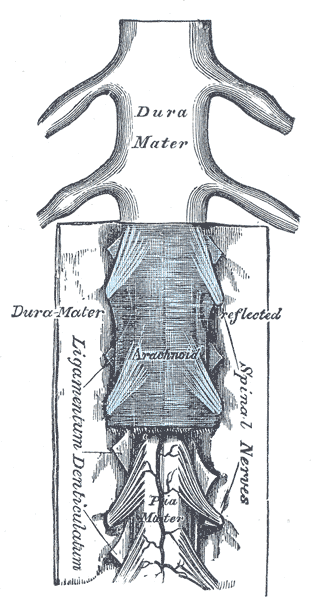
Spinal Cord and Meninges. Shown in this illustration are the spinal cord (also known as the medulla spinalis) and the meninges: the dura mater, arachnoid mater, and pia mater. Other structures labeled in this image are the spinal nerves and ligamentum denticulatum (denticulate ligament).
Henry Vandyke Carter, Public Domain, via Wikimedia Commons
(Click Image to Enlarge)
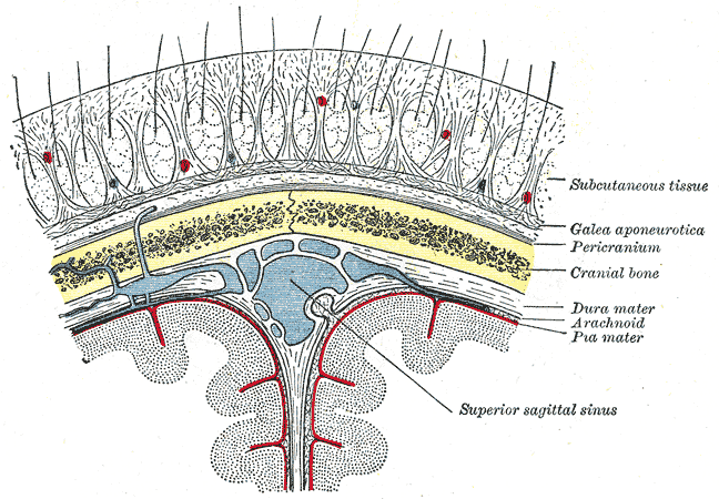
Relationship of the Meniges to the Skull and Brain. This coronal section shows the scalp, subcutaneous tissue, galea aponeurotica, pericranium, cranial bone (skull), dura mater, arachnoid mater, pia mater, superior sagittal sinus, and cerebral falx (not labeled).
Henry Vandyke Carter, Public Domain, via Wikimedia Commons
(Click Image to Enlarge)
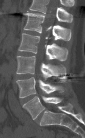
Arachnoiditis Ossificans. This image shows calcification in the posterior L1 to L2 intraspinal region.
Contributed by Steve Lange, MD
References
Todeschi J, Chibbaro S, Gubian A, Pop R, Proust F, Cebula H. Spinal adhesive arachnoiditis following the rupture of an Adamkiewicz aneurysm: Literature review and a case illustration. Neuro-Chirurgie. 2018 Jun:64(3):177-182. doi: 10.1016/j.neuchi.2017.11.003. Epub 2018 Feb 9 [PubMed PMID: 29433818]
Level 3 (low-level) evidenceKhan MU, Devlin JA, Fraser A. Adhesive arachnoiditis in mixed connective tissue disease: a rare neurological manifestation. BMJ case reports. 2016 Dec 16:2016():. doi: 10.1136/bcr-2016-217418. Epub 2016 Dec 16 [PubMed PMID: 27986694]
Level 3 (low-level) evidenceGilbert V, Chakir S, Peeters JB, Hage GE, Labidi M, Jabre R, Chaalala C, Bojanowski MW. Craniovertebral and spinal adhesive arachnoiditis: a late complication of ruptured vertebral and posterior inferior cerebellar arteries aneurysms. Neuro-Chirurgie. 2024 May:70(3):101535. doi: 10.1016/j.neuchi.2024.101535. Epub 2024 Feb 6 [PubMed PMID: 38324943]
Yokota H, Tali ET. Spinal Infections. Neuroimaging clinics of North America. 2023 Feb:33(1):167-183. doi: 10.1016/j.nic.2022.07.015. Epub [PubMed PMID: 36404042]
Werner C, Mathkour M, Scullen T, Dallapiazza RF, Dumont AS, Maulucci CM. Recurrent arachnoid cysts secondary to spinal adhesive arachnoiditis successfully treated with a ventriculoperitoneal shunt. Clinical neurology and neurosurgery. 2020 Jul:194():105835. doi: 10.1016/j.clineuro.2020.105835. Epub 2020 Apr 10 [PubMed PMID: 32305826]
Maillard J, Batista S, Medeiros F, Farid G, Santa Maria PE, Perret CM, Koester SW, Bertani R. Spinal Adhesive Arachnoiditis: A Literature Review. Cureus. 2023 Jan:15(1):e33697. doi: 10.7759/cureus.33697. Epub 2023 Jan 12 [PubMed PMID: 36788823]
Anderson TL, Morris JM, Wald JT, Kotsenas AL. Imaging Appearance of Advanced Chronic Adhesive Arachnoiditis: A Retrospective Review. AJR. American journal of roentgenology. 2017 Sep:209(3):648-655. doi: 10.2214/AJR.16.16704. Epub 2017 Jun 22 [PubMed PMID: 28639826]
Level 2 (mid-level) evidenceGuyer DW, Wiltse LL, Eskay ML, Guyer BH. The long-range prognosis of arachnoiditis. Spine. 1989 Dec:14(12):1332-41 [PubMed PMID: 2617363]
Level 2 (mid-level) evidenceJurga S, Szymańska-Adamcewicz O, Wierzchołowski W, Pilchowska-Ujma E, Urbaniak Ł. Spinal adhesive arachnoiditis: three case reports and review of literature. Acta neurologica Belgica. 2021 Feb:121(1):47-53. doi: 10.1007/s13760-020-01431-1. Epub 2020 Aug 24 [PubMed PMID: 32833147]
Level 3 (low-level) evidenceParenti V, Huda F, Richardson PK, Brown D, Aulakh M, Taheri MR. Lumbar arachnoiditis: Does imaging associate with clinical features? Clinical neurology and neurosurgery. 2020 May:192():105717. doi: 10.1016/j.clineuro.2020.105717. Epub 2020 Feb 3 [PubMed PMID: 32062307]
Warnke JP, Köppert H, Bensch-Schreiter B, Dzelzitis J, Tschabitscher M. Thecaloscopy part III: first clinical application. Minimally invasive neurosurgery : MIN. 2003 Apr:46(2):94-9 [PubMed PMID: 12761680]
Di Ieva A, Barolat G, Tschabitscher M, Rognone E, Aimar E, Gaetani P, Tancioni F, Lorenzetti M, Crotti FM, Rodriguez Baena R, Warnke JP. Lumbar arachnoiditis and thecaloscopy: brief review and proposed treatment algorithm. Central European neurosurgery. 2010 Nov:71(4):207-12. doi: 10.1055/s-0029-1243201. Epub 2009 Dec 21 [PubMed PMID: 20027540]
Singh S, Singh R, Luthra S, Singla A, Tanvir F, Antaal H, Singh A, Singh H, Singh J, Kaur MS. Evolving Radiological Approaches in the Diagnosis and Monitoring of Arachnoiditis Ossificans. Cureus. 2024 Sep:16(9):e68399. doi: 10.7759/cureus.68399. Epub 2024 Sep 1 [PubMed PMID: 39355477]
Mastorakos P, Pomeraniec IJ, Bryant JP, Chittiboina P, Heiss JD. Flexible thecoscopy for extensive spinal arachnoiditis. Journal of neurosurgery. Spine. 2022 Feb 1:36(2):325-335. doi: 10.3171/2021.4.SPINE21483. Epub 2021 Oct 1 [PubMed PMID: 34598155]
Urits I, Chesteen G, Viswanath O. Arachnoiditis Ossificans of the Lumbosacral Spine. Turkish journal of anaesthesiology and reanimation. 2019 Oct:47(5):427-428. doi: 10.5152/TJAR.2019.63239. Epub 2019 May 15 [PubMed PMID: 31572998]
Nadeem SF, Baig AN, Tariq QUA, Shamim MS. Spinal arachnoiditis and syringomyelia: Review of literature with emphasis on postinfectious inflammation and treatment. Surgical neurology international. 2022:13():299. doi: 10.25259/SNI_383_2022. Epub 2022 Jul 15 [PubMed PMID: 35928312]
Gürbüz MS, Erdoğan B, Yüksel MO, Somay H. Postlumbar puncture arachnoiditis mimicking epidural abscess. BMJ case reports. 2013 Nov 6:2013():. doi: 10.1136/bcr-2013-200169. Epub 2013 Nov 6 [PubMed PMID: 24197809]
Level 3 (low-level) evidence