 Tools
Tools
 Appendix Testis Torsion
Introduction
Torsion of testicular appendages is the most common cause of acute scrotal pain in prepubertal boys and a leading cause of pediatric orchalgia.[1] Therefore, it should be routinely considered in the differential diagnosis of any pediatric male presenting with acute scrotal symptoms. Two distinct vestigial structures may undergo torsion—the appendix testis and the appendix epididymis.
The appendix testis, also known as the hydatid of Morgagni, is a Müllerian duct remnant present in approximately 76% to 83% of testes.[2] Typically located at the superior pole of the testis between the testis and epididymis, the appendix testis is the appendage most commonly involved in torsion.[3] This structure is homologous to the female's fimbriated end of the Fallopian tube. The appendix epididymis, derived from the Wolffian (mesonephric) duct, is identified in roughly 22% to 28% of testes and lies along the head of the epididymis. This structure is sometimes regarded as a detached efferent duct.[2]
Accurate clinical and ultrasonographic differentiation of testicular appendage torsion from epididymo-orchitis, testicular torsion, and other causes of acute scrotum is essential to prevent unnecessary surgical exploration, minimize morbidity, and facilitate timely, evidence-based management.[4][5][6] Recognition of characteristic features can help establish the diagnosis and guide conservative management.
Recent pediatric data highlight the importance of standardized diagnostic algorithms, targeted parental education, and expedited referral pathways, collectively reducing preventable testicular loss and its associated medicolegal consequences.[7] Ultimately, a coordinated multidisciplinary approach remains essential to ensure timely recognition and evidence-based management of suspected testicular appendage torsion.
Etiology
Register For Free And Read The Full Article
 Search engine and full access to all medical articles
- 10 free questions in your specialty
- Free CME/CE Activities
- Free daily question in your email
- Save favorite articles to your dashboard
- Emails offering discounts
Learn more about a Subscription to StatPearls Point-of-Care
Etiology
The appendix testis and appendix epididymis are commonly pedunculated, a structural feature that predisposes them to axial rotation and subsequent torsion.[8] Although the precise etiology remains uncertain, several contributing factors have been proposed. Minor scrotal trauma, sudden cremasteric contraction during vigorous activity, hormonally driven prepubertal enlargement of the testes and appendages, and individual anatomical variations likely account for the peak incidence observed among males aged 7 to 12.[9]
A potential seasonal influence has been proposed. Several epidemiological studies have reported a higher incidence of spermatic cord and testicular appendage torsion during colder months, suggesting that lower ambient temperatures, often accompanied by an increased cremasteric reflex, may facilitate testicular appendage torsion.[10][11]
Epidemiology
Torsion of a testicular appendage can occur at any age but shows a pronounced peak between 7 and 12 years, coinciding with hormonally mediated appendiceal enlargement and accentuated cremasteric activity.[9] The appendix testis is found in about 80% (76%-83%) of males and is more likely to undergo torsion than the less common appendix epididymis (20%-24%).[12][13][14] The appendix testis accounts for 90% to 95% of all torsed testicular appendages.[15][16][17]
Contemporary series reveal that torsion of the appendix testis or epididymis accounts for more than half of all acute scrotal presentations in prepubertal boys, making it the foremost cause of pediatric orchalgia.[18][19] In a prospective study, recently updated as a systematic review involving 238 men aged 19 and younger who presented to a children's hospital with acute scrotal pain, 46% were diagnosed with torsion of the appendix testis, 35% had epididymitis, and 16% exhibited testicular torsion, indicating that a large portion of pediatric patients with an acute scrotum were likely to have torsion of a testicular appendage.[20]
Several anatomic and physiologic factors underpin this predisposition. Both testicular appendages are characteristically pedunculated with a delicate mesenteric stalk, rendering them vulnerable to axial rotation incited by minor scrotal trauma, athletic exertion, or sudden cremasteric contraction.[21] Prepubertal vascular engorgement and mucopolysaccharide accumulation increase appendiceal bulk and mobility, increasing the risk of torsion.[20]
Seasonal variation has also been documented, with epidemiologic studies reporting a higher incidence during colder months, likely due to enhanced sympathetic tone and more vigorous cremasteric reflexes at lower ambient temperatures.[7][22] Nevertheless, torsion can occur in any season or climate, and clinicians should maintain a high index of suspicion when evaluating a pediatric patient with acute scrotal pain.
Pathophysiology
The archetypal lesion is a highly mobile, pedunculated Müllerian duct remnant nourished by a tenuous arteriolar arcade that traverses a narrow mesentery. Axial rotation of this stalk, typically ≥180°, impedes venous and lymphatic egress while initially sparing arterial inflow. The resulting closed-compartment phenomenon precipitates venous congestion, elevates interstitial pressure, and leads to progressive edema. Within a few hours, the transmural pressure exceeds systolic capillary pressure, arresting arterial perfusion and culminating in hemorrhagic infarction of the appendage. Experimental models demonstrate complete ischemic necrosis within as little as 6 hours, explaining the abrupt onset and the absence of a prodrome.[23]
The ischemic appendage assumes a violaceous hue, which may be visualized through the overlying tunica vaginalis as the classic blue-dot sign—a pathognomonic but inconsistently observed physical finding (see Image. Blue-Dot Sign). Nociceptive signaling is transmitted via the genital branch of the genitofemoral nerve (L1-L2) and the anterior scrotal branch of the ilioinguinal nerve, accounting for the exquisitely localized upper-pole tenderness observed on examination.[24]
Histopathological specimens uniformly demonstrate dilated, thrombosed venules, periappendageal hemorrhage, neutrophil-predominant infiltration, and early coagulative necrosis of epithelial and stromal elements.[25] If reperfusion does not ensue, the devitalized appendage undergoes dystrophic calcification or auto-amputation, later manifesting as an intravaginal scrotolith. Reactive hydrocele formation, engorgement of the tunica vaginalis, and reflex cremasteric contraction frequently accompany the infarct, occasionally elevating the testis and simulating testicular torsion.
Critically, torsion of a testicular appendage leaves the testicular arterial pedicle and pampiniform plexus uncompromised; thus, intratesticular perfusion and spermatogenic function remain intact. Doppler ultrasonography characteristically demonstrates preserved or mildly hyperemic testicular flow with an avascular paratesticular mass—findings that permit confident, nonoperative management once the diagnosis is secured.[26][27]
History and Physical
History
Accurate bedside assessment remains the cornerstone of early recognition; nonetheless, clinical overlap with testicular torsion and epididymo-orchitis explains persistently high error rates of up to 45% of initial clinical evaluations by medical practitioners.[28] Consequently, contemporary guidelines advocate scrotal ultrasonography with color Doppler in all children presenting with acute scrotal pain, irrespective of the presumptive clinical diagnosis, due to the severe risks and sequelae of potentially misdiagnosing and undertreating a testicular torsion. However, clinically obvious cases of testicular torsion should be taken directly to surgery without waiting for a confirmatory ultrasound, as imaging may delay definitive management.
Pain from a torsion of a testicular appendage is characteristically insidious in onset and progressively intensifies over several hours, unlike the sudden, fulminant pain of acute testicular torsion. The discomfort can be focal to the superior pole of the testis or epididymal head and is rarely accompanied by systemic symptoms, such as fever, nausea, and vomiting, or lower urinary tract symptoms. A history of minor scrotal trauma or vigorous activity may be elicited but is not required.
Physical Examination
A meticulous yet gentle scrotal examination is imperative. Classical findings of a torsed testicular appendage include:
- Focal upper-pole tenderness: In approximately one-third of cases, a discrete, firm paratesticular nodule, corresponding to the infarcted testicular appendage, can be identified.
- Intact cremasteric reflex: This distinguishes a torsed testicular appendage from testicular torsion, where the reflex is typically absent.
- Blue-dot sign: A sub-capsular, cyanotic focus visible through the stretched scrotal skin overlying the superior pole of the testis. Although considered pathognomonic, its reported sensitivity is only approximately 21% (0%-52%), and rare false-positives have been documented in testicular torsion.[29]
- Absence of the contralateral bell-clapper, or an angel-wings deformity. When present, this anatomical finding heightens concern for testicular torsion.
Evaluation
Doppler ultrasonography is the first-line imaging modality recommended for evaluating acute orchalgia across all age groups due to its combined excellence in diagnostic accuracy, with an excellent sensitivity of greater than 95% and specificity of 90%, along with immediate bedside availability.[5][30][31][32][33][34] High-frequency linear array transducers (>12 MHz) provide near-histological resolution of paratesticular structures, enhancing the detection of anatomy and subtle perfusion abnormalities.[31]
- A normal testicular appendage appears as a small (≤5.6 mm) ovoid structure with absent or minimal intrinsic flow.[4][35] Often, the appendage is not directly visualized.
- Normal intratesticular vascularity and a well-perfused epididymis effectively exclude testicular torsion.
- A torsed testicular appendage demonstrates an enlarged (>5.6 mm) avascular nodule whose echogenicity evolves with time, appearing hypoechoic in the first 24 hours and becoming hyperechoic or heterogeneous thereafter.[4][5]
- Surrounding reactive hyperemia of the epididymal head and tunica vaginalis may mimic epididymitis. Therefore, correlation with the focal pain pattern and preserved intratesticular arterial flow is essential.
With progressive inflammation, the examination findings become less specific: global scrotal erythema, edema, and mixed epididymal/testicular tenderness may emerge, potentially masquerading as epididymo-orchitis. A screening urinalysis is recommended in all cases to evaluate for infectious etiologies.[4][30][36] Clinical signs alone are generally not considered sufficiently reliable to make a definitive diagnosis.
Obtaining a scrotal ultrasound may delay surgery by 1 hour or more, so obvious cases of testicular torsion should be taken directly to surgery.[37][38][39] Ultrasonography is most useful in borderline or equivocal cases, where the findings affect management and may help avoid unnecessary surgery.[39][40]
In patients with scrotal pain, epididymitis can be differentiated from a torsed testicular appendage by dysuria, diffuse testicular swelling, scrotal warmth, focal epididymal pain on palpation, increased ultrasonographic echogenicity, and greater peri-testicular perfusion on ultrasound.[4][41]
Testicular torsion is more likely to demonstrate generalized testicular swelling, a high-riding testis, a bell-clapper deformity on the contralateral testicle, onset during sleep, high white blood cell counts, lack of a cremasteric reflex, and high creatine phosphokinase compared to appendix testis torsion.[6][42][43][44] Although a combination of clinical findings is generally used, the absence of a cremasteric reflex on the affected side, by itself, has been reported to have a sensitivity of 92.7% and a specificity of 80.6%.[30][40][42][44][45] A high-riding testicle is also a highly suggestive clinical finding of testicular torsion.[40]
Childhood obesity, which is becoming a common pediatric health issue, appears to increase the risk of surgery for testicular appendage torsion.[46]
Radionuclide scrotal scintigraphy has largely been superseded by ultrasonography; the hot-dot sign, noted by the radiographer as a focal tracer uptake at the appendiceal infarct, which appears only after approximately 5 hours of ischemia and is present in less than 50% of cases.[47] Radionuclide scrotal imaging may take excessive time in the patient's workup. Given its limited sensitivity, lack of anatomic detail, and logistical delays, scintigraphy is now relegated to centers lacking immediate ultrasonography.[6]
Magnetic resonance imaging is seldom required but may be useful in equivocal cases in adolescents or adults. High-resolution T2-weighted sequences delineate an avascular, hyper-intense nodule with surrounding edema and normal testicular perfusion.[48]
Laboratory testing has a limited role in diagnosing testicular appendage torsion. A urinalysis is typically unremarkable in appendiceal torsion. Leucocytosis and elevated C-reactive protein favor an infectious process. Nonetheless, both are prudent in the initial assessment to exclude epididymo-orchitis or a urinary tract infection.
Persistent diagnostic ambiguity, particularly when intratesticular perfusion is indeterminate, mandates prompt surgical scrotal exploration, as the therapeutic window for salvage in true testicular torsion is less than 6 hours.[5][30][42][49][50] Accurate bedside assessment is essential for early recognition; however, clinical similarities with testicular torsion and epididymo-orchitis contribute to persistently high error rates of up to 45% in initial evaluations by medical practitioners.[51] Consequently, contemporary guidelines advocate scrotal ultrasonography with color Doppler in all children presenting with acute scrotal pain, irrespective of presumptive clinical diagnosis, due to the severe risks and sequelae of potentially undertreating a testicular torsion except when it is clinically prudent to go directly to surgery without delay.[39][42][52][53][54][55][56][57]
Pitfalls include a markedly swollen, avascular appendage that may simulate a pyocele or intratesticular abscess, whereas profuse periappendiceal hyperemia can be mistaken for epididymitis. When uncertainty persists, consideration of surgical exploration is warranted.
Treatment / Management
Management hinges on unequivocal exclusion of testicular torsion. Once color Doppler ultrasonography confirms preserved intratesticular perfusion and identifies a paratesticular, avascular nodule consistent with a torsed appendix, initial therapy is conservative management.[5][58]
- Analgesia and anti-inflammatory therapy: Weight-based nonsteroidal anti-inflammatory drugs (NSAIDs), such as ibuprofen 10 mg/kg every 6 hours or as per guidelines, in combination with paracetamol, constitute first-line therapy. Opioids are rarely necessary beyond the first 12 hours.
- Scrotal support and activity restriction: Bed rest, scrotal elevation, and avoidance of contact sports until tenderness resolves (typically 5-7 days).
- Ice and cold packs in the first 24 hours, followed by gentle warmth, can alleviate edema and expedite resolution.
Ice application serves a dual role—it promotes vasoconstriction to reduce swelling and offers analgesic benefit through superficial nerve desensitization. Ice is best applied in 15- to 20-minute intervals several times daily, with careful counseling on skin protection to prevent cold injury. This conservative modality complements pharmacotherapy (NSAIDs) in the outpatient setting.
Pain and swelling typically abate within 1 week; complete clinical resolution is the rule by 10 to 14 days. Routine follow-up imaging is unnecessary, provided symptoms improve steadily.[59](B2)
Indications for Surgery
- Approximately 15% to 25% of patients fail medical therapy and can benefit from scrotal exploration if experiencing prolonged pain.[30][49][58]
- Persistent or escalating pain beyond 48 hours despite adequate NSAIDs.
- Diagnostic uncertainty or inability to exclude testicular torsion on imaging.
- Large necrotic appendage (>10 mm) or sonographic features suggestive of abscess or pyocele.
- Persistent parental or patient anxiety after counseling. (B2)
Surgical resection of the testicular appendage affords rapid symptomatic relief and definitive histopathology; the testis and epididymis are otherwise left untouched, and convalescence is brief. Bilateral orchidopexy is not indicated because the underlying abnormality is confined to the testicular appendage rather than the gubernacular fixation. However, exploration and removal of the testicular appendages on the contralateral side during surgery should be considered as optional.
Some experts recommend this optional procedure due to the future likelihood of testicular appendage torsion in the opposite testis, estimated at around 4.2%.[60] Morphometric studies suggest that when a torsed testicular appendage is found on one side, anatomical factors predisposing to torsion may be present on the contralateral testis in nearly 50% of cases.[61] These findings suggest that consideration should be given to an exploration and removal of testicular appendages on the contralateral testicle when a diagnosis of a torsed testicular appendage is made.[61]
Differential Diagnosis
When evaluating an acutely painful scrotum, the medical practitioner must rapidly discriminate among 3 broad pathophysiologic categories—ischemic lesions, including torsion of the appendix testis, appendix epididymis, or testicular torsion; infective/inflammatory disorders, such as acute epididymo-orchitis and mumps orchitis; neoplasms; and traumatic or hemorrhagic insults, such as contusion, intratesticular hematoma, or testicular rupture.[50][62][63][64] Because the catastrophic loss of a viable gonad remains an ever-present threat, every acute scrotum should be managed as a potential surgical emergency until testicular torsion is definitively excluded.[42] Testicular salvage exceeds 90% when surgical detorsion is accomplished within 6 hours of symptom onset, but plummets precipitously thereafter.[42]
Testicular torsion is characterized by a sudden, severe onset of pain, often awakening the patient from sleep, whereas appendiceal torsion evolves more insidiously.[42][52] Physical examination may reveal an ipsilateral absent cremasteric reflex and a contralateral bell-clapper deformity in true testicular torsion; these findings are typically absent when only the appendage is involved.[6][42][52][65] Pain relief with scrotal elevation (Prehn sign) is notoriously unreliable and should not influence disposition or management.[42][52]
Color Doppler ultrasonography remains the imaging modality of choice for acute scrotal pain.[52] Testicular torsion demonstrates absent or attenuated intratesticular arterial flow, whereas appendiceal torsion shows preserved testicular perfusion with a focal avascular nodule.[6][42][65] Symptom onset during sleep and a high-riding, transverse testis further increase the likelihood of true testicular torsion.[4][6][42][52][65]
Acute epididymo-orchitis may mimic appendiceal torsion sonographically, as both conditions may demonstrate peri-appendiceal hyperemia.[66] However, epididymo-orchitis more frequently presents with voiding symptoms, including dysuria, frequency, and urgency, and may be preceded by a urinary tract infection.[66] Systemic manifestations such as fever or nausea occur but are less common.[30][36] Examination classically reveals an enlarged, tender epididymis with relative sparing of the testicular parenchyma; the bell-clapper deformity is absent, and the Prehn sign may or may not be positive.[4]
Torsion of an epididymal cyst may also cause acute unilateral scrotal pain in male children, but it is quite uncommon.[67][68][69][70][71] In most cases (60%), the cyst resolves on its own; treatment is conservative unless the cyst is greater than 3 cm.[67][68][69][70][71]
When diagnostic uncertainty persists, prompt surgical exploration is required as undue delay risks irreversible ischemic injury to the testis.[39] In equivocal circumstances, urgent operative intervention is the prudent course to avert adverse medicolegal and functional sequelae.[39][58]
Prognosis
The prognosis for torsion of either testicular appendage is overwhelmingly favorable, given that these structures are vestigial embryological remnants with no known physiological function. The associated pain and inflammatory response are typically self-limiting, with spontaneous resolution expected within 7 to 10 days in the majority of cases. Surgical intervention is rarely required, provided that testicular torsion has been confidently excluded and symptoms progressively improve under conservative management.
Complications
The principal clinical risk associated with torsion of a testicular appendage is misdiagnosis; most critically, the failure to identify and promptly intervene in cases of true intravaginal testicular torsion. Given the overlapping symptomatology and the potentially irreversible consequences of delayed torsion management, scrotal ultrasonography with color Doppler interrogation is strongly recommended in all presentations of acute scrotal pain.[19] This imaging modality serves as an essential adjunct to clinical assessment, enabling accurate differentiation between surgical and nonsurgical pathologies and guiding appropriate management selection.[19]
Deterrence and Patient Education
Parents and patients diagnosed with torsion of a testicular appendage should be counseled on the benign and self-limiting nature of the condition and the importance of adhering to conservative management strategies. Standard recommendations include bed rest, scrotal support or elevation, intermittent application of cold compresses, and a scheduled regimen of nonsteroidal anti-inflammatory agents and adjunctive analgesia as clinically indicated.
Patients and caregivers should be advised to monitor for symptom progression or persistence and to seek prompt reevaluation if pain worsens, new symptoms arise, or clinical improvement is not observed within 5 to 7 days. Clear guidance on red-flag symptoms, such as sudden worsening of pain, systemic symptoms, or a change in testicular position, should be provided to avoid missing an evolving case of testicular torsion.
Pearls and Other Issues
The following key points highlight important clinical insights, diagnostic considerations, and management issues related to torsion of testicular appendages.
- A torsed testicular appendage is the most common cause of an acute scrotum in prepubertal boys.
- The blue-dot sign is a classic physical examination finding unique to testicular appendix torsion. However, it is often absent and can be falsely positive in some cases of true testicular torsion. Therefore, the absence of a blue-dot sign is not diagnostic.
- Because of their lack of function and potential for future torsion, the appendix testes and epididymal appendix are commonly removed if encountered during an elective scrotal exploration for other purposes.
- Testicular appendage torsion in prepubertal boys is often misdiagnosed as epididymitis based on scrotal ultrasound imaging, as both can demonstrate epididymal hypervascularity.
- Epididymitis should be considered highly unlikely in a pre-sexual boy with no urologic abnormalities, recent catheterization, or history of urinary tract infections.
- If a testicular appendage is more than 5.6 mm on ultrasound, it should be considered suspicious for torsion of the appendage.
- Patients with testicular appendage torsion are likely younger than those with testicular torsion. These patients generally lack the angel-wing deformity and do not demonstrate the high-riding testicle position typically associated with testicular torsion.[6]
- Rare causes of an acute scrotum can include incarcerated hernias and traumatic testicular ruptures.
- There have been reported cases of simultaneous testicular torsion with testicular appendage torsion.[72]
- Removing the testicular appendages in the contralateral testis during a scrotal exploration for torsion of the testicular appendages is not required but is considered optional.
- Torsion of a testicular appendage can also occur in an undescended testis, and such cases have been reported.[73][74][75]
Enhancing Healthcare Team Outcomes
The evaluation and management of acute scrotal pain, particularly in cases of suspected torsion of the testicular appendage, require a high-functioning interprofessional team to ensure timely, accurate diagnosis and prevent catastrophic outcomes such as missed testicular torsion. Because the presentation often mimics more urgent surgical conditions, this clinical scenario demands immediate triage, efficient communication, and coordinated diagnostic efforts.
Initial assessment typically begins in the emergency department, where triage nurses play a critical role in identifying red flag symptoms such as sudden scrotal pain and the absence of systemic signs. Prompt recognition of a potential testicular torsion mandates immediate escalation to the attending clinician or emergency clinician. Rapid engagement of the interprofessional team—including emergency physicians, radiologists, urologists, pediatric urologists, advanced practice providers, and pediatric specialists—is essential to streamlining clinical decision-making.
A focused clinical examination followed by high-resolution color Doppler ultrasonography, ideally interpreted in real-time by an experienced radiologist, often provides the necessary diagnostic clarity. Radiologic evidence of preserved intratesticular blood flow with a paratesticular avascular mass helps differentiate torsion of an appendage from time-sensitive pathologies such as testicular torsion. Pharmacists may contribute by ensuring timely access to analgesics and anti-inflammatory medications during conservative management.
This interprofessional collaboration fosters efficient care pathways and supports evidence-based decision-making. Clear role delineation, shared situational awareness, and mutual respect across disciplines reduce diagnostic delay and improve the likelihood of organ preservation in true torsion while avoiding unnecessary surgical interventions in benign cases. Additionally, coordinated discharge planning and caregiver education by nursing staff ensure that patients and families are empowered to monitor for red flag symptoms, understand the benign course of appendiceal torsion, and recognize when to seek reevaluation.
By promoting cross-disciplinary communication and implementing standardized protocols for acute scrotal evaluation, the healthcare team collectively enhances diagnostic accuracy, optimizes patient outcomes, and minimizes the medicolegal and psychosocial burden of missed or delayed diagnosis.
Media
(Click Image to Enlarge)
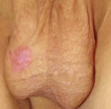
Blue-Dot Sign. The image depicts a blue-dot sign on the scrotum of a pediatric patient.
Contributed by O Chaigasame, MD
References
Laher A, Ragavan S, Mehta P, Adam A. Testicular Torsion in the Emergency Room: A Review of Detection and Management Strategies. Open access emergency medicine : OAEM. 2020:12():237-246. doi: 10.2147/OAEM.S236767. Epub 2020 Oct 12 [PubMed PMID: 33116959]
Jacob M, Barteczko K. Contribution to the origin and development of the appendices of the testis and epididymis in humans. Anatomy and embryology. 2005 Apr:209(4):287-302 [PubMed PMID: 15668777]
Level 3 (low-level) evidenceGalejs LE. Diagnosis and treatment of the acute scrotum. American family physician. 1999 Feb 15:59(4):817-24 [PubMed PMID: 10068706]
Boettcher M, Bergholz R, Krebs TF, Wenke K, Treszl A, Aronson DC, Reinshagen K. Differentiation of epididymitis and appendix testis torsion by clinical and ultrasound signs in children. Urology. 2013 Oct:82(4):899-904. doi: 10.1016/j.urology.2013.04.004. Epub 2013 Jun 2 [PubMed PMID: 23735611]
Level 2 (mid-level) evidenceLev M, Ramon J, Mor Y, Jacobson JM, Soudack M. Sonographic appearances of torsion of the appendix testis and appendix epididymis in children. Journal of clinical ultrasound : JCU. 2015 Oct:43(8):485-9. doi: 10.1002/jcu.22265. Epub 2015 Feb 20 [PubMed PMID: 25704247]
Fujita N, Tambo M, Okegawa T, Higashihara E, Nutahara K. Distinguishing testicular torsion from torsion of the appendix testis by clinical features and signs in patients with acute scrotum. Research and reports in urology. 2017:9():169-174. doi: 10.2147/RRU.S140361. Epub 2017 Aug 28 [PubMed PMID: 28920055]
Alenzi MJ, Alshalash AS, Al-Enzi AN, Al-Anazi FS, Al-Anzi NM, Alsharari KO, Alanazi AA, Alanazi SM, Thirunavukkarasu A. A Cross-Sectional Evaluation of Parents' Awareness Towards Testicular Torsion and Their Response to a Potential Torsion: A Northern Saudi Study. Patient preference and adherence. 2023:17():1671-1678. doi: 10.2147/PPA.S416024. Epub 2023 Jul 14 [PubMed PMID: 37469655]
Level 2 (mid-level) evidenceJONES P. Torsion of the testis and its appendages during childhood. Archives of disease in childhood. 1962 Apr:37(192):214-26 [PubMed PMID: 14452315]
Melekos MD, Asbach HW, Markou SA. Etiology of acute scrotum in 100 boys with regard to age distribution. The Journal of urology. 1988 May:139(5):1023-5 [PubMed PMID: 3361633]
Lyronis ID, Ploumis N, Vlahakis I, Charissis G. Acute scrotum -etiology, clinical presentation and seasonal variation. Indian journal of pediatrics. 2009 Apr:76(4):407-10. doi: 10.1007/s12098-009-0008-2. Epub 2009 Feb 10 [PubMed PMID: 19205631]
Level 2 (mid-level) evidenceMolokwu CN, Somani BK, Goodman CM. Outcomes of scrotal exploration for acute scrotal pain suspicious of testicular torsion: a consecutive case series of 173 patients. BJU international. 2011 Mar:107(6):990-3. doi: 10.1111/j.1464-410X.2010.09557.x. Epub 2010 Sep 21 [PubMed PMID: 21392211]
Level 2 (mid-level) evidenceSahni D, Jit I, Joshi K, Sanjeev. Incidence and structure of the appendices of the testis and epididymis. Journal of anatomy. 1996 Oct:189 ( Pt 2)(Pt 2):341-8 [PubMed PMID: 8886956]
Olulana DI, Egbuchulem KI, Ishola AA, Adamo OS, Ajani MA, Okeke S. CLINICO-HISTOPATHOLOGIC PRESENTATION OF A NIGERIAN CHILD WITH TORSION OF THE EPIDIDYMAL APPENDAGE: A CASE REPORT. Annals of Ibadan postgraduate medicine. 2025 Mar 31:23(1):125-128 [PubMed PMID: 40852685]
Level 3 (low-level) evidenceTanaka K, Kato H, Nikai K, Mikami T, Okazaki T. Testicular appendage torsion in children. Pediatrics international : official journal of the Japan Pediatric Society. 2022 Jan:64(1):e15010. doi: 10.1111/ped.15010. Epub [PubMed PMID: 34597452]
Rakha E, Puls F, Saidul I, Furness P. Torsion of the testicular appendix: importance of associated acute inflammation. Journal of clinical pathology. 2006 Aug:59(8):831-4 [PubMed PMID: 16569689]
Skoglund RW, McRoberts JW, Ragde H. Torsion of testicular appendages: presentation of 43 new cases and a collective review. The Journal of urology. 1970 Oct:104(4):598-600 [PubMed PMID: 5476475]
Level 3 (low-level) evidenceRuiz Montes AM, Jiménez Alvarez C, Núñez Núñez R, Cabrera R, Blesa Sánchez E. [The clinical characteristics of patients with torsion of the testicular and epididymal appendages]. Cirugia pediatrica : organo oficial de la Sociedad Espanola de Cirugia Pediatrica. 1994 Jul:7(3):140-2 [PubMed PMID: 7999519]
Mushtaq I, Fung M, Glasson MJ. Retrospective review of paediatric patients with acute scrotum. ANZ journal of surgery. 2003 Jan-Feb:73(1-2):55-8 [PubMed PMID: 12534742]
Level 2 (mid-level) evidenceKim JS, Shin YS, Park JK. Clinical features of acute scrotum in childhood and adolescence: Based on 17years experiences in primary care clinic. The American journal of emergency medicine. 2018 Jul:36(7):1302-1303. doi: 10.1016/j.ajem.2017.10.063. Epub 2017 Oct 31 [PubMed PMID: 29100785]
Qin KR, Qu LG. Diagnosing with a TWIST: Systematic Review and Meta-Analysis of a Testicular Torsion Risk Score. The Journal of urology. 2022 Jul:208(1):62-70. doi: 10.1097/JU.0000000000002496. Epub 2022 Mar 3 [PubMed PMID: 35238603]
Level 1 (high-level) evidenceTiwana MS, Leslie SW. Anatomy, Abdomen and Pelvis: Testes. StatPearls. 2026 Jan:(): [PubMed PMID: 29261881]
Kumar V, Matai P, Prabhu SP, Sundeep PT. Testicular Loss in Children Due to Incorrect Early Diagnosis of Torsion. Clinical pediatrics. 2020 May:59(4-5):436-438. doi: 10.1177/0009922820903037. Epub 2020 Feb 5 [PubMed PMID: 32019340]
Rosenberg H, Long B, Keays M. Just the facts: assessment and management of testicular torsion in the emergency department. CJEM. 2021 Nov:23(6):740-743. doi: 10.1007/s43678-021-00189-6. Epub 2021 Aug 18 [PubMed PMID: 34406643]
Babbar S. Foreword: Complementary/Alternative Medicine (Cam) In Obstetrics. Clinical obstetrics and gynecology. 2021 Sep 1:64(3):555-557. doi: 10.1097/GRF.0000000000000642. Epub [PubMed PMID: 34172616]
Makwana K, Chodavarapu H, Morones N, Chi J, Barr W, Novinbakht E, Wang Y, Nguyen PT, Jovanovic P, Cohen P, Riera CE. Sensory neurons expressing calcitonin gene-related peptide α regulate adaptive thermogenesis and diet-induced obesity. Molecular metabolism. 2021 Mar:45():101161. doi: 10.1016/j.molmet.2021.101161. Epub 2021 Jan 5 [PubMed PMID: 33412345]
Pickering JM, Hunt HW, Bhattacharya SD. Is Pathological Exam for Disorders of the Appendix Testis Necessary? The American surgeon. 2022 Apr:88(4):802-803. doi: 10.1177/00031348211054561. Epub 2021 Nov 13 [PubMed PMID: 34779270]
Fukuzawa H, Minoda N, Okamoto M, Tsuruno Y, Watanabe A. Contrast-enhanced ultrasonographic findings in torsion of the appendix testis or epididymis in children: a case series. Journal of medical ultrasonics (2001). 2024 Jan:51(1):125-131. doi: 10.1007/s10396-023-01377-w. Epub 2023 Oct 21 [PubMed PMID: 37864611]
Level 2 (mid-level) evidenceXu Y, Magnuson M, Agarwal A, Tan X, Richter CP. Infrared neural stimulation at different wavelengths and pulse shapes. Progress in biophysics and molecular biology. 2021 Jul:162():89-100. doi: 10.1016/j.pbiomolbio.2020.12.004. Epub 2020 Dec 24 [PubMed PMID: 33359901]
Wu D, Wang C, Yang X, Ye J, Mao J. Value of ultrasound in predicting the outcome of conservative treatment of testicular appendage torsion in children. The Turkish journal of pediatrics. 2025 Feb 18:67(1):69-77. doi: 10.24953/turkjpediatr.2025.5459. Epub 2025 Feb 18 [PubMed PMID: 40084724]
Sazgar M, Montazer SH, Hosseininejad SM, Jahanian F, Rezaimehr B, Behbohaninia M, Aminiahidashti H. Clinical Predictors of Testicular Torsion in Patients with Acute Scrotum; a Cross-Sectional Study. Archives of academic emergency medicine. 2022:10(1):e9. doi: 10.22037/aaem.v10i1.1484. Epub 2022 Jan 11 [PubMed PMID: 35072098]
Level 2 (mid-level) evidenceZeng C, Fang L, Li W, Chen H. Diagnostic efficacy and value of ultrasound in children's scrotal testicular torsion: A retrospective analysis. Medicine. 2024 Oct 11:103(41):e39884. doi: 10.1097/MD.0000000000039884. Epub [PubMed PMID: 39465860]
Level 2 (mid-level) evidenceNakayama A, Ide H, Osaka A, Inoue Y, Shimomura Y, Iwahata T, Kobori Y, Saito K, Soh S, Okada H. The Diagnostic Accuracy of Testicular Torsion by Doctors on Duty Using Sonographic Evaluation with Color Doppler. American journal of men's health. 2020 Sep-Oct:14(5):1557988320953003. doi: 10.1177/1557988320953003. Epub [PubMed PMID: 32993407]
Lemini R, Guanà R, Tommasoni N, Mussa A, Di Rosa G, Schleef J. Predictivity of Clinical Findings and Doppler Ultrasound in Pediatric Acute Scrotum. Urology journal. 2016 Aug 25:13(4):2779-83 [PubMed PMID: 27576885]
Liang T, Metcalfe P, Sevcik W, Noga M. Retrospective review of diagnosis and treatment in children presenting to the pediatric department with acute scrotum. AJR. American journal of roentgenology. 2013 May:200(5):W444-9. doi: 10.2214/AJR.12.10036. Epub [PubMed PMID: 23617512]
Level 2 (mid-level) evidenceJohnson KA, Dewbury KC. Ultrasound imaging of the appendix testis and appendix epididymis. Clinical radiology. 1996 May:51(5):335-7 [PubMed PMID: 8641095]
Boettcher M, Bergholz R, Krebs TF, Wenke K, Aronson DC. Clinical predictors of testicular torsion in children. Urology. 2012 Mar:79(3):670-4. doi: 10.1016/j.urology.2011.10.041. Epub [PubMed PMID: 22386422]
Level 2 (mid-level) evidenceBuch Kjeldgaard A, Kinder-Klausen MS, Nerstrøm M, Cohen J, Henriksen BM, Thorup JM. The impact of ultrasound on testicular loss in cases of testicular torsion in children. Pediatric surgery international. 2024 Mar 20:40(1):83. doi: 10.1007/s00383-024-05663-7. Epub 2024 Mar 20 [PubMed PMID: 38507099]
Level 3 (low-level) evidenceMadsen SMD, Rawashdeh YF. Assessing timeline delays associated with utilization of ultrasound diagnostics in paediatric acute scrotum, pre and per COVID-19 pandemic. Journal of pediatric urology. 2023 Oct:19(5):653.e1-653.e7. doi: 10.1016/j.jpurol.2023.07.003. Epub 2023 Jul 13 [PubMed PMID: 37544787]
Clement KD, Light A, Asif A, Chan VW, Khadhouri S, Shah TT, Banks F, Dorkin T, Driver CP, During V, Fraser N, Johnston MJ, Lucky M, Modgil V, Muneer A, Parnham A, Pearce I, Shabbir M, Shenoy M, Summerton DJ, Undre S, Williams A, MacLennan S, Kasivisvanathan V, FIX-IT collaborators. A BURST-BAUS consensus document for best practice in the conduct of scrotal exploration for suspected testicular torsion: the Finding consensus for orchIdopeXy In Torsion (FIX-IT) study. BJU international. 2022 Jun 10:130(5):662-70. doi: 10.1111/bju.15818. Epub 2022 Jun 10 [PubMed PMID: 35689399]
Level 3 (low-level) evidenceCassaro F, Arena S, Bonfiglio R, Alibrandi A, D'Antoni S, Romeo C, Impellizzeri P. Utility of Clinical Signs in the Diagnosis of Testicular Torsion in Pediatric Age: Optimization of Timing in a Time-Sensitive Pathology. Children (Basel, Switzerland). 2025 Sep 12:12(9):. doi: 10.3390/children12091220. Epub 2025 Sep 12 [PubMed PMID: 41007085]
Furugane R, Mitsunaga T, Komatsu S, Takenouchi A, Oita S, Kawaguchi Y, Kudo W, Nishimura K, Hishiki T. Clinical characteristics of pediatric acute epididymitis: a single institute review of 142 cases. Pediatric surgery international. 2025 Aug 30:41(1):278. doi: 10.1007/s00383-025-06168-7. Epub 2025 Aug 30 [PubMed PMID: 40884544]
Level 3 (low-level) evidenceSchick MA, Sternard BT. Testicular Torsion. StatPearls. 2026 Jan:(): [PubMed PMID: 28846325]
Nelson CP, Williams JF, Bloom DA. The cremasteric reflex: a useful but imperfect sign in testicular torsion. Journal of pediatric surgery. 2003 Aug:38(8):1248-9 [PubMed PMID: 12891505]
Mellick LB, Mowery ML, Al-Dhahir MA. Cremasteric Reflex. StatPearls. 2025 Jan:(): [PubMed PMID: 30020720]
Edwards LG, Ferguson C. Best evidence topic report: in emergency settings, can an absent cremasteric reflex be used to aid diagnosis of testicular torsions? Emergency medicine journal : EMJ. 2025 Jun 19:42(7):474-475. doi: 10.1136/emermed-2024-214797. Epub 2025 Jun 19 [PubMed PMID: 40537283]
Coronas-Soucheiron M, Martín-Solé O, Massaguer C, Tarrado X. Is Being Overweight a Risk Factor for Torsion of the Appendix Testis in Children?: A Paired Case-Control Study. Pediatric emergency care. 2024 Apr 1:40(4):279-282. doi: 10.1097/PEC.0000000000002994. Epub 2023 Jun 5 [PubMed PMID: 37276066]
Level 2 (mid-level) evidenceMelloul M, Paz A, Lask D, Luttwak Z, Mukamel E. The pattern of radionuclide scrotal scan in torsion of testicular appendages. European journal of nuclear medicine. 1996 Aug:23(8):967-70 [PubMed PMID: 8753687]
Level 2 (mid-level) evidencePark SJ, Kim HL, Yi BH. Sonography of intrascrotal appendage torsion: varying echogenicity of the torsed appendage according to the time from onset. Journal of ultrasound in medicine : official journal of the American Institute of Ultrasound in Medicine. 2011 Oct:30(10):1391-6 [PubMed PMID: 21968490]
Saxena AK, Castellani C, Ruttenstock EM, Höllwarth ME. Testicular torsion: a 15-year single-centre clinical and histological analysis. Acta paediatrica (Oslo, Norway : 1992). 2012 Jul:101(7):e282-6. doi: 10.1111/j.1651-2227.2012.02644.x. Epub 2012 Mar 24 [PubMed PMID: 22385478]
Level 2 (mid-level) evidenceVelasquez J, Boniface MP, Mohseni M. Acute Scrotum Pain. StatPearls. 2025 Jan:(): [PubMed PMID: 29262236]
Lim SH, Yang DM, Kim HC, Kim SW, Kim H, Lee DI, Moon SK, Park SJ. Ultrasonography of intrascrotal torsed appendages: size and interval between symptom onset and the ultrasonographic examination according to echogenicity. Ultrasonography (Seoul, Korea). 2023 Apr:42(2):259-264. doi: 10.14366/usg.22169. Epub 2022 Dec 5 [PubMed PMID: 36935598]
Agrawal AM, Tripathi PS, Shankhwar A, Naveen C. Role of ultrasound with color Doppler in acute scrotum management. Journal of family medicine and primary care. 2014 Oct-Dec:3(4):409-12. doi: 10.4103/2249-4863.148130. Epub [PubMed PMID: 25657954]
Expert Panel on Urological Imaging, Gerena M, Allen BC, Turkbey B, Barker SJ, Costa DN, Flink C, Meyers ML, Ramasamy R, Rosario J, Sharma A, Whitworth P 3rd, Williams WL, Oto A. ACR Appropriateness Criteria® Acute Onset of Scrotal Pain-Without Trauma, Without Antecedent Mass: 2024 Update. Journal of the American College of Radiology : JACR. 2024 Nov:21(11S):S364-S371. doi: 10.1016/j.jacr.2024.08.011. Epub [PubMed PMID: 39488348]
Hamel C, Avard B, Brahm G, Fung D, Martens B, Michaud A, Miller L, Sala E, Wallis CJD, Fung C. Canadian Association of Radiologists Genitourinary Imaging Referral Guideline. Canadian Association of Radiologists journal = Journal l'Association canadienne des radiologistes. 2025 Feb:76(1):25-32. doi: 10.1177/08465371241261317. Epub 2024 Jul 25 [PubMed PMID: 39054585]
Friedman AA, Palmer LS, Maizels M, Bittman ME, Avarello JT. Pediatric acute scrotal pain: A guide to patient assessment and triage. Journal of pediatric urology. 2016 Apr:12(2):72-5. doi: 10.1016/j.jpurol.2016.03.003. Epub 2016 Mar 11 [PubMed PMID: 27036070]
Ota K, Fukui K, Oba K, Shimoda A, Oka M, Ota K, Sakaue M, Takasu A. The role of ultrasound imaging in adult patients with testicular torsion: a systematic review and meta-analysis. Journal of medical ultrasonics (2001). 2019 Jul:46(3):325-334. doi: 10.1007/s10396-019-00937-3. Epub 2019 Mar 7 [PubMed PMID: 30847624]
Level 1 (high-level) evidenceExpert Panel on Urological Imaging:, Wang CL, Aryal B, Oto A, Allen BC, Akin O, Alexander LF, Bardo DME, Chong J, Froemming AT, Fulgham PF, Heller MT, Maranchie JK, Mody RN, Patel BN, Schieda N, Turkbey IB, Venkatesan AM, Yoo DC, Lockhart ME. ACR Appropriateness Criteria(®) Acute Onset of Scrotal Pain-Without Trauma, Without Antecedent Mass. Journal of the American College of Radiology : JACR. 2019 May:16(5S):S38-S43. doi: 10.1016/j.jacr.2019.02.016. Epub [PubMed PMID: 31054757]
Holland JM, Graham JB, Ignatoff JM. Conservative management of twisted testicular appendages. The Journal of urology. 1981 Feb:125(2):213-4 [PubMed PMID: 7206059]
Syed MK, Al Faqeeh AA, Othman A, Hussein AA, Hussain S, Almas T, Alsufyani R, Alaeddin H, Syed S, Syed SK. The Spectrum of Testicular Pathologies Upon Scrotal Exploration for Acute Scrotum: A Retrospective Analysis. Cureus. 2020 Oct 16:12(10):e10984. doi: 10.7759/cureus.10984. Epub 2020 Oct 16 [PubMed PMID: 33209540]
Level 2 (mid-level) evidenceKoh YH, Granger J, Cundy TP, Boucaut HA, Goh DW. Testicular Appendage Torsion-To Explore the Other Side or Not? Urology. 2020 Jul:141():130-134. doi: 10.1016/j.urology.2020.02.033. Epub 2020 Apr 10 [PubMed PMID: 32283168]
Barbosa RG, Favorito LA, Sampaio FJB. Morphometric study applied to testicular and epididymis hydatids torsion. Scientific reports. 2024 Feb 8:14(1):3249. doi: 10.1038/s41598-024-52734-9. Epub 2024 Feb 8 [PubMed PMID: 38332206]
Vasdev N, Chadwick D, Thomas D. The acute pediatric scrotum: presentation, differential diagnosis and management. Current urology. 2012 Sep:6(2):57-61. doi: 10.1159/000343509. Epub 2012 Sep 27 [PubMed PMID: 24917714]
Günther P, Rübben I. The acute scrotum in childhood and adolescence. Deutsches Arzteblatt international. 2012 Jun:109(25):449-57; quiz 458. doi: 10.3238/arztebl.2012.0449. Epub 2012 Jun 22 [PubMed PMID: 22787516]
Benn M, Leslie SW. Ultrasound of the Urinary Tract. StatPearls. 2026 Jan:(): [PubMed PMID: 30571002]
Zvizdic Z, Aganovic A, Milisic E, Jonuzi A, Zvizdic D, Vranic S. The Role of Demographic and Clinical Characteristics in Distinguishing Testicular Torsion from Torsion of the Appendix Testis: A Single-center Retrospective Study. Prague medical report. 2023:124(3):255-264. doi: 10.14712/23362936.2023.20. Epub [PubMed PMID: 37736949]
Level 2 (mid-level) evidenceRupp TJ, Leslie SW. Epididymitis. StatPearls. 2026 Jan:(): [PubMed PMID: 28613565]
Messina M,Fusi G,Ferrara F,Bindi E,Pellegrino C,Molinaro F,Angotti R, A rare cause of acute scrotum in a child: torsion of an epididymal cyst. Case report and review of the literature. La Pediatria medica e chirurgica : Medical and surgical pediatrics. 2019 Jun 21; [PubMed PMID: 31232012]
Level 3 (low-level) evidenceWang Y, Wang K, Yu L, Ma X. A rare case of scrotal emergency: torsion of epididymal cyst-a case report and literature review. Frontiers in pediatrics. 2023:11():1245842. doi: 10.3389/fped.2023.1245842. Epub 2024 Jan 9 [PubMed PMID: 38264504]
Level 3 (low-level) evidenceVafadar M, Rakhshankhah N, Salek M, Zarei E. Torsion of epididymal Cyst as a cause of acute Scrotum in a child. Urology case reports. 2023 May:48():102417. doi: 10.1016/j.eucr.2023.102417. Epub 2023 May 3 [PubMed PMID: 37215055]
Level 3 (low-level) evidenceIlangovan G, M M, Mounika V, Khan MA. Torsion of Epididymal Cyst: A Case Report With Review of Literature. Cureus. 2023 Dec:15(12):e51158. doi: 10.7759/cureus.51158. Epub 2023 Dec 27 [PubMed PMID: 38283501]
Level 3 (low-level) evidenceAmeli M,Boroumand-Noughabi S,Gholami-Mahtaj L, A 14-Year-Old Boy with Torsion of the Epididymal Cyst. Case reports in urology. 2015; [PubMed PMID: 26798545]
Level 3 (low-level) evidenceVasconcelos-Castro S, Soares-Oliveira M, Campos JM, Estevão-Costa J. Simultaneous Testicular and Appendix Testis Torsion: A Novel Clinically Relevant Association. Pediatric emergency care. 2021 Dec 1:37(12):e868-e869. doi: 10.1097/PEC.0000000000001784. Epub [PubMed PMID: 30839439]
Hajji F, Ameur A. Torsion of the appendix testis in an undescended testicle. The Pan African medical journal. 2015:22():265. doi: 10.11604/pamj.2015.22.265.7493. Epub 2015 Nov 20 [PubMed PMID: 26958128]
Malik MIK, Abbas J, Sabri S, Michael G, Ellenbogen S. Torsion of undescended abdominal testicle in a 16-year-old and its management. BMJ case reports. 2020 Jan 29:13(1):. doi: 10.1136/bcr-2019-232179. Epub 2020 Jan 29 [PubMed PMID: 32001538]
Level 3 (low-level) evidenceKrukowski ZH, Auld CD. Torsion of the appendix epididymis in a maldescended testis. British journal of urology. 1983 Apr:55(2):244-5 [PubMed PMID: 6132646]