 Tools
Tools
 Anatomy, Thorax, Heart Aorta
Introduction
The aorta is the largest blood vessel in the human body. The vessel originates from the left ventricle of the heart anterior to the pulmonary artery, then arches posteriorly and descends through the posterior mediastinum. The aorta continues to the level of the L4 vertebral body, where it bifurcates into the right and left common iliac arteries. As the main artery of the systemic circulation, the aorta distributes oxygenated blood throughout the body. The following discussion focuses on the thoracic portion of the aorta, which includes the ascending aorta, aortic arch, and descending thoracic aorta until the vessel crosses the diaphragm and becomes the abdominal aorta. The thoracic aorta supplies oxygenated blood to multiple structures, including the head, neck, upper extremities, and structures within the thorax.
Structure and Function
Register For Free And Read The Full Article
 Search engine and full access to all medical articles
- 10 free questions in your specialty
- Free CME/CE Activities
- Free daily question in your email
- Save favorite articles to your dashboard
- Emails offering discounts
Learn more about a Subscription to StatPearls Point-of-Care
Structure and Function
The thoracic aorta originates from the left ventricle and is guarded by the aortic valve. Just above the aortic valve cusps, the aorta gives rise to the right and left main coronary arteries, which travel within the coronary grooves of the heart and perfuse the myocardium. The initial portion of the aorta, which ascends posterior to the sternum, is referred to as the ascending aorta and extends approximately to the level of the T4 vertebral body. At this level, the vessel becomes the aortic arch and begins to curve posteriorly and to the left of the vertebral bodies within the posterior mediastinum. The aortic arch begins and ends at the level of the second rib and lies within the superior mediastinum. On chest radiography, the aortic arch is visible as the aortic knob. The aortic arch gives rise to 3 branches, including:
-
Brachiocephalic artery: The brachiocephalic artery, also known as the innominate artery, is the first vessel to branch from the aortic arch. The artery travels superiorly and toward the right before bifurcating into the right subclavian artery and right common carotid artery.
-
Left common carotid artery: The left common carotid artery is the second vessel to branch from the aortic arch.
-
Left subclavian artery: The left subclavian artery is the third vessel to branch from the aortic arch.
The final section of the aortic arch, located just distal to the origin of the left subclavian artery, is known as the aortic isthmus. Mild aortic narrowing occurs near the ligamentum arteriosum, a remnant of the ductus arteriosus. At the level of the T4 vertebral body, the vessel continues as the descending thoracic aorta and travels inferiorly to the aortic hiatus of the diaphragm at T12, where it exits the thorax. The descending thoracic aorta gives rise to multiple vessels before exiting the thoracic cavity, including arteries that supply the pericardium, bronchi, mediastinum, and esophagus. Additional branches include the superior phrenic, posterior intercostal, and subcostal arteries.
Embryology
The aorta develops during the third gestational week.[1] The ascending aorta and aortic arch develop independently from 2 different embryologic structures. The ascending aorta develops from the truncus arteriosus, a component of the developing heart. The truncus arteriosus is a single outflow tract that forms during cardiac development and originates from both the left and right ventricles. The aorticopulmonary septum then divides the truncus arteriosus into 2 separate outflow tracts, which later become the pulmonary artery and ascending aorta.
The aortic arch forms through the development of the branchial arch arteries. Six branchial arch arteries, also known as the pharyngeal arch vessels, develop into the following structures. These develop into the following:
- First branchial arch artery: Forms the maxillary and external carotid arteries.
- Second branchial arch artery: Forms the stapedial arteries.
- Third branchial arch artery: Contributes to the formation of the common carotid arteries and the proximal internal carotid arteries.
- Fourth branchial arch artery: Forms the main portion of the aortic arch and the proximal right subclavian artery. The left subclavian artery develops from the left seventh intersegmental artery, one of a series of arteries arising from the dorsal aorta.
- Fifth branchial arch artery: Does not form a persistent vessel and subsequently regresses.
- Sixth branchial arch artery: Contributes to the formation of the pulmonary arteries and the ductus arteriosus.[2][3]
Surgical Considerations
Coarctation of the Aorta
Coarctation of the aorta is a congenital condition characterized by aortic narrowing that obstructs distal blood flow. The narrowing most commonly occurs near the ductus arteriosus and is classified as preductal, ductal, or postductal coarctation.
Preductal coarctation: Preductal coarctation occurs proximal to the ductus arteriosus. This form most often results from a congenital cardiac anomaly that decreases blood flow to the left side of the heart and aorta, leading to hypoplastic development.
Ductal coarctation: Ductal coarctation occurs at the insertion of the ductus arteriosus. This form most often becomes apparent at birth when the ductus arteriosus closes.
Postductal coarctation: Postductal coarctation occurs distal to the ductus arteriosus and is the most common form in adults. Patients often present with hypertension in the upper extremities and weak pulses in the lower extremities. The difference in perfusion results from increased blood flow through the aortic arch vessels and decreased blood flow to the descending aorta distal to the stenotic segment. Collateral vessels may enlarge to improve blood delivery to the descending aorta. Collateral circulation most commonly extends from the subclavian artery to the internal thoracic artery, anterior intercostal arteries, posterior intercostal arteries, and finally the descending thoracic aorta. Dilation of the intercostal arteries may produce the characteristic finding of rib notching on chest radiography.[4][5]
Coarctation of the aorta can cause substantial morbidity in patients with severe narrowing and is associated with poor outcomes among patients who survive beyond 1 year of age without treatment.[6] Treatment options include surgical repair, balloon angioplasty, and endovascular stent placement. Although these options are available, the decision on an optimal treatment strategy can be complicated. No universally accepted treatment algorithm exists for these patients.[7] Management is at the physician's discretion, with age at presentation and complexity of coarctation influencing treatment choice.
Clinical Significance
Aortic Aneurysm
A thoracic aortic aneurysm refers to dilation of the thoracic aorta, often involving the proximal ascending segment. In adults, chronic hypertension is a common underlying factor. In younger adults, the most common underlying factor is a connective tissue disorder such as Marfan syndrome or Ehlers-Danlos syndrome. An aortic aneurysm is clinically significant because progressive dilation weakens the aortic wall, increasing the risk of aortic rupture or dissection. For this reason, elective repair is often recommended once an aneurysm has reached a diameter of 5.5 cm or greater.[8][9] Elective repair can reduce the risk of rupture and restore survival rates to nearly those of the general population.
Aortic Dissection
Aortic dissection refers to a disruption of the innermost layer of the aorta that allows blood to tunnel through the middle layer of the aortic wall. The initial tear most often occurs in the proximal portion of the ascending aorta, just distal to the aortic valve. Aortic dissections can then propagate distally or proximally through the aortic wall. Acute aortic dissection is a life-threatening emergency that may require urgent surgical repair to increase the likelihood of survival. Nearly 40% of patients with acute aortic dissection die immediately. Of those who survive the initial event, approximately 80% die within 2 weeks.[10]
Patent Ductus Arteriosus
The ductus arteriosus is a communication between the pulmonary artery and the aortic arch during fetal development. The vessel usually closes by the second day after birth in full-term infants. Persistence of this communication beyond 2 days is termed patent ductus arteriosus. Depending on the size of the persistent opening, patent ductus arteriosus may cause failure to thrive and heart failure.[1] Other aberrant conditions of development seen in the aorta are hypoplastic ascending aorta, interrupted aortic arch, right aortic arch, and double aortic arch.[1]
Media
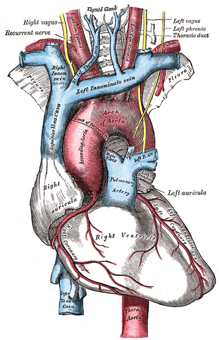
(Click Image to Enlarge)
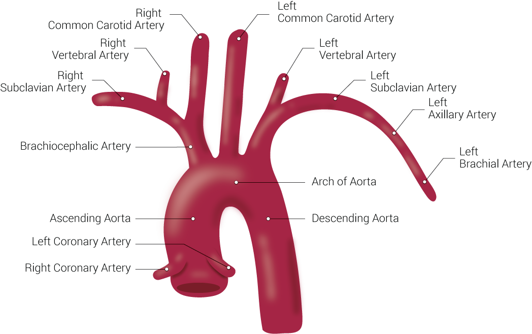
Branches of the Aorta. This illustration includes the right common carotid artery, right vertebral artery, right subclavian artery, brachiocephalic artery, ascending aorta, left coronary artery, right coronary artery, left common carotid artery, left vertebral artery, left subclavian artery, left axillary artery, left brachial artery, arch of aorta, and descending aorta.
Illustrated by B Palmer
(Click Image to Enlarge)
References
Kau T, Sinzig M, Gasser J, Lesnik G, Rabitsch E, Celedin S, Eicher W, Illiasch H, Hausegger KA. Aortic development and anomalies. Seminars in interventional radiology. 2007 Jun:24(2):141-52. doi: 10.1055/s-2007-980040. Epub [PubMed PMID: 21326792]
Dungan DH, Heiserman JE. The carotid artery: embryology, normal anatomy, and physiology. Neuroimaging clinics of North America. 1996 Nov:6(4):789-99 [PubMed PMID: 8824131]
Bamforth SD, Chaudhry B, Bennett M, Wilson R, Mohun TJ, Van Mierop LH, Henderson DJ, Anderson RH. Clarification of the identity of the mammalian fifth pharyngeal arch artery. Clinical anatomy (New York, N.Y.). 2013 Mar:26(2):173-82. doi: 10.1002/ca.22101. Epub 2012 May 23 [PubMed PMID: 22623372]
Level 3 (low-level) evidenceStout KK, Daniels CJ, Aboulhosn JA, Bozkurt B, Broberg CS, Colman JM, Crumb SR, Dearani JA, Fuller S, Gurvitz M, Khairy P, Landzberg MJ, Saidi A, Valente AM, Van Hare GF. 2018 AHA/ACC Guideline for the Management of Adults With Congenital Heart Disease: A Report of the American College of Cardiology/American Heart Association Task Force on Clinical Practice Guidelines. Circulation. 2019 Apr 2:139(14):e698-e800. doi: 10.1161/CIR.0000000000000603. Epub [PubMed PMID: 30586767]
Level 1 (high-level) evidenceKaraosmanoglu AD, Khawaja RD, Onur MR, Kalra MK. CT and MRI of aortic coarctation: pre- and postsurgical findings. AJR. American journal of roentgenology. 2015 Mar:204(3):W224-33. doi: 10.2214/AJR.14.12529. Epub [PubMed PMID: 25714305]
Campbell M. Natural history of coarctation of the aorta. British heart journal. 1970 Sep:32(5):633-40 [PubMed PMID: 5470045]
Torok RD, Campbell MJ, Fleming GA, Hill KD. Coarctation of the aorta: Management from infancy to adulthood. World journal of cardiology. 2015 Nov 26:7(11):765-75. doi: 10.4330/wjc.v7.i11.765. Epub [PubMed PMID: 26635924]
Coady MA, Rizzo JA, Hammond GL, Kopf GS, Elefteriades JA. Surgical intervention criteria for thoracic aortic aneurysms: a study of growth rates and complications. The Annals of thoracic surgery. 1999 Jun:67(6):1922-6; discussion 1953-8 [PubMed PMID: 10391339]
Coady MA, Rizzo JA, Hammond GL, Mandapati D, Darr U, Kopf GS, Elefteriades JA. What is the appropriate size criterion for resection of thoracic aortic aneurysms? The Journal of thoracic and cardiovascular surgery. 1997 Mar:113(3):476-91; discussion 489-91 [PubMed PMID: 9081092]
Coady MA, Rizzo JA, Goldstein LJ, Elefteriades JA. Natural history, pathogenesis, and etiology of thoracic aortic aneurysms and dissections. Cardiology clinics. 1999 Nov:17(4):615-35; vii [PubMed PMID: 10589336]