 Tools
Tools
 Anti-NMDAR Encephalitis
Introduction
Anti-N-methyl-D-aspartate (anti-NMDA) receptor encephalitis is an autoimmune encephalitis characterized by complex neuropsychiatric manifestations and the presence of Immunoglobulin G (IgG) antibodies directed against the NMDA receptor (NR1) subunit within the central nervous system (CNS). These pathogenic antibodies can be detected in serum or cerebrospinal fluid (CSF). [1][2] Anti-NMDA receptor encephalitis represents the best-known and likely the most common form of immune-mediated limbic encephalitis.
Patients most commonly present with acute or subacute neuropsychiatric symptoms. Clinical manifestations encompass a broad spectrum of neurological abnormalities, including cognitive and behavioral changes, autonomic disturbances, altered levels of consciousness, stereotypic movements, seizures, and coma. Confirmation of the diagnosis relies on detection of CSF IgG antibodies targeting the GluN1 subunit of the NMDA receptor. Early recognition of the condition, timely diagnostic evaluation, and prompt initiation of immunotherapy can improve clinical outcomes.[3]
Etiology
Register For Free And Read The Full Article
 Search engine and full access to all medical articles
- 10 free questions in your specialty
- Free CME/CE Activities
- Free daily question in your email
- Save favorite articles to your dashboard
- Emails offering discounts
Learn more about a Subscription to StatPearls Point-of-Care
Etiology
Anti-NMDA receptor encephalitis develops through the formation of IgG antibodies, particularly IgG1 and IgG3 subclasses, that bind to the NR1 subunit. This antibody interaction causes internalization of NMDA (glutamate) receptors, resulting in reduced neuronal Ca2+ influx and decreased receptor-dependent synaptic currents.[4] These pathogenic antibody-mediated effects contribute to the neurological dysfunction characteristic of this autoimmune encephalitis.
In some patients, antibody production occurs in association with an underlying ovarian teratoma and, less commonly, other tumors. Viral encephalitis, particularly herpes simplex virus (HSV) encephalitis, may also contribute to NMDAR antibody development, with antibody production occurring over the subsequent 3 weeks and followed by the emergence of autoimmune encephalitis.[5] In most cases, the specific trigger responsible for antibody formation remains unknown. Regardless of the initiating factor, the disease process ultimately involves excessive production of autoantibodies by intrathecal plasma cells, contributing to ongoing immune-mediated neuronal dysfunction.
Epidemiology
Although anti-NMDAR encephalitis remains a rare disorder, the estimated incidence reaches approximately 1.5 cases per million population.[6] Anti-NMDAR encephalitis represents the best-known and likely the most common form of autoimmune encephalitis.[7] Data from the California Encephalitis Project demonstrated that the incidence of anti-NMDAR encephalitis exceeded the incidence of any individual viral encephalitis.[8]
Reported cases have occurred across a broad age range, including infants as young as 2 months and adults up to 85 years of age. Females develop anti-NMDAR encephalitis approximately 4 times more frequently than males. Young adult females between 25 and 35 years of age represent the population most commonly affected.
Pathophysiology
Autoimmune encephalitis was previously classified as paraneoplastic or nonparaneoplastic, depending on whether tumor-associated antibodies were present. These paraneoplastic antibodies include anti-neuronal nuclear antibody type 1 (anti-Hu), anti-Ri, or anti-Yo. With a better understanding of the pathophysiology of different autoimmune encephalitis, a more nuanced, pathophysiologically based classification is used.
The modern-day classification of immune encephalitis is based on the targets of the antibodies. The classical paraneoplastic encephalitis, as noted above, is mediated predominantly by T cells, with cytotoxic T cells demonstrated in pathological specimens. These immune responses result from molecular mimicry between neuronal tissue antigens and tumor antigens. The antibodies themselves are not pathogenic. These antibodies direct their activity towards intracellular constituents.
The other class of autoimmune encephalitis consists of antibodies directed against synaptic or cell-surface antigens, eg, anti-NMDAR, anti-GAD, and anti-VGKC antibody-mediated encephalitides. These are real antibody- or B-cell-mediated autoimmune encephalitis with real pathogenic antibodies.[9] Anti-NMDAR antibody-mediated encephalitis is a B-cell-mediated autoimmune encephalitis with an actual pathogenic antibody that can be removed by plasma exchange, resulting in improvement of the underlying pathology. The anti-NMDR antibody binds to the NR1 subunit, leading to internalization of NMDA (glutamate) receptors, reduced neuronal Ca2+ influx, and decreased receptor-dependent synaptic currents.[4]
History and Physical
The clinical presentation of anti-NMDAR encephalitis has been categorized into 5 distinct phases: the prodromal phase, psychotic phase, unresponsive phase, hyperkinetic phase, and recovery phase.[10]
Prodromal and Psychotic Phases
The disease typically begins with a prodromal phase resembling a common viral infection. Within weeks to a few months, usually less than 3 months, patients rapidly develop complex neuropsychiatric manifestations during the psychotic phase. Clinical presentations vary between children and adults. Adults more commonly present with psychiatric symptoms, whereas children frequently exhibit movement disorders or seizures as initial manifestations. Acute or subacute behavioral changes represent the most common presenting features among adult patients. No specific psychiatric phenotype defines anti-NMDAR encephalitis; however, patients without a prior psychiatric diagnosis may rapidly develop a range of positive and negative psychiatric symptoms, including visual or auditory hallucinations, acute schizoaffective episodes, depression, mania, addictive behaviors, and eating disorders (see Image. Clinical Features of Psychotic Phase Anti-NMDAR Encephalitis).
The rapid onset of symptoms contrasts with the slower progression typically observed in primary psychiatric disorders. Anti-NMDAR antibodies have been identified in 3% to 4% of patients initially diagnosed with schizophrenia.[11] Many patients require psychiatric inpatient admission because of severe behavioral symptoms and may receive symptomatic treatment for weeks before the diagnosis is recognized. Although some patients demonstrate neurological features at initial presentation, others develop these manifestations within several weeks. Importantly, some individuals exhibit intolerance to neuroleptic medications and may develop hyperthermia, muscle rigidity, rhabdomyolysis, or coma following neuroleptic exposure.
Unlike schizophrenia, which more commonly presents with predominant positive symptoms, patients with anti-NMDAR encephalitis frequently exhibit both positive and negative symptoms. The neuropsychiatric spectrum includes apathy, anxiety, fluctuating sensorium, bizarre behaviors, hypersexuality, wandering, aphasia, amnesia, apraxia, sleep-wake cycle disruption with severe insomnia, and delusions. Many patients, particularly children and adult men, also experience seizures, which may present as focal or generalized events. Seizures can become resistant to antiepileptic drugs (AEDs) and may progress to status epilepticus or refractory status epilepticus.
Unresponsive Phase
The psychotic phase may progress to the unresponsive phase, characterized by mutism, reduced motor activity, and catatonia. The subsequent hyperkinetic phase features autonomic instability and prominent movement disorders. Significant autonomic dysfunction, including labile blood pressure and heart rate, cardiac arrhythmias, temperature instability, and central hypoventilation, may require intensive care unit (ICU) admission, mechanical ventilation, and, in some cases, cardiac pacemaker placement.[12]
Hyperkinetic and Recovery Phases
The characteristic movement disorder during the hyperkinetic phase includes oro-lingual dyskinesias with lip-smacking, tongue protrusion, and jaw movements. However, patients may develop a broad range of abnormal movements, including automatisms, dyskinesia, dystonia, choreoathetosis, myorhythmia, blepharospasm, oculogyric crisis, and hemiballismus. Following appropriate immunotherapy and supportive care, patients may progress into the recovery phase after months of treatment. Recovery of language abilities and behavioral symptoms generally occurs later in the recovery process.
Rarely, patients with anti-NMDAR encephalitis develop demyelinating disorders, including neuromyelitis optica spectrum disorder, concurrently with, before, or after the onset of encephalitis. These cases may involve disease-specific antibody production, including aquaporin-4 or myelin oligodendrocyte glycoprotein antibodies.[13][14]
Clinical Feature Distinctions in Pediatric and Adult Populations
Clinical manifestations also differ between pediatric and adult populations. Compared with adults, children with anti-NMDAR encephalitis more frequently present with seizures, movement disorders, irritability, speech dysfunction, and sleep disturbances. Adults, particularly young women, more commonly demonstrate prominent psychiatric symptoms and have a stronger association with ovarian teratoma. Autonomic instability and central hypoventilation generally occur more frequently or with greater severity in adults, whereas pediatric cases demonstrate a lower association with paraneoplastic disease and may occur following herpes simplex virus encephalitis.[15]
Evaluation
Routine laboratory studies in patients with anti-NMDAR encephalitis typically produce nonspecific findings, making a high index of clinical suspicion essential for timely diagnosis. Detection of anti-NMDAR IgG antibodies in the CSF through indirect immunofluorescence assay is the gold-standard diagnostic evaluation for this disorder. Although anti-NMDAR antibodies may also be detected in serum, the clinical significance of isolated serum positivity remains uncertain unless accompanied by supportive electroencephalography (EEG) or magnetic resonance imaging (MRI) abnormalities.[16] Antibody titers generally demonstrate higher concentrations in the CSF, and some patients receive a diagnosis based on positive CSF testing despite concurrent negative serum results. CSF analysis may also reveal inflammatory changes, including lymphocytic pleocytosis, elevated protein levels, and oligoclonal bands.[1] Antibody titers correlate with disease severity and clinical manifestations, providing additional information regarding disease activity.
Brain MRI findings may appear normal, particularly during the early stages of anti-NMDAR encephalitis. When abnormalities occur, MRI commonly demonstrates T2-weighted and fluid-attenuated inversion recovery (FLAIR) signal hyperintensities, most frequently involving the medial temporal lobes, especially the hippocampus. Diffusion restriction may also occur. Cerebellar atrophy has been reported as a rare imaging finding and represents the only recognized irreversible radiologic abnormality associated with this encephalitis.[17]
EEG findings may provide additional supportive information, although most abnormalities remain nonspecific. A characteristic interictal EEG pattern known as extreme delta brushes, consisting of bursts of beta activity superimposed on diffuse delta activity, has been described in anti-NMDAR encephalitis and resembles a pattern commonly observed in healthy premature neonates. Other EEG abnormalities, including generalized theta and delta slowing, subclinical seizures, and nonconvulsive status epilepticus, frequently occur but generally reflect nonspecific features of encephalopathy or encephalitis.
Following confirmation of anti-NMDAR encephalitis, clinicians should perform a comprehensive evaluation for an underlying malignancy. Diagnostic approaches commonly include whole-body computed tomography (CT), abdominal MRI, and transvaginal ultrasonography to identify associated tumors. Transvaginal ultrasonography represents the most important screening test for young women with anti-NMDAR encephalitis because of the strong association with ovarian teratomas. When initial imaging studies fail to identify an underlying tumor, additional evaluation with positron emission tomography (PET) scans and exploratory laparotomy may be considered. [18] Patients with negative initial malignancy screening require continued surveillance, with repeat abdominal and pelvic MRI performed every 6 months for at least 4 years to monitor for delayed tumor detection.[19]
Treatment / Management
Early recognition and prompt treatment significantly improve clinical outcomes in patients with anti-NMDAR encephalitis.[1] Patients who undergo surgical resection of an associated ovarian teratoma experience lower relapse rates than those who do not receive tumor removal.[20] Treatment should begin as soon as clinical suspicion reaches a reasonable threshold after collection of serum and CSF samples for confirmatory autoimmune encephalitis testing, rather than waiting for definitive antibody results.[21](A1)
Immunotherapies
First-line therapy
First-line therapy includes rapid initiation of immunomodulatory and immunosuppressive treatment with corticosteroids, intravenous immunoglobulin (IVIG), and plasmapheresis (PLEX), combined with tumor resection when an underlying neoplasm has been identified and comprehensive supportive care.[22][23] A prospective cohort study demonstrated that prednisone tapering beyond 6 months failed to improve modified Rankin Scale (mRS) scores, Clinical Assessment Scale in Autoimmune Encephalitis (CASE) scores, relapse risk, or seizure freedom compared with shorter tapering regimens and produced a higher incidence of adverse events, particularly weight gain. [24](B2)
Controlled studies have not established the optimal sequence for initiating first-line immunotherapies or determined whether combination therapy should routinely begin at treatment onset. Similarly, standardized recommendations regarding the ideal sequence of sequential immunotherapies remain unavailable. PLEX frequently serves as an early therapeutic intervention because of its ability to rapidly remove pathogenic autoantibodies, and recent retrospective studies have suggested that administering PLEX before IVIG may improve clinical outcomes. Approximately 50% of patients respond to first-line therapy alone.
Second-line therapies
Patients who fail to demonstrate meaningful clinical improvement after 4 weeks of first-line treatment commonly receive second-line therapies, including rituximab, cyclophosphamide, azathioprine, or mycophenolate mofetil (see Table 1). Some experts advocate early rituximab administration as part of first-line treatment. A multicenter cohort study found that a single course of rituximab prolonged the time to first relapse. Among patients receiving repeat dosing, the greatest benefit appeared around 6 months, without clear evidence of sustained benefit through 12 months.[25] Another study reported that rituximab initiation within less than 60 days of disease onset reduced relapse risk.[26]
Table 1. Therapy for Anti-NMDAR Encephalitis
| Proposed Therapy for Anti-NMDAR Encephalitis | |
| First-Line Therapy |
Initiate after tumor resection:
|
| Second-Line Therapy |
If disease is refractory for >10 days:
|
Management of Refractory Disease
Patients with refractory disease may benefit from additional therapies, including bortezomib (a proteasome inhibitor), alemtuzumab (a humanized monoclonal antibody targeting CD52), intrathecal methotrexate, or tocilizumab (a monoclonal antibody directed against the interleukin-6 receptor). A prospective multicenter cohort also demonstrated faster clinical improvement and more favorable outcomes among patients with first-line refractory anti-NMDAR encephalitis treated with ofatumumab, without reports of serious treatment-associated adverse events.[27](B2)
Seizure Management
Acute seizure management often presents significant challenges and generally requires AEDs in combination with immunotherapy. Seizures usually resolve as encephalitis improves, and most patients do not develop chronic epilepsy. A retrospective series found similar effectiveness among valproate, levetiracetam, and carbamazepine, although carbamazepine produced fewer adverse effects.[28] Gradual reduction of AED therapy during follow-up remains appropriate, and most patients discontinue antiepileptic medications within 2 years without seizure recurrence.
Management of Behavioral and Movement Disorders
Antipsychotic medications frequently address severe behavioral manifestations; however, clinicians should remain vigilant for neuroleptic malignant syndrome, which may complicate treatment (see Table 2).[29] Benzodiazepines and electroconvulsive therapy have demonstrated utility in managing catatonia. Movement disorders associated with anti-NMDAR encephalitis often prove difficult to control and may require high-dose sedative medications, botulinum toxin, or tetrabenazine. Severe disease frequently necessitates intensive care unit (ICU) management to provide airway protection, monitor altered cognition, control dyskinesias and seizures, manage abnormal behavior, and treat temperature instability, heart rate variability, and cardiac arrhythmias.[30](B3)
Table 2. Symptomatic Management of Anti-NMDAR Encephalitis
| Symtoms | First-Line Therapy | Second-Line Therapy | Refractory Therapy |
| Agitation | Trihexyphenidyl (0.02-0.06 mg/kg or 1 mg 1-2 times daily) and low-dose opioids fentanyl 1 µg/kg/hour infusion or bolus | Dexmedetomidine infusion (0.2 µg-1.5 µg/kg/min) | Propofol 10-30 µg/kg/min |
| Catatonia, Autonomic Dysfunction, Bradycardia | Glycopyrrolate (0.004-0.01 mg/kg every 4-8 hours as needed; maximum dosage, 0.1-0.2 mg/dose, 0.8 mg/day) | Theophylline (begin oral 300 mg/day divided every 4-6 hours up to a maximum dose of 600 mg/24 hours) | Electroconvulsive therapy |
| Hypoventalation | Tracheostomy (if ventilated for >4 hours) | ||
| Additional Consideration |
Gastric-tube therapy Early physical therapy |
HSV Encephalitis Treatment
HSV encephalitis remains the most common sporadic form of encephalitis. Patients presenting with clinical features consistent with acute encephalitis should receive empiric intravenous acyclovir while awaiting HSV polymerase chain reaction (PCR) results. Continuation or discontinuation of acyclovir depends on PCR findings. Clinicians should also recognize that early recurrence of encephalitic symptoms within 2 to 3 weeks following HSV encephalitis frequently reflects secondary anti-NMDAR encephalitis rather than recurrent viral infection.
HSV demonstrates a strong affinity for limbic gray matter, which contains a high density of NMDA receptors. Viral injury may promote release of NMDA receptor antigens, triggering autoantibody formation and the subsequent development of secondary autoimmune encephalitis. Early recognition of this postinfectious complication supports timely immunotherapy and improves the likelihood of favorable neurological recovery.
| Pause and Reflect |
A 27-year-old woman with no psychiatric history presents with a 10-day history of anxiety, hallucinations, insomnia, and bizarre behavior. During hospitalization, she develops generalized seizures, orolingual dyskinesias, fluctuating consciousness, and autonomic instability. Brain MRI is unremarkable, and EEG demonstrates generalized slowing. Cerebrospinal fluid analysis reveals lymphocytic pleocytosis, while anti-NMDA receptor antibody results remain pending.
|
Differential Diagnosis
Differential diagnoses that share clinical features with anti-NMDAR encephalitis include:
- Another autoimmune encephalitis
- Primary psychiatric disorder
- Viral encephalitis
- Neuroleptic malignant syndrome
- Catatonia
- Acute disseminated encephalomyelitis
- Mitochondrial encephalitis
- Cerebral space-occupying lesions
- Exposure to drugs, toxins, or withdrawal symptoms
Prognosis
Several independent factors predict worse clinical outcomes in patients with anti-NMDAR encephalitis. These predictors include admission to the ICU, treatment delays exceeding 4 weeks, lack of clinical improvement after 4 weeks of therapy, abnormal brain MRI findings, and a cerebrospinal fluid white blood cell count greater than 20 cells/µL. Each variable contributes 1 point to a validated functional scoring system designed to estimate long-term neurological recovery.
Higher cumulative scores correlate with poorer functional status at 1 year after disease onset. However, clinicians should avoid using this scoring system to direct treatment decisions or establish a definitive prognosis. A substantial proportion of patients continue to recover beyond the first year, with approximately 1-third of those demonstrating poor functional status at 1 year achieving meaningful improvement by 2 years.[31][3] These findings emphasize the importance of continued treatment, comprehensive rehabilitation, and long-term follow-up despite early indicators of severe disease.
Complications
Overall, 20% of patients develop focal deficits or die from anti-NMDAR encephalitis. Approximately 10% of patients may relapse within 2 years of initial presentation but usually with a less severe presentation.[22] More commonly reported long-term deficits involve attention, memory, and executive functions.
Deterrence and Patient Education
Deterrence of complications associated with anti-NMDAR encephalitis relies on early recognition, timely diagnostic evaluation, and prompt initiation of appropriate treatment. Clinicians should maintain a high index of suspicion when patients present with rapidly progressive psychiatric symptoms, cognitive changes, seizures, abnormal movements, autonomic instability, or altered consciousness, particularly when symptoms develop over days to weeks rather than following the slower progression typical of primary psychiatric disorders. Delayed diagnosis may result in prolonged neurological dysfunction, increased intensive care requirements, preventable complications, and poorer functional recovery. Recognizing characteristic clinical patterns and considering autoimmune encephalitis early can reduce unnecessary psychiatric-only management and facilitate appropriate referral to neurology and other specialists.
Patients with suspected anti-NMDAR encephalitis should undergo lumbar puncture with CSF antibody testing to confirm the diagnosis. CSF testing remains the preferred diagnostic approach because antibody concentrations are typically higher in CSF than serum, and some patients may have positive CSF results despite negative serum testing. Clinicians should emphasize that treatment decisions should not be postponed while awaiting antibody confirmation when clinical suspicion remains high. Immunotherapy with corticosteroids, intravenous immunoglobulin, and/or plasma exchange may be initiated after appropriate diagnostic samples are collected, as earlier treatment is associated with improved outcomes. Evaluation for underlying malignancy, particularly ovarian teratoma in young women, should occur promptly because tumor removal can reduce disease activity and relapse risk.
Patient and family education plays a critical role throughout the course of anti-NMDAR encephalitis because the disease can involve dramatic behavioral changes, psychiatric symptoms, impaired communication, seizures, and prolonged recovery. Families should be informed that symptoms, eg, agitation, hallucinations, memory impairment, abnormal movements, or personality changes may result from autoimmune brain inflammation rather than a primary psychiatric condition. Education should address the importance of adherence to immunotherapy, recognition of treatment-related adverse effects, seizure precautions, infection prevention, and the need for ongoing neurological, cognitive, and psychological support. Families should also understand that recovery may require months of rehabilitation, with improvements in language, behavior, and cognitive function often occurring gradually.
Long-term prevention of complications requires coordinated follow-up among neurology, primary care clinicians, advanced practitioners, psychiatry, nursing, pharmacy, rehabilitation specialists, and oncology when appropriate. Continued surveillance for relapse, monitoring of cognitive and functional recovery, management of medication effects, and reinforcement of safety strategies help optimize outcomes. Patients should receive individualized care plans that incorporate shared decision-making, clear communication among healthcare professionals, and access to rehabilitation and psychosocial resources. These strategies support recovery, reduce preventable complications, and improve quality of life after acute anti-NMDAR encephalitis.
Enhancing Healthcare Team Outcomes
Anti-NMDA receptor encephalitis is a severe but potentially reversible autoimmune encephalitis caused by pathogenic IgG antibodies targeting the NR1 subunit of NMDA receptors, resulting in receptor internalization, impaired synaptic signaling, and neuronal dysfunction. Patients often progress through prodromal, psychiatric, unresponsive, hyperkinetic, and recovery phases, with manifestations including behavioral changes, psychosis, seizures, cognitive impairment, movement disorders, autonomic instability, catatonia, and coma. Diagnosis requires high clinical suspicion and confirmation with cerebrospinal fluid anti-NMDA receptor IgG antibody testing, supported by inflammatory cerebrospinal fluid findings, electroencephalography, magnetic resonance imaging, and evaluation for associated malignancy, particularly ovarian teratoma. Early treatment with immunotherapy, tumor removal when indicated, seizure management, and supportive critical care improves outcomes.
Interprofessional collaboration is essential to reduce diagnostic delays, prevent complications, and optimize recovery. Neurologists coordinate diagnostic confirmation, immunotherapy selection, seizure management, and long-term neurological follow-up, while psychiatrists help differentiate autoimmune encephalitis from primary psychiatric disorders and manage behavioral symptoms safely. Primary care clinicians and advanced practitioners support early recognition, referral, medication monitoring, preventive care, and continuity after hospitalization. Nurses provide close monitoring of neurological status, autonomic instability, medication effects, safety risks, and patient and family education. Pharmacists assist with immunotherapy management, drug interactions, adverse effect monitoring, and medication optimization. Critical care specialists, oncologists, rehabilitation professionals, and other team members contribute expertise in intensive management, tumor evaluation, functional recovery, and long-term support. Shared decision-making, coordinated communication, and structured follow-up strategies promote patient-centered care and improve quality and safety across the continuum of treatment.
Media
(Click Image to Enlarge)
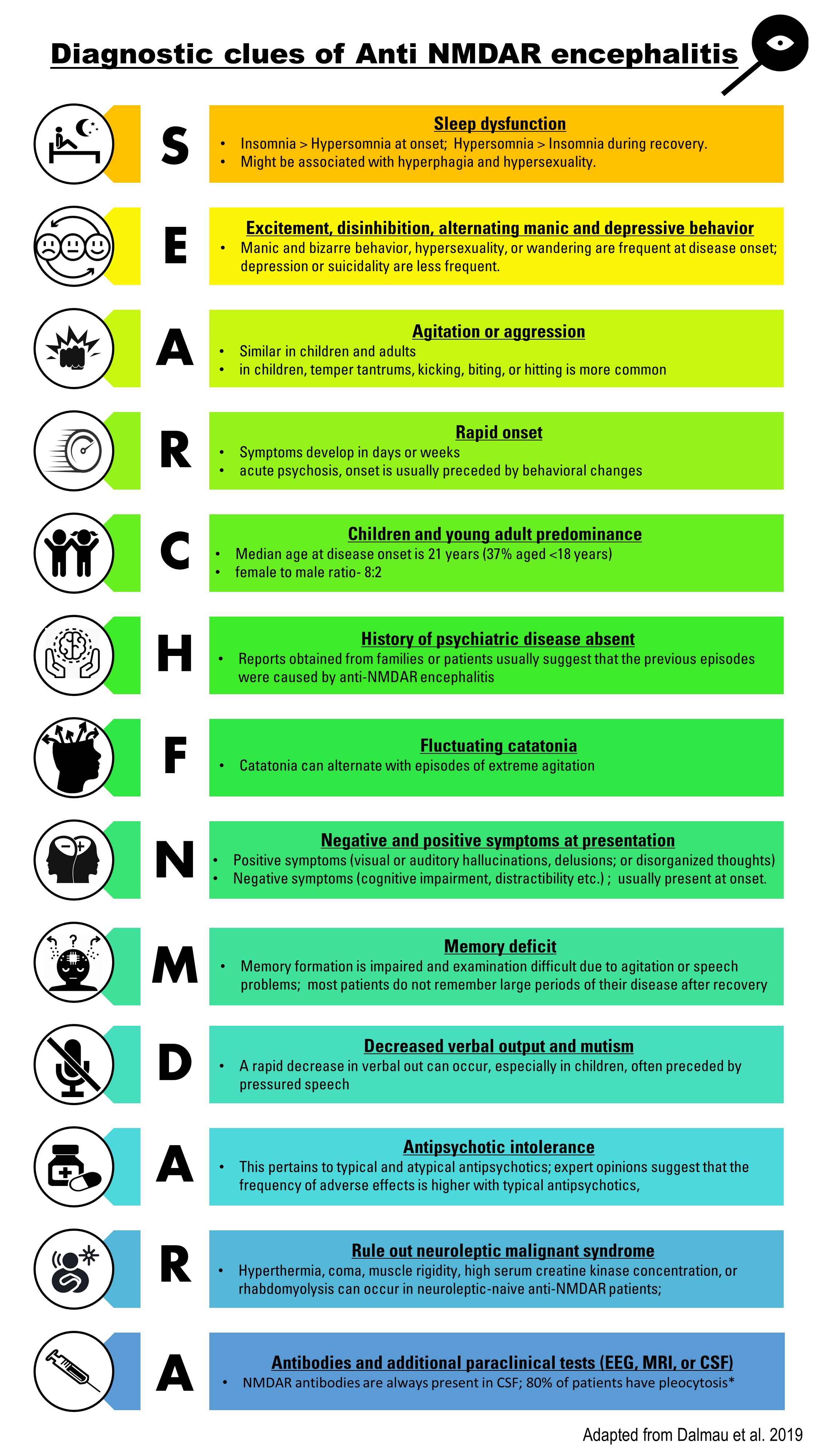
Clinical Features of Psychotic Phase Anti-NMDAR Encephalitis. Findings used to help diagnose anti-NMDAR encephalitis in patients presenting with new acute onset of psychosis.
Contributed by R Agarwal, MBBS. Adapted from Dalmau J, Armangué T, Planagumà J, et al. An update on anti-NMDA receptor encephalitis for neurologists and psychiatrists: mechanisms and models. Lancet Neurol. 2019;18(11):1045-1057. doi: 10.1016/S1474-4422(19)30244-3.
References
Dalmau J, Gleichman AJ, Hughes EG, Rossi JE, Peng X, Lai M, Dessain SK, Rosenfeld MR, Balice-Gordon R, Lynch DR. Anti-NMDA-receptor encephalitis: case series and analysis of the effects of antibodies. The Lancet. Neurology. 2008 Dec:7(12):1091-8. doi: 10.1016/S1474-4422(08)70224-2. Epub 2008 Oct 11 [PubMed PMID: 18851928]
Level 3 (low-level) evidencePădureanu V, Dop D, Pădureanu R, Pîrșcoveanu DFV, Olaru G, Streata I, Bugă AM. Anti-NMDA Receptor Encephalitis: A Narrative Review. Brain sciences. 2025 May 18:15(5):. doi: 10.3390/brainsci15050518. Epub 2025 May 18 [PubMed PMID: 40426689]
Level 3 (low-level) evidenceBrenner J, Ruhe CJ, Kulderij I, Bastiaansen AEM, Crijnen YS, Kret CN, Verkoelen JCP, Tolido AAG, Thomassen B, Kersten LP, de Bruijn MAAM, Olijslagers SHC, Mandarakas MR, Kerstens J, van Steenhoven RW, de Vries JM, Veenbergen S, Schreurs MWJ, Neuteboom RF, Sillevis Smitt PAE, van den Berg E, Titulaer MJ. Long-Term Cognitive, Functional, and Patient-Reported Outcomes in Patients With Anti-NMDAR Encephalitis. Neurology. 2024 Dec 24:103(12):e210109. doi: 10.1212/WNL.0000000000210109. Epub 2024 Nov 20 [PubMed PMID: 39566012]
Hughes EG, Peng X, Gleichman AJ, Lai M, Zhou L, Tsou R, Parsons TD, Lynch DR, Dalmau J, Balice-Gordon RJ. Cellular and synaptic mechanisms of anti-NMDA receptor encephalitis. The Journal of neuroscience : the official journal of the Society for Neuroscience. 2010 Apr 28:30(17):5866-75. doi: 10.1523/JNEUROSCI.0167-10.2010. Epub [PubMed PMID: 20427647]
Level 3 (low-level) evidenceArmangue T, Spatola M, Vlagea A, Mattozzi S, Cárceles-Cordon M, Martinez-Heras E, Llufriu S, Muchart J, Erro ME, Abraira L, Moris G, Monros-Giménez L, Corral-Corral Í, Montejo C, Toledo M, Bataller L, Secondi G, Ariño H, Martínez-Hernández E, Juan M, Marcos MA, Alsina L, Saiz A, Rosenfeld MR, Graus F, Dalmau J, Spanish Herpes Simplex Encephalitis Study Group. Frequency, symptoms, risk factors, and outcomes of autoimmune encephalitis after herpes simplex encephalitis: a prospective observational study and retrospective analysis. The Lancet. Neurology. 2018 Sep:17(9):760-772. doi: 10.1016/S1474-4422(18)30244-8. Epub 2018 Jul 23 [PubMed PMID: 30049614]
Level 2 (mid-level) evidenceCeanga M, Chung HY, Geis C. Anti-NMDA receptor encephalitis: epidemiological differences and common challenges. Annals of translational medicine. 2020 Jun:8(11):716. doi: 10.21037/atm.2020.02.34. Epub [PubMed PMID: 32617336]
Level 2 (mid-level) evidenceDalmau J, Armangué T, Planagumà J, Radosevic M, Mannara F, Leypoldt F, Geis C, Lancaster E, Titulaer MJ, Rosenfeld MR, Graus F. An update on anti-NMDA receptor encephalitis for neurologists and psychiatrists: mechanisms and models. The Lancet. Neurology. 2019 Nov:18(11):1045-1057. doi: 10.1016/S1474-4422(19)30244-3. Epub 2019 Jul 17 [PubMed PMID: 31326280]
Gable MS, Sheriff H, Dalmau J, Tilley DH, Glaser CA. The frequency of autoimmune N-methyl-D-aspartate receptor encephalitis surpasses that of individual viral etiologies in young individuals enrolled in the California Encephalitis Project. Clinical infectious diseases : an official publication of the Infectious Diseases Society of America. 2012 Apr:54(7):899-904. doi: 10.1093/cid/cir1038. Epub 2012 Jan 26 [PubMed PMID: 22281844]
van Coevorden-Hameete MH, Titulaer MJ, Schreurs MW, de Graaff E, Sillevis Smitt PA, Hoogenraad CC. Detection and Characterization of Autoantibodies to Neuronal Cell-Surface Antigens in the Central Nervous System. Frontiers in molecular neuroscience. 2016:9():37. doi: 10.3389/fnmol.2016.00037. Epub 2016 May 31 [PubMed PMID: 27303263]
Iizuka T, Sakai F, Ide T, Monzen T, Yoshii S, Iigaya M, Suzuki K, Lynch DR, Suzuki N, Hata T, Dalmau J. Anti-NMDA receptor encephalitis in Japan: long-term outcome without tumor removal. Neurology. 2008 Feb 12:70(7):504-11 [PubMed PMID: 17898324]
Level 3 (low-level) evidenceLuykx JJ, Visscher R, Winter-van Rossum I, Waters P, de Witte LD, Fleischhacker WW, Lin BD, de Boer N, van der Horst M, Yeeles K, Davidson M, Pollak TA, Hasan A, Lennox BR. Clinical symptoms and psychosocial functioning in patients with schizophrenia spectrum disorders testing seropositive for anti-NMDAR antibodies: a case-control comparison with patients testing negative. The lancet. Psychiatry. 2024 Oct:11(10):828-838. doi: 10.1016/S2215-0366(24)00249-9. Epub [PubMed PMID: 39300641]
Level 2 (mid-level) evidenceTang E, Tang P, Wang W, Wang L. Anti-NMDA receptor encephalitis associated with severe sinoatrial node dysfunction: three case reports and a review of the literature. Neurological sciences : official journal of the Italian Neurological Society and of the Italian Society of Clinical Neurophysiology. 2026 Jun 10:47(7):. pii: 561. doi: 10.1007/s10072-026-09154-6. Epub 2026 Jun 10 [PubMed PMID: 42265428]
Level 3 (low-level) evidenceTitulaer MJ, Höftberger R, Iizuka T, Leypoldt F, McCracken L, Cellucci T, Benson LA, Shu H, Irioka T, Hirano M, Singh G, Cobo Calvo A, Kaida K, Morales PS, Wirtz PW, Yamamoto T, Reindl M, Rosenfeld MR, Graus F, Saiz A, Dalmau J. Overlapping demyelinating syndromes and anti–N-methyl-D-aspartate receptor encephalitis. Annals of neurology. 2014 Mar:75(3):411-28 [PubMed PMID: 24700511]
Level 3 (low-level) evidenceHaruna K, Ishii J, Ota K, Yoshimura H, Fujiwara S, Kawamoto M. Clinical feature of anti-myelin oligodendrocyte glycoprotein antibody-associated cerebral cortical encephalitis with cerebrospinal fluid anti-N-methyl-D-aspartate receptor antibody co-positivity: a single center case series. Immunological medicine. 2026 Jun 15:():1-11. doi: 10.1080/25785826.2026.2688713. Epub 2026 Jun 15 [PubMed PMID: 42296189]
Level 2 (mid-level) evidenceFlorance NR, Davis RL, Lam C, Szperka C, Zhou L, Ahmad S, Campen CJ, Moss H, Peter N, Gleichman AJ, Glaser CA, Lynch DR, Rosenfeld MR, Dalmau J. Anti-N-methyl-D-aspartate receptor (NMDAR) encephalitis in children and adolescents. Annals of neurology. 2009 Jul:66(1):11-8. doi: 10.1002/ana.21756. Epub [PubMed PMID: 19670433]
Blackman G, Lim MF, Pollak T, Al-Diwani A, Symmonds M, Mazumder A, Carter B, Irani S, David A. The clinical relevance of serum versus CSF NMDAR autoantibodies associated exclusively with psychiatric features: a systematic review and meta-analysis of individual patient data. Journal of neurology. 2022 Oct:269(10):5302-5311. doi: 10.1007/s00415-022-11224-6. Epub 2022 Jul 5 [PubMed PMID: 35790561]
Level 1 (high-level) evidenceIizuka T, Kaneko J, Tominaga N, Someko H, Nakamura M, Ishima D, Kitamura E, Masuda R, Oguni E, Yanagisawa T, Kanazawa N, Dalmau J, Nishiyama K. Association of Progressive Cerebellar Atrophy With Long-term Outcome in Patients With Anti-N-Methyl-d-Aspartate Receptor Encephalitis. JAMA neurology. 2016 Jun 1:73(6):706-13. doi: 10.1001/jamaneurol.2016.0232. Epub [PubMed PMID: 27111481]
Lagarde S, Lepine A, Caietta E, Pelletier F, Boucraut J, Chabrol B, Milh M, Guedj E. Cerebral (18)FluoroDeoxy-Glucose Positron Emission Tomography in paediatric anti N-methyl-D-aspartate receptor encephalitis: A case series. Brain & development. 2016 May:38(5):461-70. doi: 10.1016/j.braindev.2015.10.013. Epub 2015 Nov 2 [PubMed PMID: 26542469]
Level 2 (mid-level) evidenceStaley EM, Jamy R, Phan AQ, Figge DA, Pham HP. N-Methyl-d-aspartate Receptor Antibody Encephalitis: A Concise Review of the Disorder, Diagnosis, and Management. ACS chemical neuroscience. 2019 Jan 16:10(1):132-142. doi: 10.1021/acschemneuro.8b00304. Epub 2018 Aug 31 [PubMed PMID: 30134661]
Erlebach R, Brandi G. Effect and timing of operative treatment for teratoma associated N-Methyl-d-Aspartate receptor-antibody encephalitis: A systematic review with meta-analysis. Journal of neuroimmunology. 2023 Sep 15:382():578153. doi: 10.1016/j.jneuroim.2023.578153. Epub 2023 Jul 22 [PubMed PMID: 37499300]
Level 1 (high-level) evidenceBreese EH, Dalmau J, Lennon VA, Apiwattanakul M, Sokol DK. Anti-N-methyl-D-aspartate receptor encephalitis: early treatment is beneficial. Pediatric neurology. 2010 Mar:42(3):213-4. doi: 10.1016/j.pediatrneurol.2009.10.003. Epub [PubMed PMID: 20159432]
Level 3 (low-level) evidenceTitulaer MJ, McCracken L, Gabilondo I, Armangué T, Glaser C, Iizuka T, Honig LS, Benseler SM, Kawachi I, Martinez-Hernandez E, Aguilar E, Gresa-Arribas N, Ryan-Florance N, Torrents A, Saiz A, Rosenfeld MR, Balice-Gordon R, Graus F, Dalmau J. Treatment and prognostic factors for long-term outcome in patients with anti-NMDA receptor encephalitis: an observational cohort study. The Lancet. Neurology. 2013 Feb:12(2):157-65. doi: 10.1016/S1474-4422(12)70310-1. Epub 2013 Jan 3 [PubMed PMID: 23290630]
Level 2 (mid-level) evidenceZhang L, Wu MQ, Hao ZL, Chiang SM, Shuang K, Lin MT, Chi XS, Fang JJ, Zhou D, Li JM. Clinical characteristics, treatments, and outcomes of patients with anti-N-methyl-d-aspartate receptor encephalitis: A systematic review of reported cases. Epilepsy & behavior : E&B. 2017 Mar:68():57-65. doi: 10.1016/j.yebeh.2016.12.019. Epub 2017 Jan 19 [PubMed PMID: 28109991]
Level 3 (low-level) evidenceCai L, Li G, Abdulaziz AT, Gong X, Liu X, Kong X, Guo K, Li A, Li J, Zhou D, Hong Z. Efficacy and safety of different oral prednisone tapering courses in adult anti-NMDAR encephalitis: A multicenter prospective cohort study. Epilepsia. 2024 Nov:65(11):3199-3215. doi: 10.1111/epi.18107. Epub 2024 Sep 26 [PubMed PMID: 39324872]
Seery N, Wesselingh R, Beech P, McLaughlin L, Rushen T, Halliday AJ, Ter Horst L, Griffith SP, Forcadela M, Tan TH, Kazzi C, Nesbitt C, Broadley J, Buzzard K, Duncan A, D'Souza WJ, Tran Y, Van Der Walt A, Skinner G, Taylor BV, Swayne A, Brodtmann A, Gillis D, Butler EG, Kalincik T, Seneviratne UK, Macdonell RA, Blum S, Ramanathan S, Malpas CB, Reddel SW, Hardy TA, O'Brien TJ, Sanfilippo PG, Butzkueven H, Monif M, Australian Autoimmune Encephalitis Consortium. Rituximab Use for Relapse Prevention in Anti-NMDAR Antibody-Mediated Encephalitis: A Multicenter Cohort Study. Neurology(R) neuroimmunology & neuroinflammation. 2025 Jul:12(4):e200395. doi: 10.1212/NXI.0000000000200395. Epub 2025 May 30 [PubMed PMID: 40446185]
Ciano-Petersen NL, Villagrán-García M, Muñiz-Castrillo S, Farina A, Vogrig A, Goncalves D, Nicole F, Rogemond V, Picard G, Aubart M, Psimaras D, Oliver B, Serrano-Castro PJ, Joubert B, Honnorat J. Relapses in Anti-NMDAR Encephalitis: Clinical Characterization and Predictive Features. Neurology(R) neuroimmunology & neuroinflammation. 2025 Jul:12(4):e200421. doi: 10.1212/NXI.0000000000200421. Epub 2025 Jun 18 [PubMed PMID: 40532146]
Guo K, Peng F, Liu J, Long Y, Guo S, Wang H, Yu G, Zhang Y, Han X, Tu E, Zheng Y, Huang J, Zhou Y, An D, Lin G, Wang B, Wang Y, Yang P, Jiang Y, Cui B, Yang Z, Du M, Jiang M, Qin L, Kong X, Gong X, Liu X, Cai L, Li J, Zhou D, Hong Z. Efficacy and Safety of Ofatumumab Treatment for Anti-NMDA Receptor Autoimmune Encephalitis (OFF-AE): A Prospective, Multicenter Cohort Study. Annals of neurology. 2025 Jul:98(1):80-92. doi: 10.1002/ana.27218. Epub 2025 Feb 24 [PubMed PMID: 39991981]
Level 2 (mid-level) evidencede Bruijn MAAM, van Sonderen A, van Coevorden-Hameete MH, Bastiaansen AEM, Schreurs MWJ, Rouhl RPW, van Donselaar CA, Majoie MHJM, Neuteboom RF, Sillevis Smitt PAE, Thijs RD, Titulaer MJ. Evaluation of seizure treatment in anti-LGI1, anti-NMDAR, and anti-GABA(B)R encephalitis. Neurology. 2019 May 7:92(19):e2185-e2196. doi: 10.1212/WNL.0000000000007475. Epub 2019 Apr 12 [PubMed PMID: 30979857]
Wang HY, Li T, Li XL, Zhang XX, Yan ZR, Xu Y. Anti-N-methyl-D-aspartate receptor encephalitis mimics neuroleptic malignant syndrome: case report and literature review. Neuropsychiatric disease and treatment. 2019:15():773-778. doi: 10.2147/NDT.S195706. Epub 2019 Apr 2 [PubMed PMID: 31040676]
Level 3 (low-level) evidenceNeyens RR, Gaskill GE, Chalela JA. Critical Care Management of Anti-N-Methyl-D-Aspartate Receptor Encephalitis. Critical care medicine. 2018 Sep:46(9):1514-1521. doi: 10.1097/CCM.0000000000003268. Epub [PubMed PMID: 29927776]
Balu R, McCracken L, Lancaster E, Graus F, Dalmau J, Titulaer MJ. A score that predicts 1-year functional status in patients with anti-NMDA receptor encephalitis. Neurology. 2019 Jan 15:92(3):e244-e252. doi: 10.1212/WNL.0000000000006783. Epub 2018 Dec 21 [PubMed PMID: 30578370]