 Tools
Tools
 Anatomy, Shoulder and Upper Limb, Forearm Anterior Interosseous Nerve
Introduction
The anterior interosseous nerve is predominantly motor. This nerve is a branch of the median nerve, which is formed from the roots of the C5, C6, C7, C8, and T1 spinal nerves. These nerve roots form the brachial plexus, which divides to form the medial and lateral cords before converging to form the median nerve. The anterior interosseous nerve branches from the median nerve at the radiohumeral joint line, approximately 4 to 6 cm distal to the medial epicondyle and 5 to 8 cm distal to the lateral epicondyle. The nerve then passes between the 2 heads of pronator teres, runs along the volar surface of the flexor digitorum profundus, courses along the interosseous membrane between the ulna and radius and between the flexor digitorum profundus and flexor pollicis longus, branches to innervate these muscles, and ends in the pronator quadratus near the wrist joint. The anterior interosseous nerve innervates 3 muscles: flexor digitorum profundus, flexor pollicis longus, and pronator quadratus.[1]
Structure and Function
Register For Free And Read The Full Article
 Search engine and full access to all medical articles
- 10 free questions in your specialty
- Free CME/CE Activities
- Free daily question in your email
- Save favorite articles to your dashboard
- Emails offering discounts
Learn more about a Subscription to StatPearls Point-of-Care
Structure and Function
The AIN provides motor innervation to deep forearm muscles, including flexor digitorum profundus (radial half/index and middle finger), flexor pollicis longus, and pronator quadratus. It also provides some sensation to the distal radioulnar and carpal joints and joint capsule. The AIN does not provide cutaneous innervation.[1]
Physiologic Variants
Martin-Gruber anastomosis occurs in 10% to 15% of all forearms (motor nerve connection between the median and ulnar nerves). In 50% of these cases, the nerve communication from the median nerve arises from the AIN branch. Therefore, palsy of the AIN could lead to palsy of the intrinsic muscles of the hand normally supplied by the ulnar nerve.[2]
Surgical Considerations
AIN Compressive Neuropathy
The surgical decompression of the AIN is indicated only if a prolonged period of nonoperative treatment fails. The technique may include the release of the superficial arch of flexor digitorum superficialis and lacertus fibrosus, detachment of superficial head of pronator teres, ligation of any crossing vessels, and removal of any space-occupying lesion.[3]
Pediatric Supracondylar Fracture
Closed reduction and percutaneous pinning are indicated in type II and III supracondylar fractures, flexion type, or medial column collapse. The urgency of this surgery depends on the clinical presentation. Of note, an isolated AIN injury is not an urgent surgical consideration and can be done electively.[4]
Clinical Significance
Clinical Examination
The anterior interosseous nerve examination is performed by testing the motor function of the flexor digitorum profundus, flexor pollicis longus, and pronator quadratus. The flexor digitorum profundus tendon is tested by asking the patient to bend the tips of the second and third digits while stabilizing the proximal interphalangeal joints. Uninjured patients can flex the distal interphalangeal joints. The flexor pollicis longus can be tested by instructing the patient to bend the interphalangeal joint of the first digit. The pronator quadratus is tested by positioning the patient's arm at the side with the elbow flexed to 90 degrees and the forearm in supination. The examiner then asks the patient to pronate the forearm while providing resistance. As with all orthopedic examinations, the affected extremity should be compared with the unaffected extremity.[5]
AIN Syndrome
Anterior interosseous nerve syndrome typically presents with forearm pain and partial or complete dysfunction of muscles innervated by the anterior interosseous nerve, including the flexor pollicis longus, flexor digitorum profundus to the second digit and third digit, and pronator quadratus. The anterior interosseous nerve may become entrapped at any of the following locations: the tendinous edge of the deep head of pronator teres (most common cause), flexor digitorum superficialis arcade, edge of the lacertus fibrosus, accessory head of the flexor pollicis longus (Gantzer muscle), accessory muscle from the flexor digitorum superficialis to the flexor digitorum profundus, and aberrant muscles (flexor carpi radialis brevis, palmaris profundus). However, transient neuritis is the most common cause. Complete anterior interosseous nerve palsy typically causes partial or complete loss of motor function in the flexor digitorum profundus to the second digit, flexor digitorum profundus to the third digit, flexor pollicis longus, and pronator quadratus. The function of the flexor digitorum profundus to the third digit may be preserved because of cross-innervation by the ulnar nerve. The patient may demonstrate grip and pinch weakness, specifically with flexion of the first, second, and third digits. The patient may be unable to make an okay sign (Kiloh-Nevin sign) because of an inability to flex the first digit interphalangeal joint and the distal interphalangeal joint of the second digit, indicating complete loss of function of the flexor pollicis longus and flexor digitorum profundus to the second digit. This weakness can be further evaluated by asking the patient to pinch a sheet of paper between the first and second digits using only the fingertips while the examiner slowly pulls the paper away. Patients with anterior interosseous nerve syndrome may compensate for flexor pollicis longus and flexor digitorum profundus to the second digit weakness by using intrinsic hand muscles innervated by the ulnar nerve. The patient may also demonstrate weakness with resisted pronation with the elbow maximally flexed, which tests the pronator quadratus. Patients with Martin-Gruber anastomosis can present with weakness of the intrinsic hand muscles typically supplied by the ulnar nerve. The anterior interosseous nerve does not provide sensory innervation to the skin, so isolated anterior interosseous nerve palsy does not cause sensory deficits.
The differential diagnosis for patients presenting with suspected anterior interosseous nerve syndrome includes trauma, flexor pollicis longus tendon rupture, proximal sites of nerve compression (ie, cervical nerves, brachial plexus), thoracic outlet syndrome, pronator syndrome, carpal tunnel syndrome, and Parsonage-Turner syndrome. Parsonage-Turner syndrome can present as bilateral anterior interosseous nerve neuropathy caused by viral brachial neuritis. Parsonage-Turner syndrome is more likely if the motor loss is preceded by intense shoulder pain and a viral prodrome. Flexor pollicis longus rupture is typically seen in patients with rheumatoid arthritis or a history of trauma and is distinguished from anterior interosseous nerve compressive neuropathy with passive flexion and extension of the wrist. An intact flexor pollicis longus tendon causes the first digit interphalangeal joint and second digit distal interphalangeal joint to flex during this motion. Electromyography and nerve conduction studies can help rule out other, more proximal lesions and assess the severity of anterior interosseous nerve compressive neuropathy. Imaging is typically not helpful for the diagnosis and treatment of anterior interosseous nerve syndrome. The majority of patients improve with nonoperative treatment, including rest and splinting in 90 degrees flexion for 8 to 12 weeks. Surgical decompression of the anterior interosseous nerve is indicated if nonoperative treatment fails.[3][6][7]
Pronator Teres Syndrome
AIN syndrome should be distinguished from pronator teres syndrome, a distinct but similar proximal median neuropathy. Pronator teres syndrome is characterized by vague forearm pain associated with paresthesia of the median nerve. This is distinguished from AIN neuropathy, which is a pure motor palsy.[5]
Pediatric Supracondylar Fractures
Anterior interosseous nerve neuropraxia is the most common nerve palsy associated with pediatric supracondylar fractures. Anterior interosseous nerve palsy is found in 7.6% to 8.6% of supracondylar fractures and alone accounts for 47% of all nerve-related complications and 70% of median nerve injuries. The mechanism of injury is typically a fall onto an outstretched hand. Patients most often present with pain and refusal to move the elbow. Physical examination findings consistent with a supracondylar fracture include gross deformity, swelling, ecchymosis in the antecubital fossa, and limited active elbow motion. Patients with anterior interosseous nerve neuropraxia are unable to flex the interphalangeal joint of the first digit and the distal interphalangeal joint of the second digit (inability to make an okay sign). The mechanism of injury for the anterior interosseous nerve in supracondylar fractures is tenting of the nerve on the fracture or entrapment in the fracture site. Radiography findings indicative of a pediatric supracondylar fracture include the posterior fat pad sign, a lucency on a lateral view along the posterior distal humerus and olecranon fossa that is highly suggestive of occult fracture around the elbow. Other findings include displacement of the anterior humeral line and alteration of the Baumann angle. Nearly all cases of neuropraxia following supracondylar humerus fractures resolve spontaneously. Operative treatment is indicated based on the severity of the fracture.[4][8]
Media
(Click Image to Enlarge)
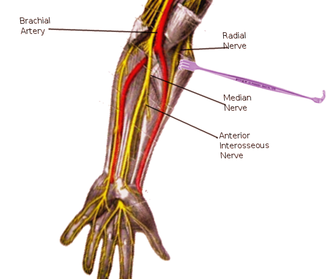
Anterior Interosseous Nerve. The Image shows the anterior interosseous nerve and surrounding nerves.
Contributed by S Bhimji, MD
References
Caetano EB, Vieira LA, Sabongi Neto JJ, Caetano MBF, Sabongi RG. Anterior interosseous nerve: anatomical study and clinical implications. Revista brasileira de ortopedia. 2018 Sep-Oct:53(5):575-581. doi: 10.1016/j.rboe.2018.07.010. Epub 2018 Aug 2 [PubMed PMID: 30245997]
Nakashima T. An anatomic study on the Martin-Gruber anastomosis. Surgical and radiologic anatomy : SRA. 1993:15(3):193-5 [PubMed PMID: 8235961]
Seror P. Anterior interosseous nerve lesions. Clinical and electrophysiological features. The Journal of bone and joint surgery. British volume. 1996 Mar:78(2):238-41 [PubMed PMID: 8666633]
Barrett KK, Skaggs DL, Sawyer JR, Andras L, Moisan A, Goodbody C, Flynn JM. Supracondylar humeral fractures with isolated anterior interosseous nerve injuries: is urgent treatment necessary? The Journal of bone and joint surgery. American volume. 2014 Nov 5:96(21):1793-7. doi: 10.2106/JBJS.N.00136. Epub [PubMed PMID: 25378506]
Level 2 (mid-level) evidenceRodner CM, Tinsley BA, O'Malley MP. Pronator syndrome and anterior interosseous nerve syndrome. The Journal of the American Academy of Orthopaedic Surgeons. 2013 May:21(5):268-75. doi: 10.5435/JAAOS-21-05-268. Epub [PubMed PMID: 23637145]
Chi Y, Harness NG. Anterior interosseous nerve syndrome. The Journal of hand surgery. 2010 Dec:35(12):2078-80. doi: 10.1016/j.jhsa.2010.08.018. Epub 2010 Oct 20 [PubMed PMID: 20961706]
Level 3 (low-level) evidenceKILOH LG, NEVIN S. Isolated neuritis of the anterior interosseous nerve. British medical journal. 1952 Apr 19:1(4763):850-1 [PubMed PMID: 14916168]
Vincelet Y, Journeau P, Popkov D, Haumont T, Lascombes P. The anatomical basis for anterior interosseous nerve palsy secondary to supracondylar humerus fractures in children. Orthopaedics & traumatology, surgery & research : OTSR. 2013 Sep:99(5):543-7. doi: 10.1016/j.otsr.2013.04.002. Epub 2013 Aug 2 [PubMed PMID: 23916783]