 Tools
Tools
 Anterior Cerebral Artery Stroke
Introduction
Infarcts involving the territory of the anterior cerebral artery (ACA) account for approximately 20% of all ischemic strokes (see Image. Anterior Cerebral Artery Stroke). The risk factors and etiology of strokes in this vascular territory mimic those of the other principal cerebral arteries, including hypertension, dyslipidemias, diabetes mellitus, smoking, atherosclerosis, and cardioembolism. The unusual clinical syndromes and many being silent probably result in the underdiagnosis of strokes involving the ACA or its branches.[1]
The ACA emerges from the anterior clinoid segment of the internal carotid artery. It then continues anteromedially towards the longitudinal fissure. Near this point, the anterior communicating artery (ACoA) forms, creating an anastomosis between both ACA’s. Each ACA then advances between the 2 cerebral hemispheres and over the callosal sulcus in a posterior direction towards the parieto-occipital sulcus. Superficial and deep branches emerge along its course. These include Heubner’s, orbitofrontal, frontopolar, anterior internal frontal, middle internal frontal, posterior internal frontal, paracentral, superior parietal, inferior parietal, pericallosal, and callosomarginal arteries. The ACA itself often divides into 5 segments, usually labeled as A1 through A5, or as proximal (A1), ascending (A2, A3), and horizontal segments.[2][3][4]
A significant feature of the ACA is its robust anastomotic complex; this may account for the low rate of infarcts in this vascular distribution.[1] Notably, infarctions simultaneously affecting both cerebral hemispheres may also be present among ACA stroke cases. These are rare and typically occur due to clinically significant anatomical variations at any point along the ACA’s course. The most recognizable patterns are the azygos, bihemispheric, and ACA with a hypoplastic or absent A1 segment.[5]
Etiology
Register For Free And Read The Full Article
 Search engine and full access to all medical articles
- 10 free questions in your specialty
- Free CME/CE Activities
- Free daily question in your email
- Save favorite articles to your dashboard
- Emails offering discounts
Learn more about a Subscription to StatPearls Point-of-Care
Etiology
Hypertension, hypercholesterolemia, diabetes mellitus, and smoking are known risk factors frequently found in stroke patients. These underlie various processes that ultimately result in atherosclerosis of both large and small arteries. Atrial fibrillation is another significant risk factor, its frequency surpassing that of dyslipidemia among patients with ACA stroke in one study.[6]
Atherosclerosis, particularly in the Asian population, is a primary cause of ischemic stroke, which results in stroke secondary to either local branch occlusion by plaque, artery-to-artery embolism, or in situ thrombosis, with the latter considered to be the most prevalent in ACA infarction.[3][4][7]
Cardioembolism from different sources, including atrial fibrillation, intracardiac thrombus, valve disease, and tumors, constitutes a significant cause of ACA infarction. Some reports suggest that cardiac emboli are more frequently the cause of ACA infarcts than of MCA or posterior cerebral artery (PCA) infarcts.[4][6] A hypoplastic or absent A1 segment is thought to facilitate embolic strokes due to increased vascular flow through the unique proximal section that branches off distally into the bilateral ACAs.[5]
Another significant mechanism of ACA stroke is arterial dissection. Although rarely reported in Western populations, other sources report a high prevalence among Japanese patients. Those with stroke secondary to arterial dissection also tend to be younger.[4][8] Less common mechanisms have been described, including vasculitis and coagulation disorders. Vasospasm is another cause. Reported triggers include subarachnoid hemorrhage and pituitary apoplexy. This mechanism has correlated with both unilateral and bilateral ACA infarcts. Some cases have been reported that have no known etiology.[1][3][6][7][8][9]
Distal vessel occlusion secondary to lost or fragmented thrombi associated with the use of intravenous thrombolysis and mechanical thrombectomy is another potential mechanism of ACA stroke. One study evaluating the frequency of ACA embolism in 105 patients undergoing mechanical thrombectomy of occlusions of the M1 segment of the MCA identified 12 new ACA emboli (11.4% of studied cases). ACA infarcts were identified on follow-up imaging in 5.7% of patients. The significance of this particular mechanism of infarction lies in its potential for adverse outcomes secondary to distal occlusions following otherwise effective recanalization of the affected vessel.[10]
Epidemiology
Infarctions of the ACA and its branches are infrequent, accounting for 0.3% to 4.4% of stroke cases reported in different series. Overall, the studies show that males are affected more frequently than females. The mean patient age reported in some ACA infarct studies ranged from 59 to 74.4 years. One study addressed the increased prevalence of ACA infarcts among those older than 85, which was similar to findings in other stroke studies, including all vascular territories. Left-sided ACA infarcts are more frequent.[1][3][6][7][11] Prior stroke history tends to be more frequent among patients with ACA strokes compared to patients with strokes in other vascular territories.[12]
History and Physical
Patients presenting with an acute stroke of the ACA will have varied presentations depending on the size of the infarct and the involved branches of the ACA. The size of the infarct will also influence the clinical presentation. Most commonly, patients present with motor deficits, typically involving the lower extremity contralateral to the infarct site. This finding is present in 86.3% to 90% of patients.[3][6][7] Heubner’s artery and medial striate artery infarcts are associated with contralateral face and arm weakness, resulting from damage to the anteromedial caudate nucleus, anterior limb of the internal capsule, and anterior perforated substance.[4]
A syndrome characterized by homolateral ataxia and crural hemiparesis has been reported as another distinct phenomenon of ACA infarction, attributed to damage to both the corticopontine fibers and the lower-limb strip.[13] Case reports of isolated unilateral axial weakness have been attributed to hypotonia of the paravertebral muscles secondary to strokes involving the primary motor cortex.[14] Other motor disorders related to ACA infarction include hypometria, bradykinesia, global akinesia, loss of reciprocal coordination, Parkinsonian gait, tremor, dystonia, and motor neglect.[11][15] The alien hand syndrome, in which 1 hand appears to be independent and which the patient cannot control, may present in infarcts involving the corpus callosum, frontal lobe, or posterolateral parietal lobe.[16] Abdominal myoclonus has been reported.[17]
Isolated sensory deficits are less common, occurring in 0% to 25% of subjects. When present, they always correlate with a weak extremity.[7][11] One case of an ACA infarct in the lower-limb sensory homunculus has been reported, presenting exclusively with sensory disturbances.[18] Abulia, agitation, motor perseveration, memory impairments, emotional lability, and anosognosia are among the neuropsychological features associated with ACA infarction.[7]
Altered consciousness and speech disorders have been identified as independent variables in 43% of ACA infarction when compared to MCA infarcts.[6] Speech disorders associated with ACA infarction include transcortical motor aphasia, with reports of it occurring following a period of muteness. Damage to the supplementary motor area correlates with these disorders. Another speech disorder found is transcortical mixed aphasia.[3][11] A case has been reported in which the patient's reading and writing changed direction, from right to left, following a stroke involving the left supplementary motor area.[19]
Bilateral ACA infarction is rare. A study involving 48 patients with ACA infarction had only 2 cases, with a mean age at presentation of 40.[3] The most consistent findings in one study, which included patients with bilateral ACA infarction, were frontal disinhibition signs such as enhanced glabellar tap, utilization behavior, forced grasping, snout, and other primitive reflexes. The prefrontal cortex was not always affected in these cases.[15] Paraparesis and akinetic mutism were also documented in the context of bilateral ACA stroke.[3] The presence of an azygos variant ACA can present with bihemispheric involvement, and 1 case has been reported where the presentation mimicked that of basilar artery infarction with a fatal outcome.[20] Headaches also correlate with ACA infarction, specifically in instances of arterial dissection.[4]
Evaluation
Once an acute ischemic stroke is suspected, the standard evaluation includes performing routine airway, breathing, and circulation assessment, checking blood glucose, performing a validated stroke severity scale assessment, and obtaining an accurate, focused history regarding the time of symptom onset or last known well or at baseline. The standardized National Institutes of Health Stroke Scale (NIHSS) can quantify the severity of stroke symptoms. The NIHSS is the preferred scoring system, and scores range from 0 to 42. Higher scores portend a greater disability; however, the definition of "disabling" depends on age, occupation, underlying life-limiting comorbidities, and advance directives.
The crucial step in the evaluation of stroke patients is to obtain brain imaging to ascertain the type and characteristics of the stroke. A noncontrast computed tomography (CT) of the head is the imaging modality of choice. Ischemic changes may be classified as acute, subacute, or chronic, depending on the time at which they present after stroke onset. A CT scan can also rule out intracranial hemorrhage.[21] If an intracranial hemorrhage is present, aneurysmal rupture should be investigated.[3] ACA strokes could be missed on imaging studies, depending on their location or size. One case series found that 37.5% (6 of 16) of ACA infarcts evaluated by CT were identifiable only after using contrast injection or angiography. If the area of hypodensity is small and localized over a sulcus, the infarct could be overlooked.[1][14] Noncontrast head CT should be followed promptly by CT angiography of the head and neck to expedite identification of large-vessel intracranial occlusion (see Image. Left Anterior Cerebral Artery Stenosis).
The finding of a hyperdense lesion in the ACA on CT scan can aid in the diagnosis of stroke in its acute phase. The frequency of this sign in ACA infarcts is similar to that in the territories of the middle cerebral artery and the posterior circulation.[22] As in strokes involving other areas of the brain, magnetic resonance imaging (MRI) is also of critical value in the diagnosis of ACA strokes. MRI with diffusion-weight imaging (DW-MRI) facilitates the demarcation of ischemic boundaries in the territory of the ACA.[3][21] MR angiography can be a helpful adjunct in the evaluation of stroke mechanisms.[7] The goal for completing a head CT or MRI is 25 minutes or less after patient arrival. The National Institutes of Neurological Disorders and Stroke (NINDS) established time frame goals in the evaluation of stroke patients: door to physician less than 10 minutes, door to stroke team less than 15 minutes, door to CT scan less than 25 minutes, and door to drug less than 60 minutes.[23]
Along with accurate history and early imaging, laboratory studies, including capillary blood glucose, complete blood count with platelets, chemistries, coagulation studies, hemoglobin A1c, lipid panel, and markers of hypercoagulability or inflammation, can be useful in identifying the risk factors or establishing the etiology of stroke. The medication checklist is an integral part of the evaluation, particularly for recent anticoagulant use, as contraindications to thrombolytic therapy should be assessed promptly.
Cardiac sources of embolism can be evaluated as part of the work-up with electrocardiographic monitoring and echocardiogram. When an embolic etiology is suspected, and a specific cause is otherwise not clearly identifiable, as in younger patients without established cardiovascular disease risk factors, the Risk of Paradoxical Embolism (RoPE) score can be used to estimate the probability that the embolic stroke could be attributed to a patent foramen ovale (PFO) when found.[24]
Treatment / Management
Acute Stroke Management
Pulse oximetry can guide the use of supplemental oxygen to maintain oxygen saturation greater than 94%. Hyperoxia should be avoided as it may be detrimental in stroke. Hypertension is common in an acute ischemic stroke. Low blood pressure, however, may indicate poor coronary or cerebral perfusion. A blood pressure above 220/120 mm Hg requires treatment in patients who are not candidates for fibrinolysis.[25] For a patient who is a potential candidate for fibrinolysis, immediate reduction of blood pressure below 185/110 mm Hg is required. Usually, titratable short-acting intravenous hypotensive agents are recommended to avoid tanking the blood pressure. The most common agents include labetalol, nicardipine, clevidipine, hydralazine, and enalaprilat.[25]
Thrombolysis
For those who present within the therapeutic window, the decision to treat with intravenous recombinant tissue plasminogen activator or tenecteplase (within 4.5 hours of symptom onset), or to perform endovascular treatment with mechanical thrombectomy, should be made. Major modern stroke guidelines no longer set a strict upper age limit for intravenous thrombolysis (IVT); treatment decisions are now individualized based on overall health, stroke severity, and time from onset, not just chronological age. Only patients with disabling symptoms are considered eligible for thrombolytic treatment.
Eligibility and absolute and relative contraindications should be assessed rapidly. Randomized controlled trials have shown that intravenous administration of recombinant tissue plasminogen activator (alteplase) reduces functional disability, with an absolute risk reduction of 7% to 13% compared with placebo.[25] The ACT randomized clinical trial (n=1600 patients with large vessel occlusion, including ACA/MCA/basilar) found that tenecteplase (0.25/mg/kg max 25/mg) given as a single 5-second IV bolus was noninferior to alteplase (0.9/mg/kg max 90/mg) for the primary outcome of modified Rankin Scale (mRS) score of 0 to 1 at 90 days, with similar rates of mRS of 0 to 2, mortality, and symptomatic intracerebral hemorrhage.[26](A1)
Unfortunately, over half of patients arrive after this time window has closed and are not eligible for thrombolysis. Treatment delays may result from failure to attribute the patient's symptoms to a stroke, and the risk of harm increases with time elapsed from symptom onset.[25] This situation could be of particular concern in ACA strokes, given their sometimes atypical presentation. Furthermore, IVT alone has low recanalization rates in proximal ACA occlusions and may cause distal embolization, potentially reducing the efficacy of endovascular thrombectomy (EVT).[27]
Mechanical thrombectomy
Endovascular treatment with mechanical thrombectomy is another proven treatment modality in the management of patients with acute stroke suffering a large vessel occlusion, although treatment efficacy is highly time-dependent. The procedure is available at tertiary hospitals and requires a stroke team with the expertise to perform timely imaging and interventions. Studies have concluded that while recanalization rates are high, and complication rates are low, the outcomes can be otherwise unsatisfactory and potentially associated with increased morbidity and mortality. The negative outcomes can be attributed to larger infarct volumes and longer times to recanalization.[28][29][30] However, thrombectomy outcomes appear comparable to those of medical treatment, regardless of whether thrombolysis was used.[31](A1)
New guidelines recommend that in patients with acute ischemic stroke within 6 to 24 hours from last known well and who have large vessel occlusion in the anterior circulation, obtaining CT perfusion (CTP), DW-MRI, or MRI perfusion is recommended to aid in selection for mechanical thrombectomy. However, this is only with the strict application of imaging or other eligibility criteria from randomized controlled trials (RCTs) showing benefit in selecting patients for mechanical thrombectomy. The DAWN trial used clinical imaging mismatch (imaging from CTP or DW-MRI and NIHSS scoring) as criteria to select patients with anterior circulation large vessel occlusion (LVO) for mechanical thrombectomy between 6 and 24 hours from last known well. The trial demonstrated an overall functional benefit at 90 days in the treatment group (modified Rankin score [mRS] 0 to 2, 49% versus 13%; adjusted difference, 33%; 95% confidence interval [CI], 21 to 44; probability of superiority greater than 0.999).
The DEFUSE 3 (Endovascular Therapy Following Imaging Evaluation for Ischemic Stroke 3) trial used perfusion core mismatch and maximum core size as criteria in selecting patients with an acute ischemic stroke due to internal carotid artery (ICA) or middle cerebral artery (MCA) occlusion presenting 6 to 16 hours from the last time seen normal. This trial also showed an outcome benefit at 90 days in the treated group (mRS score 0 to 2: 44.6% versus 16.7%; risk ratio [RR], 2.67; 95% CI, 1.60 to 4.48; P > 0.0001). DAWN and DEFUSE 3 are the only trials to show a benefit of mechanical thrombectomy when performed more than 6 hours after symptom onset. Only the criteria from these trials should be used for patient selection for those who might benefit from mechanical thrombectomy.[25] Clinicians should be aware that most of the patients involved in the DAWN and DEFUSE 3 trials had middle cerebral artery occlusions. The multicenter STAR (Stroke Thrombectomy and Aneurysm) trial showed that mechanical thrombectomy for ACA occlusions is safe and efficacious, with the following benefits:
-
Recanalization (modified TICI 2b/3) was achieved in about 80% of patients
-
90-day mortality around 19%
-
Postprocedural complications occurred in approximately 17% of cases
-
Symptomatic intracranial hemorrhage (sICH) rates vary by embolus type (primary isolated ACA versus combined ACA) [32]
(A1)
Compared with more proximal large-vessel occlusions (MCA/ICA), ACA occlusions tend to have lower short-term good functional outcomes and a higher risk of sICH.[32](A1)
ACA stroke can occur following an anterior communicating artery aneurysm rupture, either due to vasospasm or due to inadvertent surgical clipping of the ACA branches or perforator vessels. Intraoperative indocyanine green video angiography can reduce the complications from improper clipping.[33](B2)
The combination of high-risk echocardiographic PFO features and a patient's RoPE score can help further establish stroke causality with the PFO-Associated Stroke Causal Likelihood (PASCAL) classification. In patients younger than the age of 60, candidacy for secondary stroke prevention with PFO closure may be considered if they fall within the "probable" or "possible" categories.[24]
Recurrent Stroke Prevention
Beyond the acute management of stroke, the use of antihypertensives, dual antiplatelet therapy, anticoagulants, and carotid endarterectomy should be used to prevent recurrent events. Antiplatelet therapy or anticoagulants are not recommended within 24 hours after thrombolysis. Administration of a glycoprotein IIb/IIIa receptor inhibitor is not recommended, and a recent Cochrane review showed that these agents correlated with a high risk of intracranial hemorrhage.
Dual antiplatelet therapy (aspirin and clopidogrel) is recommended to start within 24 hours for 21 days in patients with minor stroke for early secondary stroke prevention. The CHANCE trial showed that the primary outcome of a recurrent stroke at 90 days favored dual antiplatelet therapy over aspirin alone (HR 0.68; 95% CI, 0.57 to 0.81, P < 0.0001). Ticagrelor over aspirin in acute stroke treatment is not recommended. According to the SOCRATES trial, with the primary outcome of time to the composite endpoint of stroke, myocardial infarction, or death up to 90 days, ticagrelor was not found to be superior to aspirin (hazard ratio [HR], 0.89; 95% CI, 0.78-1.01; P = 0.07). However, ticagrelor is a reasonable alternative in patients with contraindications to aspirin. The efficacy of tirofiban and eptifibatide is currently unknown.[23][25] Optimizing risk factors is essential for secondary prevention of stroke to improve outcomes following the principal event.[25]
Differential Diagnosis
The differential of stroke in general, as well as one that involves the ACA, includes metabolic, hypoglycemia, infectious (fever, sepsis), cardiovascular (eg, syncope), hypertensive emergency, seizures with Todd paralysis, migraines, tumors, abscess, neuromuscular, and varied neuropsychiatric conditions. Clinicians should adopt strategies to reduce the likelihood of missing the diagnosis in a narrow time window in stroke cases, given the time-sensitive nature of its acute treatment. A suggested approach includes suspecting stroke in the context of acute-onset neurological symptoms, increased clinician awareness of uncommon stroke syndromes, and the performance of a systematic neurological exam to better determine the nature of the problem.[16][34]
Prognosis
In-hospital mortality of ACA stroke patients can range between 0% and 7.8%. This is lower than the 17.3% found for patients with MCA stroke in a study evaluating patients with ACA stroke. Case series show a favorable prognosis for patients, with up to 68% of patients in one series having a modified Rankin scale score of 2 or less at discharge.[3][4][6] Successful vascular recanalization, whether pharmacologic or interventional, is associated with reduced in-hospital mortality and improved functional outcomes.[35]
With regard to specific deficits, studies suggest that aphasia from ACA infarcts tends to improve within a short period, in contrast to that resulting from MCA territory lesions. Infarct size has been found to correlate poorly with functional recovery.[7][11] One case of akinetic mutism reversal with L-DOPA therapy has been reported.[36] Generally, patients with major neurological deficits have a high risk of poor outcomes, regardless of whether or not IVT is administered.
Complications
Many patients with large infarctions are at a high risk of developing brain edema. Early transfer of patients at risk to an institution with neurosurgical expertise should be considered.[25] Recurrent seizures after stroke should receive therapy with antiseizure medications; however, prophylactic use of these drugs is not recommended.[25]
Complications related to IV alteplase administration are intracranial hemorrhage and angioedema. If the patient develops a headache, nausea, vomiting, or new or worsening neurological deficits, cerebral hemorrhage should be suspected; IV alteplase should be discontinued immediately, and a stat head CT scan should be obtained. In the event of signs or symptoms of angioedema, maintaining airway patency should be the primary goal, along with discontinuation of alteplase and consideration of IV methylprednisolone and diphenhydramine. Epinephrine and icatibant, a selective bradykinin B2 receptor antagonist, and plasma-derived C1 esterase inhibitor can be therapeutic considerations for severe angioedema secondary to thrombolysis.[25]
Postoperative and Rehabilitation Care
The recommendation is for early rehabilitation in environments with organized, interprofessional stroke care to improve outcomes for patients with stroke.[25]
Deterrence and Patient Education
Patients with risk factors for stroke, including hypertension, hypercholesterolemia, diabetes mellitus, atrial fibrillation, and smoking, should be educated on the signs and symptoms of stroke. Additionally, they should be encouraged to call 911 if they ever develop stroke symptoms, as patients who utilize emergency medical services frequently have better outcomes. Lifestyle modification, eg, weight loss, limiting carbohydrates and sodium in the diet, and tobacco cessation, can lower the risk of stroke. Patients who have had a stroke should be made aware of the importance of medication adherence and the consequences of inadequate treatment.
Enhancing Healthcare Team Outcomes
ACA stroke accounts for approximately 20% of ischemic strokes and frequently presents with atypical neurologic, behavioral, or speech disturbances that contribute to delayed recognition and underdiagnosis. Common etiologies include atherosclerosis, cardioembolism, arterial dissection, vasospasm, and anatomic vascular variants. Patients often present with contralateral lower-extremity weakness, abulia, aphasia, gait abnormalities, or rare syndromes such as alien hand syndrome or akinetic mutism. Rapid evaluation with neurologic assessment, noncontrast head CT, vascular imaging, and magnetic resonance imaging supports timely diagnosis and treatment selection. Evidence-based management includes intravenous thrombolysis, mechanical thrombectomy for eligible large vessel occlusions, blood pressure optimization, secondary stroke prevention, and monitoring for complications such as intracranial hemorrhage, cerebral edema, and recurrent stroke. Early recognition and treatment substantially improve neurologic recovery and functional outcomes.
Interprofessional collaboration improves patient-centered outcomes by reducing delays in diagnosis, expediting reperfusion therapy, and coordinating longitudinal stroke care. Emergency medical services personnel use validated prehospital stroke scales, establish last-known-well time, initiate rapid transport, and provide advance notification to stroke teams to shorten door-to-imaging and treatment intervals. Emergency physicians, neurologists, radiologists, and advanced practitioners collaborate to interpret imaging, determine thrombolytic or thrombectomy eligibility, and guide acute management. Nurses perform neurologic monitoring, administer medications, assess for complications, reinforce patient education, and support transitions of care. Pharmacists evaluate contraindications, manage thrombolytic and antihypertensive therapies, and reduce medication-related adverse events. Primary care clinicians coordinate long-term vascular risk reduction, medication adherence, smoking cessation, diabetes and hypertension management, and follow-up care. Rehabilitation specialists, speech therapists, and case managers support recovery, functional assessment, discharge planning, and caregiver education. In hospitals without imaging interpretation expertise, teleradiology systems within a telestroke network are useful for rapidly interpreting images for IVT decision-making. Administration of fibrinolysis, guided by telestroke consultation, may be as safe and beneficial as at stroke centers. Telestroke systems are also useful for triaging patients who may be eligible for interfacility transfer for consideration of mechanical thrombectomy.[23][25]
Media
(Click Image to Enlarge)
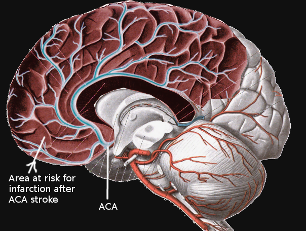
Anterior Cerebral Artery Stroke. Infarcts involving the territory of the anterior cerebral artery (ACA) account for approximately 20% of all ischemic strokes. The ACA itself often divides into 5 segments, usually labeled as A1 through A5, or as proximal (A1), ascending (A2, A3), and horizontal segments.
Contributed by S Bhimji, MD
(Click Image to Enlarge)
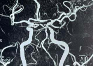
Left Anterior Cerebral Artery Stenosis. Stenosis of the first segment of the left anterior cerebral artery.
Contributed by S Munakomi, MD
References
Kubis N, Guichard JP, Woimant F. Isolated anterior cerebral artery infarcts: A series of 16 patients. Cerebrovascular diseases (Basel, Switzerland). 1999 May-Jun:9(3):185-7 [PubMed PMID: 10207214]
Level 3 (low-level) evidenceChandra A, Li WA, Stone CR, Geng X, Ding Y. The cerebral circulation and cerebrovascular disease I: Anatomy. Brain circulation. 2017 Apr-Jun:3(2):45-56. doi: 10.4103/bc.bc_10_17. Epub 2017 Jul 18 [PubMed PMID: 30276305]
Kumral E, Bayulkem G, Evyapan D, Yunten N. Spectrum of anterior cerebral artery territory infarction: clinical and MRI findings. European journal of neurology. 2002 Nov:9(6):615-24 [PubMed PMID: 12453077]
Toyoda K. Anterior cerebral artery and Heubner's artery territory infarction. Frontiers of neurology and neuroscience. 2012:30():120-2. doi: 10.1159/000333607. Epub 2012 Feb 14 [PubMed PMID: 22377877]
Krishnan M, Kumar S, Ali S, Iyer RS. Sudden bilateral anterior cerebral infarction: unusual stroke associated with unusual vascular anomalies. Postgraduate medical journal. 2013 Feb:89(1048):120-1. doi: 10.1136/postgradmedj-2011-130588. Epub 2012 Sep 6 [PubMed PMID: 22955997]
Level 3 (low-level) evidenceArboix A, García-Eroles L, Sellarés N, Raga A, Oliveres M, Massons J. Infarction in the territory of the anterior cerebral artery: clinical study of 51 patients. BMC neurology. 2009 Jul 9:9():30. doi: 10.1186/1471-2377-9-30. Epub 2009 Jul 9 [PubMed PMID: 19589132]
Level 2 (mid-level) evidenceKang SY, Kim JS. Anterior cerebral artery infarction: stroke mechanism and clinical-imaging study in 100 patients. Neurology. 2008 Jun 10:70(24 Pt 2):2386-93. doi: 10.1212/01.wnl.0000314686.94007.d0. Epub [PubMed PMID: 18541871]
Hensler J, Jensen-Kondering U, Ulmer S, Jansen O. Spontaneous dissections of the anterior cerebral artery: a meta-analysis of the literature and three recent cases. Neuroradiology. 2016 Oct:58(10):997-1004 [PubMed PMID: 27516097]
Level 3 (low-level) evidenceMohindra S, Kovai P, Chhabra R. Fatal Bilateral ACA Territory Infarcts after Pituitary Apoplexy: A Case Report and Literature Review. Skull base : official journal of North American Skull Base Society ... [et al.]. 2010 Jul:20(4):285-8. doi: 10.1055/s-0030-1249243. Epub [PubMed PMID: 21311623]
Level 3 (low-level) evidenceKurre W, Vorlaender K, Aguilar-Pérez M, Schmid E, Bäzner H, Henkes H. Frequency and relevance of anterior cerebral artery embolism caused by mechanical thrombectomy of middle cerebral artery occlusion. AJNR. American journal of neuroradiology. 2013 Aug:34(8):1606-11. doi: 10.3174/ajnr.A3462. Epub 2013 Mar 7 [PubMed PMID: 23471019]
Level 2 (mid-level) evidenceNagaratnam N, Davies D, Chen E. Clinical effects of anterior cerebral artery infarction. Journal of stroke and cerebrovascular diseases : the official journal of National Stroke Association. 1998 Nov-Dec:7(6):391-7 [PubMed PMID: 17895117]
Cho H, Kim T, Kim YD, Na S, Choi YH, Song IU, Chung SW, Koo J, Kwon H, Park JH, Im H. A clinical study of 288 patients with anterior cerebral artery infarction. Journal of neurology. 2022 Jun:269(6):2999-3005. doi: 10.1007/s00415-021-10904-z. Epub 2021 Nov 16 [PubMed PMID: 34783885]
Bogousslavsky J, Martin R, Moulin T. Homolateral ataxia and crural paresis: a syndrome of anterior cerebral artery territory infarction. Journal of neurology, neurosurgery, and psychiatry. 1992 Dec:55(12):1146-9 [PubMed PMID: 1479393]
Level 3 (low-level) evidenceHonig A, Eliahou R, Auriel E. Confined anterior cerebral artery infarction manifesting as isolated unilateral axial weakness. Journal of the neurological sciences. 2017 Feb 15:373():18-20. doi: 10.1016/j.jns.2016.11.061. Epub 2016 Nov 25 [PubMed PMID: 28131184]
Kobayashi S, Maki T, Kunimoto M. Clinical symptoms of bilateral anterior cerebral artery territory infarction. Journal of clinical neuroscience : official journal of the Neurosurgical Society of Australasia. 2011 Feb:18(2):218-22. doi: 10.1016/j.jocn.2010.05.013. Epub 2010 Dec 14 [PubMed PMID: 21159512]
Level 3 (low-level) evidenceEdlow JA, Selim MH. Atypical presentations of acute cerebrovascular syndromes. The Lancet. Neurology. 2011 Jun:10(6):550-60. doi: 10.1016/S1474-4422(11)70069-2. Epub [PubMed PMID: 21601162]
Nokhrina K, Wold J, Raghavan M, Lamotte G. A Case Report of Abdominal Myoclonus Following Anterior Cerebral Artery Stroke. Tremor and other hyperkinetic movements (New York, N.Y.). 2025:15():64. doi: 10.5334/tohm.1119. Epub 2025 Dec 17 [PubMed PMID: 41426499]
Level 3 (low-level) evidenceNishida Y, Irioka T, Sekiguchi T, Mizusawa H. Pure sensory infarct in the territories of anterior cerebral artery. Neurology. 2010 Jul 20:75(3):287. doi: 10.1212/WNL.0b013e3181e8e906. Epub [PubMed PMID: 20644157]
Level 3 (low-level) evidenceReuter L, Carvalho G, Reuter A, Caldeira P. Reading and writing from right to left after anterior cerebral artery stroke. Dementia & neuropsychologia. 2024:18():e20230044. doi: 10.1590/1980-5764-DN-2023-0044. Epub 2024 Apr 15 [PubMed PMID: 38628560]
Macedo M, Florêncio A, Frias C, Nzwalo H, Fidalgo AP. Extensive and bilateral anterior cerebral stroke mimicking basilar artery occlusion. Revista de neurologia. 2023 May 16:76(10):341. doi: 10.33588/rn.7610.2022230. Epub [PubMed PMID: 37165531]
Birenbaum D, Bancroft LW, Felsberg GJ. Imaging in acute stroke. The western journal of emergency medicine. 2011 Feb:12(1):67-76 [PubMed PMID: 21694755]
Jensen UR, Weiss M, Zimmermann P, Jansen O, Riedel C. The hyperdense anterior cerebral artery sign (HACAS) as a computed tomography marker for acute ischemia in the anterior cerebral artery territory. Cerebrovascular diseases (Basel, Switzerland). 2010:29(1):62-7. doi: 10.1159/000256648. Epub 2009 Nov 10 [PubMed PMID: 19907164]
Level 2 (mid-level) evidenceJauch EC, Saver JL, Adams HP Jr, Bruno A, Connors JJ, Demaerschalk BM, Khatri P, McMullan PW Jr, Qureshi AI, Rosenfield K, Scott PA, Summers DR, Wang DZ, Wintermark M, Yonas H, American Heart Association Stroke Council, Council on Cardiovascular Nursing, Council on Peripheral Vascular Disease, Council on Clinical Cardiology. Guidelines for the early management of patients with acute ischemic stroke: a guideline for healthcare professionals from the American Heart Association/American Stroke Association. Stroke. 2013 Mar:44(3):870-947. doi: 10.1161/STR.0b013e318284056a. Epub 2013 Jan 31 [PubMed PMID: 23370205]
Kent DM, Wang AY. Patent Foramen Ovale and Stroke: A Review. JAMA. 2025 Oct 28:334(16):1463-1473. doi: 10.1001/jama.2025.10946. Epub [PubMed PMID: 40720119]
Powers WJ, Rabinstein AA, Ackerson T, Adeoye OM, Bambakidis NC, Becker K, Biller J, Brown M, Demaerschalk BM, Hoh B, Jauch EC, Kidwell CS, Leslie-Mazwi TM, Ovbiagele B, Scott PA, Sheth KN, Southerland AM, Summers DV, Tirschwell DL, American Heart Association Stroke Council. 2018 Guidelines for the Early Management of Patients With Acute Ischemic Stroke: A Guideline for Healthcare Professionals From the American Heart Association/American Stroke Association. Stroke. 2018 Mar:49(3):e46-e110. doi: 10.1161/STR.0000000000000158. Epub 2018 Jan 24 [PubMed PMID: 29367334]
Bala F, Singh N, Buck B, Ademola A, Coutts SB, Deschaintre Y, Khosravani H, Appireddy R, Moreau F, Phillips S, Gubitz G, Tkach A, Catanese L, Dowlatshahi D, Medvedev G, Mandzia J, Pikula A, Shankar JJ, Williams H, Field TS, Manosalva Alzate A, Siddiqui M, Zafar A, Imoukhoude O, Hunter G, Alhabli I, Benali F, Horn M, Hill MD, Shamy M, Sajobi TT, Swartz RH, Menon BK, Almekhlafi M. Safety and Efficacy of Tenecteplase Compared With Alteplase in Patients With Large Vessel Occlusion Stroke: A Prespecified Secondary Analysis of the ACT Randomized Clinical Trial. JAMA neurology. 2023 Aug 1:80(8):824-832. doi: 10.1001/jamaneurol.2023.2094. Epub [PubMed PMID: 37428494]
Level 1 (high-level) evidenceAmerican College of Emergency Physicians Clinical Policies Subcommittee (Writing Committee) on Thrombolytics, Lo BM, Carpenter CR, Milne K, Panagos P, Haukoos JS, Diercks DB, Members of the American College of Emergency Physicians (ACEP) Clinical Policies Committee (Oversight Committee), Diercks DB, Anderson JD, Byyny R, Carpenter CR, Friedman BW, Gemme SR, Gerardo CJ, Godwin SA, Hatten BW, Haukoos JS, Kaji A, Kwok H, Lo BM, Mace SE, Mattu A, Promes SB, Shah KH, Shih RD, Silvers SM, Slivinski A, Smith MD, Thiessen MEW, Thompson JT, Tomaszewski CA, Trent SA, Valente JH, Westafer LM, Wall SP, Yu Y, Lin MP, Finnell JT, Schulz T, Vandertulip K. Clinical Policy: Use of Thrombolytics for the Management of Acute Ischemic Stroke in the Emergency Department. Annals of emergency medicine. 2024 Dec:84(6):e57-e86. doi: 10.1016/j.annemergmed.2024.07.023. Epub [PubMed PMID: 39578010]
Musuka TD, Wilton SB, Traboulsi M, Hill MD. Diagnosis and management of acute ischemic stroke: speed is critical. CMAJ : Canadian Medical Association journal = journal de l'Association medicale canadienne. 2015 Sep 8:187(12):887-93. doi: 10.1503/cmaj.140355. Epub 2015 Aug 4 [PubMed PMID: 26243819]
Uno J, Kameda K, Otsuji R, Ren N, Nagaoka S, Kazushi M, Ikai Y, Gi H. Mechanical Thrombectomy for Acute Anterior Cerebral Artery Occlusion. World neurosurgery. 2018 Dec:120():e957-e961. doi: 10.1016/j.wneu.2018.08.196. Epub 2018 Sep 3 [PubMed PMID: 30189299]
Bhatia S, Nouduri ST, Sankar A, Kann MR, Hardi A, Hudson JS, Al-Bayati AR, Nogueira RG, Lang MJ, Gross BA. Endovascular thrombectomy for acute ischemic stroke with primary occlusion of the anterior cerebral artery: A meta-analysis. Journal of stroke and cerebrovascular diseases : the official journal of National Stroke Association. 2025 Feb:34(2):108208. doi: 10.1016/j.jstrokecerebrovasdis.2024.108208. Epub 2024 Dec 22 [PubMed PMID: 39719206]
Level 1 (high-level) evidenceMeyer L, Stracke P, Broocks G, Elsharkawy M, Sporns P, Piechowiak EI, Kaesmacher J, Maegerlein C, Hernandez Petzsche MR, Zimmermann H, Naziri W, Abdullayev N, Kabbasch C, Diamandis E, Thormann M, Maus V, Fischer S, Möhlenbruch M, Weyland CS, Ernst M, Jamous A, Meila D, Miszczuk M, Siebert E, Lowens S, Krause LU, Yeo L, Tan B, Gopinathan A, Arenillas-Lara JF, Navia P, Raz E, Shapiro M, Arnberg F, Zeleňák K, Martínez-Galdámez M, Alexandrou M, Kastrup A, Papanagiotou P, Kemmling A, Dorn F, Psychogios M, Andersson T, Chapot R, Fiehler J, Hanning U, TOPMOST Study Group. Thrombectomy versus Medical Management for Isolated Anterior Cerebral Artery Stroke: An International Multicenter Registry Study. Radiology. 2023 Apr:307(2):e220229. doi: 10.1148/radiol.220229. Epub 2023 Feb 14 [PubMed PMID: 36786705]
Dabhi N, Mastorakos P, Sokolowski J, Kellogg RT, Park MS. Mechanical Thrombectomy for the Treatment of Anterior Cerebral Artery Occlusion: A Systematic Review of the Literature. AJNR. American journal of neuroradiology. 2022 Dec:43(12):1730-1735. doi: 10.3174/ajnr.A7690. Epub 2022 Nov 3 [PubMed PMID: 36328405]
Level 1 (high-level) evidenceRoessler K, Krawagna M, Dörfler A, Buchfelder M, Ganslandt O. Essentials in intraoperative indocyanine green videoangiography assessment for intracranial aneurysm surgery: conclusions from 295 consecutively clipped aneurysms and review of the literature. Neurosurgical focus. 2014 Feb:36(2):E7. doi: 10.3171/2013.11.FOCUS13475. Epub [PubMed PMID: 24484260]
Level 2 (mid-level) evidenceAllen CM. Differential diagnosis of acute stroke: a review. Journal of the Royal Society of Medicine. 1984 Oct:77(10):878-81 [PubMed PMID: 6387114]
Neal JO, Hu S, Reid J, Matheson K, Gubitz G, Simpkin W, Christian C, Phillips S. Mortality After Total Anterior Circulation Stroke: A 25-Year Observational Study. The Canadian journal of neurological sciences. Le journal canadien des sciences neurologiques. 2023 Jul:50(4):535-541. doi: 10.1017/cjn.2022.262. Epub 2022 Jun 20 [PubMed PMID: 35722755]
Level 2 (mid-level) evidenceGuery D, Ong E, Nighoghossian N. Akinetic mutism reversibility after L-dopa therapy in unilateral left anterior cerebral artery infarction. Neurocase. 2017 Apr:23(2):171-172. doi: 10.1080/13554794.2017.1319493. Epub 2017 Apr 27 [PubMed PMID: 28447507]
Level 3 (low-level) evidence