 Tools
Tools
 Anatomy, Shoulder and Upper Limb, Hand Adductor Pollicis
Introduction
The musculature of the hand is complex, with several superimposed layers of muscles, tendons, and fascial compartments. Complex interactions between these groups modulate specific movements; therefore, understanding their role in hand biomechanics can greatly enhance clinical evaluation and diagnosis for clinicians. The first digit is one of the most important structures of the hand. The first digit’s versatility of movement, compared with the other digits, makes the hand capable of exceptional dexterity. Because the first digit is oriented perpendicular to the other digits, its movements are modulated by a diverse set of specific muscles within the hand. Many muscles contribute to its movements, and one of the most important is the adductor pollicis.
The adductor pollicis muscle is an intrinsic muscle of the hand that lies in the deepest muscular plane of the palm, within the adductor compartment. This triangular, 2-headed muscle is unique. The oblique head originates at the capitate and at the bases of the second and third metacarpals, whereas the transverse head originates from the volar aspect of the third metacarpal. These 2 heads then merge while the fibers travel laterally, forming the tendon of the adductor pollicis, which often contains a sesamoid bone. The tendon then inserts on the medial aspect of the proximal phalanx of the first digit and the extensor hood.
Structure and Function
Register For Free And Read The Full Article
 Search engine and full access to all medical articles
- 10 free questions in your specialty
- Free CME/CE Activities
- Free daily question in your email
- Save favorite articles to your dashboard
- Emails offering discounts
Learn more about a Subscription to StatPearls Point-of-Care
Structure and Function
The main function of the adductor pollicis is to adduct the first digit. Because of the altered plane of the first digit in relation to the palm and other digits, adduction refers to bringing the first digit into a position of opposition at the center of the palm. Additionally, the adductor pollicis can bring the first digit alongside the palm and second digit. The ability to adduct the first digit is critical for hand dexterity. Pinching and gripping are the 2 principal movements that require intact first digit adduction.
Embryology
Beginning on day 26, week 4 of the fetal period, the upper extremity begins to form as a limb bud composed of mesodermal tissue from somites and the lateral plate. The limb forms in a proximal-to-distal direction and is controlled by several signaling centers that produce specific factors controlling differentiation. The signaling centers include the apical ectodermal ridge, which controls proximal-to-distal signaling by inducing differentiation of the underlying mesoderm and programmed cell death in interdigital tissue; the zone of polarizing activity, which controls radioulnar limb formation by producing sonic hedgehog protein; and the Wnt pathway, which regulates the ventral-to-dorsal limb axis.[1][2] Disruptions to any one of these 3 processes can ultimately lead to developmental anomalies of the upper limb.
At about day 36, chondrogenic formations of the digits arise, initiating the formation of hand structures. During the sixth and seventh weeks, early muscle masses begin to form throughout the upper extremity and hand. During this period, the intrinsic hand muscles begin to form, including development of the adductor pollicis. During week 7, interdigital apoptosis occurs, allowing the formation of separate digits, followed by ossification of the upper extremity bones in week 8. The majority of upper extremity development is complete by week 8; therefore, developmental abnormalities of the upper extremity are likely to arise in weeks 3 through 8.
Blood Supply and Lymphatics
The deep palmar arterial arch is the main blood supply to the adductor pollicis. The adductor pollicis is an important anatomic landmark for the radial artery. As the radial artery enters the hand, it passes through the 2 heads of the first dorsal interosseous muscle and then proceeds anteromedially between the 2 heads of the adductor pollicis. After passing between the 2 heads, the artery enters the deep compartment of the hand, forming the deep palmar arch. Superficial lymphatic channels that travel alongside the basilic and cephalic veins drain the hand to the cubital, epitrochlear, and supratrochlear lymph nodes of the elbow. These nodes then drain into the deep brachial and deltopectoral lymph nodes and ultimately terminate at the infraclavicular and axillary lymph nodes.
Nerves
The adductor pollicis is innervated by the deep branch of the ulnar nerve (C8 to T1). Understanding the ulnar nerve pathway is important for assessing clinical signs and symptoms of neuromuscular upper-extremity injuries. The ulnar nerve is the terminal branch of the medial cord of the brachial plexus, and the nerve courses distally, running posterior to the medial epicondyle of the elbow at the cubital tunnel. Once in the forearm, the ulnar nerve pierces the 2 heads of the flexor carpi ulnaris, giving off several branches to innervate 2 forearm muscles. The nerve then traverses the wrist by traveling superficial to the flexor retinaculum and enters the hand via the Guyon canal. Once in the hand, the ulnar nerve divides into superficial and deep branches. The deep branch specifically travels between the muscles of the hypothenar eminence to the deep compartment of the palm to join the deep palmar arterial arch. In addition to the adductor pollicis, the deep branch also innervates the hypothenar muscles, the medial 2 lumbricals, the interossei of the hand, and the palmaris brevis.
Muscles
Nine total muscles contribute to the diverse movements of the first digit. These muscles can be subdivided into 2 groups by the location of the muscle fibers in relation to the first digit: extrinsic muscles of the forearm and intrinsic muscles of the hand. The 4 extrinsic muscles influencing first digit movement include the abductor pollicis longus, which abducts and extends the metacarpophalangeal joint; the extensor pollicis brevis, which extends the metacarpophalangeal joint; the extensor pollicis longus, which extends the metacarpophalangeal and interphalangeal joints; and the flexor pollicis longus, which flexes the metacarpophalangeal and interphalangeal joints.
Five intrinsic muscles facilitate first-digit movement. The first group of intrinsic muscles is the thenar eminence, a group of 3 muscles at the base of the first digit that includes the abductor pollicis brevis, which abducts the metacarpophalangeal joint and flexes the interphalangeal joint; flexor pollicis brevis, which flexes the metacarpophalangeal joint; and the opponens pollicis, which opposes and adducts the first digit. The final 2 intrinsic muscles are the first dorsal interosseous and the adductor pollicis, both of which contribute to first-digit adduction.
Physiologic Variants
The 2 heads of the adductor pollicis can be subdivided into 9 different fascicles. The presence of 9 different fascicles creates considerable variation in exact origins and insertions.[3] Each head of the muscle contains 2 layers of fascicles. The transverse head contains 2 palmar and 2 dorsal fascicles, whereas the oblique head contains 3 dorsal and 2 palmar fascicles. These 9 fascicles create several minor anatomic differences that contribute to variation in the biomechanical properties of individual first digits.
Surgical Considerations
Surrogate Marker for Malnutrition
The thickness of the adductor pollicis muscle (TAPM) has been proposed recently as a relatively accurate marker of malnutrition in surgical patients.[4][5] Because malnutrition is a significant cause of morbidity and mortality in surgical patients, using this measurement may help diagnose malnutrition and improve outcomes. Currently, the data are not sufficient to support routine use of this measurement, although it remains under investigation as a potential option for assessing malnutrition moving forward.
Thumb-in-Palm Deformity
One of the most common manifestations of cerebral palsy in the upper extremity is the thumb-in-palm deformity. This is characterized by a fixed contraction of the thumb at the metacarpophalangeal joint into the palm. Because of the dynamic interplay of many muscles on thumb positioning and movement, correcting significant deformity poses a unique surgical challenge. The 2 main muscle issues that lead to the thumb-in-palm deformity in cerebral palsy are as follows:
- Spasticity and contraction of thenar adduction muscles
- Weakness of voluntary control of thenar abduction muscles [6]
Mild thumb-in-palm deformities can be initially treated with a combination of botulinum toxin injections, occupational therapy, and splinting.[7] Indications for surgery include functional impairment of the grasp and pinch functions secondary to the deformity. The surgical procedure typically is performed in children aged 7 to 10 years because patients in this age group usually can complete postoperative rehabilitation. Contraindications to deformity correction include insufficient cognitive ability to participate in postoperative rehabilitation.[8]
The most common type of thumb-in-palm deformity is a type 1 deformity, in which the first digit is adducted across the palm.[6] The main surgical plan in the correction of this deformity is 3-tiered:
- Release or lengthen spastic muscles: Releasing the adductor pollicis muscle in the palm can achieve this.[9]
- Augment the weak or flaccid muscles: Rerouting of the extensor pollicis longus to the first dorsal compartment increases the strength of the muscle.[10]
- Stabilize the joint if unstable.
Several other surgical techniques are available to correct this particular deformity, along with additional different subtypes of the thumb-in-palm deformity. This surgery, combined with a patient dedicated to post-operative rehabilitation, typically has a very good prognosis regarding regaining dexterity.
Clinical Significance
Ulnar Nerve Entrapment
Assessment of the adductor pollicis function is often used as a diagnostic measure for potential ulnar nerve entrapment at the elbow. These patients typically present with intrinsic muscle atrophy, leading to weakened pinch and grasp strength, difficulty turning keys, and difficulty with activities requiring manual dexterity. Clinically, patients may display compensatory thumb IP joint flexion (via flexor pollicis longus, innervated by the anterior interosseous nerve) in the setting of compromised thumb adduction strength (ie, positive Froment sign). Contralateral comparison is facilitated by asking the patient to grasp a sheet of paper by opposite ends simultaneously.[11][12]
Thumb Collateral Ligament Injury [13]
Injury to the ulnar collateral ligament of the thumb occurs secondary to hyperabduction or hyperextension mechanisms imposed at the metacarpalphalangeal joint. Thumb ulnar collateral ligament–based injuries range from purely ligamentous injuries to nondisplaced or displaced avulsion fractures of the base of the proximal phalanx of the thumb. In displaced injuries, surgery is required secondary to the so-called Stener lesion, which constitutes a bony or ligamentous injury that has displaced above the adductor aponeurosis. These injuries are often called by their respective eponyms, skier thumb for acute injuries or gamekeeper thumb for chronic injuries, and these specific injuries cannot heal without surgical repair.
Neuromuscular Blockade Monitoring
Assessment of adductor pollicis movements with ulnar nerve stimulation is a common method for monitoring neuromuscular blockade in anesthesia.[14] Clinicians must monitor neuromuscular blockade levels to avoid complications related to ventilatory support. For example, assessing the level of neuromuscular blockade before extubation is critical because the patient must have adequate diaphragmatic strength before respiratory support is removed. The test is performed by placing electrodes on the anterior forearm and delivering electrical stimulation to the nerve. Because the ulnar nerve innervates additional muscles besides the adductor pollicis, the palm and other 4 digits are typically taped down to isolate first digit movements. An accelerometer is then attached to the first digit to objectively record the kinematic responses to the specific nerve stimuli delivered through the electrodes. Anesthesiologists can deliver customized signals through the electrodes, and the accelerometer recordings can guide subsequent medication and treatment delivery based on the level of neuromuscular blockade.
Media
(Click Image to Enlarge)
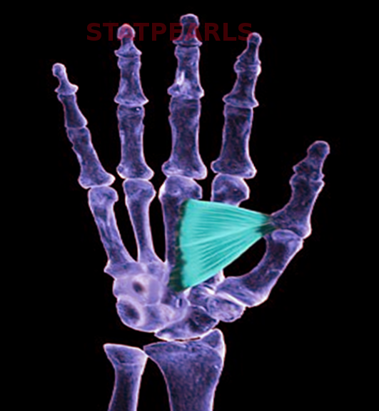
Adductor Pollicis.
Contributed by S Bhimji, MD
References
Guéro S. Developmental biology of the upper limb. Hand surgery & rehabilitation. 2018 Oct:37(5):265-274. doi: 10.1016/j.hansur.2018.03.007. Epub 2018 Jul 21 [PubMed PMID: 30041930]
Daluiski A, Yi SE, Lyons KM. The molecular control of upper extremity development: implications for congenital hand anomalies. The Journal of hand surgery. 2001 Jan:26(1):8-22 [PubMed PMID: 11172363]
Level 3 (low-level) evidenceVan Sint Jan S, Rooze M. Anatomical variations of the intrinsic muscles of the thumb. The Anatomical record. 1994 Jan:238(1):131-46 [PubMed PMID: 8116886]
Caporossi FS, Caporossi C, Borges Dock-Nascimento D, de Aguilar-Nascimento JE. Measurement of the thickness of the adductor pollicis muscle as a predictor of outcome in critically ill patients. Nutricion hospitalaria. 2012 Mar-Apr:27(2):490-5. doi: 10.1590/S0212-16112012000200021. Epub [PubMed PMID: 22732973]
Level 2 (mid-level) evidenceBragagnolo R, Caporossi FS, Dock-Nascimento DB, de Aguilar-Nascimento JE. [Adductor pollicis muscle thickness: a fast and reliable method for nutritional assessment in surgical patients]. Revista do Colegio Brasileiro de Cirurgioes. 2009 Oct:36(5):371-6 [PubMed PMID: 20069147]
Level 2 (mid-level) evidenceHouse JH, Gwathmey FW, Fidler MO. A dynamic approach to the thumb-in palm deformity in cerebral palsy. The Journal of bone and joint surgery. American volume. 1981 Feb:63(2):216-25 [PubMed PMID: 7462278]
Van Heest AE. Applications of botulinum toxin in orthopedics and upper extremity surgery. Techniques in hand & upper extremity surgery. 1997 Mar:1(1):27-34 [PubMed PMID: 16609526]
Van Heest AE. Surgical technique for thumb-in-palm deformity in cerebral palsy. The Journal of hand surgery. 2011 Sep:36(9):1526-31. doi: 10.1016/j.jhsa.2011.06.014. Epub 2011 Aug 4 [PubMed PMID: 21816546]
Luis-Alejandro G, Bárbara G. Adductor Tenotomy Combined with Palmar Capsulodesis for Spastic Thumb-in-Palm Deformity in Cerebral Palsy: Description of a Surgical Technique and Clinical Results. The journal of hand surgery Asian-Pacific volume. 2017 Sep:22(3):315-319. doi: 10.1142/S0218810417500368. Epub [PubMed PMID: 28774242]
Manske PR. Redirection of extensor pollicis longus in the treatment of spastic thumb-in-palm deformity. The Journal of hand surgery. 1985 Jul:10(4):553-60 [PubMed PMID: 4020069]
Goldman SB, Brininger TL, Schrader JW, Koceja DN. Erratum to "A Review of Clinical Tests and Signs for the Assessment of Ulnar Neuropathy" [J Hand Ther 2009;22(3):209-220]. Journal of hand therapy : official journal of the American Society of Hand Therapists. 2010 Jan-Mar:23(1):96. doi: 10.1016/j.jht.2009.10.009. Epub 2010 Jan 29 [PubMed PMID: 30011625]
Goldman SB, Brininger TL, Schrader JW, Curtis R, Koceja DM. Analysis of clinical motor testing for adult patients with diagnosed ulnar neuropathy at the elbow. Archives of physical medicine and rehabilitation. 2009 Nov:90(11):1846-52. doi: 10.1016/j.apmr.2009.06.007. Epub [PubMed PMID: 19887207]
Tsiouri C, Hayton MJ, Baratz M. Injury to the ulnar collateral ligament of the thumb. Hand (New York, N.Y.). 2009 Mar:4(1):12-8. doi: 10.1007/s11552-008-9145-8. Epub 2008 Oct 31 [PubMed PMID: 18975032]
Ortega R, Brull SJ, Prielipp R, Gutierrez A, De La Cruz R, Conley CM. Monitoring Neuromuscular Function. The New England journal of medicine. 2018 Jan 25:378(4):e6. doi: 10.1056/NEJMvcm1603741. Epub [PubMed PMID: 29365307]