 Tools
Tools
 Acute Myocardial Infarction
Introduction
Acute myocardial infarction (AMI) is one of the leading causes of death in the developed world. The prevalence of the disease approaches 3 million people worldwide, with more than 1 million deaths in the United States annually. AMI can be divided into 2 categories: non–ST-segment elevation myocardial infarction (NSTEMI) and ST-segment elevation myocardial infarction (STEMI). Unstable angina resembles an NSTEMI, but normal cardiac markers distinguish it.[1][2][3]
Myocardial infarction (MI) causes permanent damage to the heart muscle due to inadequate oxygen supply. MI can impair diastolic and systolic function, increasing the risk of arrhythmias. Additionally, an MI can lead to various serious complications. Prompt reperfusion and restoration of blood flow to the heart are crucial. Early treatment within 6 hours of symptom onset significantly improves prognosis.
An MI is diagnosed when 2 of the following criteria are met:
- Symptoms of ischemia
- New ST-segment changes or a left bundle branch block (LBBB)
- Presence of pathological Q waves on electrocardiogram (ECG)
- New regional wall motion abnormality on imaging study
- Presence of an intracoronary thrombus at autopsy or angiography
Myocardial infarction (MI), colloquially known as “heart attack,” is defined as myocardial cell death because of prolonged inadequate oxygen supply to a portion of the myocardium, most commonly occur due to acute atherothrombotic coronary artery disease, such as plaque rupture with superimposed thrombosis which is called type 1 MI.Another mechanism include mismatch between the demand and the supply without acute atherothrombosis which is called type 2 MI, coronary artery spasm, embolism, spontaneous coronary artery dissection, and MI with nonobstructive coronary arteries (MINOCA), which accounts for 6–10% of cases and is more prevalent in women. Most myocardial infarctions are due to underlying coronary artery disease, the leading cause of death in the United States. Myocardial infarction may be “silent” and go undetected, or it could present with chest discomfort or pressure that can radiate to the neck, jaw, shoulder, or arm. In addition to the history and physical exam, myocardial ischemia may be associated with ECG changes and elevated biochemical markers such as cardiac troponins.
Etiology
Register For Free And Read The Full Article
 Search engine and full access to all medical articles
- 10 free questions in your specialty
- Free CME/CE Activities
- Free daily question in your email
- Save favorite articles to your dashboard
- Emails offering discounts
Learn more about a Subscription to StatPearls Point-of-Care
Etiology
Acute myocardial infarction occurs due to decreased coronary blood flow, leading to insufficient oxygen supply to the heart and cardiac ischemia. Decreased coronary blood flow is multifactorial. Atherosclerotic plaques classically rupture and lead to thrombosis, contributing to acutely decreased blood flow in the coronary. Other etiologies of myocardial ischemia include coronary artery embolism, which accounts for 2.9% of patients, cocaine-induced ischemia, coronary dissection, and coronary vasospasm.[4][5]
Nonmodifiable Risk Factors
- Sex
- Age
- Family history
- Male pattern baldness
Modifiable Risk Factors
- Smoking
- Dyslipidemia
- Diabetes mellitus
- Hypertension
- Obesity
- Sedentary lifestyle
- Poor oral hygiene
- Presence of peripheral vascular disease
- Elevated levels of homocysteine
Other Causes of MI
- Trauma
- Vasculitis
- Drug use (cocaine)
- Coronary artery anomalies
- Coronary artery emboli
- Aortic dissection
- Excess demand on the heart (hyperthyroidism, anemia)
As stated above, myocardial infarction is closely associated with coronary artery disease. INTERHEART is an international multi-center case-control study which delineated the following modifiable risk factors for coronary artery disease:
- Smoking
- Abnormal lipid profile/blood apolipoprotein (raised ApoB/ApoA1)
- Hypertension
- Diabetes mellitus
- Abdominal obesity (waist/hip ratio) (greater than 0.90 for males and greater than 0.85 for females)
- Psychosocial factors such as depression, loss of the locus of control, global stress, financial stress, and life events including marital separation, job loss, and family conflicts
- Lack of daily consumption of fruits or vegetables
- Lack of physical activity
- Alcohol consumption (weaker association, protective)
The INTERHEART study showed that all the above risk factors were significantly associated with acute myocardial infarction except for alcohol consumption, which showed a weaker association. Smoking and abnormal apolipoprotein ratio showed the strongest association with acute myocardial infarction. The increased risk associated with diabetes and hypertension were found to be higher in women, and the protective effect of exercise and alcohol was also found to be higher in women.
Other risk factors include a moderately high level of plasma homocysteine, which is an independent risk factor of MI. Elevated plasma homocysteine is potentially modifiable and can be treated with folic acid, vitamin B6, and vitamin B12.
Some non-modifiable risk factors for myocardial infarction include advanced age, male gender (males tend to have myocardial infarction earlier in life), genetics (there is an increased risk of MI if a first-degree relative has a history of cardiovascular events before the age of 50). The role of genetic loci that increase the risk for MI is under active investigation.
Epidemiology
Approximately 70% of fatal AMI cases are attributed to occlusion caused by atherosclerotic plaques. As atherosclerosis is the predominant cause of AMI, risk-factors for atherosclerotic disease are often mitigated in disease prevention. Modifiable risk factors account for 90% of AMI cases in men and 94% in women. These modifiable risk factors include cigarette smoking, physical inactivity, hypertension, obesity, elevated cholesterol levels (particularly LDL), and high triglyceride levels. In contrast, age, sex, and family history are nonmodifiable risk factors for atherosclerosis and AMI.[6][7]
The most common cause of death and disability in the USA and worldwide is coronary artery disease. According to the American College of Cardiology and American Heart Association, recent data from 2015–2018 estimate that 20.1 million Americans aged 20 years or older have coronary artery disease , corresponding to a prevalence of 7.2%. The prevalence is higher in men (8.3%) than in women (6.2%) across all age groups. The prevalence of myocardial infarction (MI) in U.S. adults aged 20 years or older is 3.1%, with 8.8 million affected individuals (4.3% of men and 2.1% of women). Mortality from CAD and MI has decreased significantly over recent decades, Despite this fact CAD remains the most common cause of death in the U.S. In 2019, ischemic heart disease accounted for approximately 9.1 million deaths worldwide.
Prevalence of MI in the US Sub-populations
Non-Hispanic Whites
- 4.4% (Male)
- 2.0% (Female)
Non-Hispanic Blacks
- 3.9% (Male)
- 2.3% (Female)
Hispanics
- 3.7% (Male)
- 2.1% (Female)
Non-Hispanic Asians
- 2.7% (Male)
- 0.7% (Female)
Based on the Atherosclerosis Risk in Communities Study (ARIC) performed by National Heart, Lung, and Blood Institute (NHLBI) collected between 2005 and 2014, the estimated annual incidence is 605,000 new MIs and 200,000 recurrent MIs.[8]
The ARIC study also found that the average age at first MI is 65.6 years for males and 72.0 years for females. In the past decades, several studies have shown a declining incidence of MI in the United States.
Pathophysiology
The rupture of an atherosclerotic plaque initiates an inflammatory response of monocytes and macrophages, leading to thrombus formation and platelet aggregation. This process decreases oxygen delivery through the coronary artery, resulting in inadequate oxygenation of the myocardium (see Image. Specimen Showing MI). The subsequent inability to produce ATP in the mitochondria triggers an ischemic cascade, ultimately leading to apoptosis (cell death) of the endocardium or myocardial infarction.
With some exceptions due to genetic variation, coronary arteries exhibit unique and diagnostic territorial distributions. For example, the left anterior descending coronary artery supplies blood flow to the interventricular septum, anterolateral wall, and ventricular apex. The left circumflex artery supplies blood to the inferolateral wall. The right coronary artery supplies the right ventricle. The inferior wall is supplied either by the left circumflex or right coronary artery.[9]
The acute occlusion of one or multiple large epicardial coronary arteries, typically due to atherothrombotic events following plaque rupture or erosion, can lead to acute myocardial infarction.Ischemic injury initiates ultrastructural changes within 10–15 minutes, including sarcolemmal disruption and myofibril relaxation, followed by mitochondrial alterations. The prolonged ischemia ultimately results in coagulative necrosis of myocardial tissue. Necrosis progresses in a wavefront from the subendocardium to the subepicardium over several hours, with the subepicardium often spared initially due to more robust collateral circulation. Depending on the territory affected by the infarction, the cardiac function is compromised. Due to the negligible regeneration capacity of the myocardium, the infarcted area heals by scar formation, and often, the heart is remodeled, characterized by dilation, segmental hypertrophy of remaining viable tissue, and cardiac dysfunction.
Histopathology
The histological changes observed in MI evolve throughout the disease course. At time 0, no microscopic histologic changes are observed. Within 0.5 to 4 hours, light microscopy reveals the waviness of fibers at the tissue periphery along with glycogen depletion. At 4 to 12 hours, the myocardium undergoes coagulation necrosis and edema. At 12 to 24 hours, the gross specimen appears dark and mottled, with contraction band necrosis and neutrophil predominance observed on histopathology.
Subsequently, nuclei reduction occurs between 1 to 3 days, and then, within the next 3 to 7 days, macrophages emerge to clear apoptotic cells. Granulation tissue appears at 7 to 10 days, and collagen type I deposition occurs after 10 days. Ultimately, after 2 months, the myocardium undergoes scarring.
History and Physical
The history and physical examination alone may not always diagnose AMI definitively. The history should focus on the onset, quality, and associated symptoms. Recent studies suggest that diaphoresis and bilateral arm radiating pain are more commonly associated with MI in men. Other associated symptoms may include the following:
- Lightheadedness
- Anxiety
- Cough
- Choking sensation
- Diaphoresis
- Wheezing
- Irregular heart rate
The physical examination should include the patient’s vital signs and appearance, including diaphoresis, pulmonary evaluation, and cardiac auscultation. Specific vital aspects of the examination are as follows:
-
Heart rate: Tachycardia, atrial fibrillation, or ventricular arrhythmia may be present, indicating cardiac electrical disturbances.
-
Pulses: Unequal pulses may be observed if the patient has an aortic dissection, indicating a potential arterial problem.
-
Blood pressure: Blood pressure is typically high in AMI but may become hypotensive if the patient is in shock due to compromised cardiac function.
-
Respiratory findings: Tachypnea and fever may be present, indicating an inflammatory response.
-
Neck veins: Distended neck veins may be observed, suggesting right ventricular failure and increased central venous pressure.
-
Cardiac findings: The heart may exhibit lateral displacement of the apical impulse, a soft S1 sound, a palpable S4 sound, and a new mitral regurgitation murmur. A loud holosystolic murmur radiating to the sternum may indicate ventricular septal rupture.
-
Pulmonary findings: Wheezing and rales may be heard if the patient has developed pulmonary edema, indicating fluid accumulation in the lungs.
-
Extremities: Edema or cyanosis may be present in the extremities, with a sensation of coldness due to compromised circulation.
The imbalance between oxygen supply and the demand leads to myocardial ischemia and can sometimes lead to myocardial infarction. Clinical diagnosis of MI depends on a combination of patient history, electrocardiographic changes and elevated cardiac biomarkers, especially high sensitivity troponin, which is the preferred marker for early and accurate detection of myocardial injury. Most of the time patients with MI present with chest discomfort described as pressure, heaviness, tightness or squeezing in nature, typically retrosternal and may radiate to the shoulders, arms, neck, jaw or upper abdomen. The discomfort is usually diffuse, not localized, and not affected by position or palpation. Symptoms may be provoked by exertion or emotional stress and relieved by rest, but can also occur at rest, especially in acute coronary syndromes. Dyspnea, fatigue, nausea, diaphoresis, palpitations, lightheadedness, and syncope are known to be ischemic equivalents., and these presentations are more common in women, older adults, and patients with diabetes.The MI can also present atypically with subtle findings such as palpitations, or more dramatic manifestations, such as cardiac arrest. The MI can sometimes present with no symptoms.
Evaluation
Early and prompt ECG testing should be employed in all patients presenting with chest pain. Women may experience atypical symptoms such as abdominal pain or dizziness and may present without chest pain (see Image. Myocardial Infarction Warning Signs in Women). Older patients often present with shortness of breath rather than chest pain in cases of MI. All of the aforementioned presentations should prompt ECG testing.[10][11][12]
The ECG is a highly specific diagnostic tool for MI, with a specificity of 95% to 97%. However, its sensitivity is relatively low, at approximately 30%. Right-sided, posterior lead placement and repeat ECG testing can increase ECG sensitivity. For example, peaked T-waves on ECG, known as "hyperacute T waves," often indicate early ischemia and will progress to ST elevation. When present, ST-elevation findings greater than 2 mm in 2 contiguous leads on ECG (inferior: leads II, III, aVF; septal: equal V1, V2; anterior: V3, V4; lateral: I, aVL, V5, V6) are indicative of a STEMI (see Image. ECG With Pardee Waves Indicating AMI). Often, ST depressions are visualized in opposite anatomical regions of the myocardium.
Diagnosing STEMI using the ECG can be challenging, particularly in patients with a left bundle branch block and pacemakers. However, specific criteria have been identified to aid in the diagnosis. Sgarbossa criteria suggest isolated ST elevations in lead aVR can indicate left main coronary artery occlusion in the appropriate clinical setting. Wellens criteria noted deeply biphasic T waves in leads V2 and V3, often predictive of an impending proximal left anterior descending artery occlusion, potentially leading to devastating anterior wall myocardial infarction.
Patients presenting with symptoms of MI may not exhibit diagnostic ST-elevation ECG abnormalities. In cases in which patients have typical chest pain but no obvious ST elevation, further investigation for NSTEMI is warranted. Subtle abnormalities on ECG, such as ST depressions and T-wave changes, may be present. Serial ECGs can be helpful in looking for dynamic changes. ECG findings without acute changes or any abnormalities are common in NSTEMI.
Diagnostic guidelines can aid practitioners in determining whether additional testing helps identify patients with NSTEMI. Considering the limited sensitivity of ECG in STEMI detection, troponin levels have become an almost universally employed diagnostic tool for patients with a clinical history suggestive of MI. The HEART score has been validated and popularized. The HEART score incorporates the clinician's suspicion, patient risk factors, ECG findings, and troponin levels to assess the patient's risk level.
Laboratory Studies
A cardiac troponin test should be the only cardiac marker test ordered. CDC, lipid profile, renal function, and metabolic panel are relevant labs.
Cardiac biomarkers are helpful in the diagnosis of AMI, particularly NSTEMI. Troponin, creatine kinase-MB (CK-MB), and LDH are cardiac markers observed.
A troponin test is the most specific lab test for early detection of AMI; levels of the troponin isoforms I and T are measured. Troponin levels peak at 12 hours and remain elevated for 7 days. High-sensitivity troponin has been approved for use in the United States after being heavily studied and utilized in Europe. Although it is more sensitive than conventional troponin, it is also less specific. Thus, potential challenges include numerous false-positive interpretations.[3]
The level of CK-MB, an isoenzyme of creatine kinase usually found in the myocardium, reaches its peak at 10 hours and normalizes within 2 to 3 days. CK-MB is also not clinically utilized due to its low specificity, rapid rise, and normalization.
LDH levels reach their peak after 72 hours and return to normal within 10 to 14 days. In clinical practice, LDH is not used to diagnose acute MI.
B-type natriuretic peptide (BNP) should not be ordered as a marker for MI; instead, it is more valuable for risk stratification, particularly in patients with MI who subsequently develop heart failure.
Cardiac Imaging
Cardiac angiography is used to perform the percutaneous coronary intervention (PCI) procedure or determine obstructions in the coronary vessels.
An echocardiogram is used to assess wall motion, degree of valve abnormality, ischemic mitral regurgitation, and presence of cardiac tamponade.
The three components in the evaluation of the MI are clinical features, ECG findings, and cardiac biomarkers.
ECG
The resting 12 lead ECG is the first-line diagnostic tool for the diagnosis of acute coronary syndrome (ACS). It should be obtained and interpreted by a trained clinician within 10 minutes of the patient’s arrival in the emergency department. ECG plays an essential role in identifying ST-segment elevation MI and other high risk patterns that require immediate reperfusion therapy.Acute MI is often associated with dynamic changes in the ECG waveform. Serial ECG monitoring can provide important clues to the diagnosis if the initial EKG is non-diagnostic at initial presentation. Serial or continuous ECG recordings may help determine reperfusion or re-occlusion status. A large and prompt reduction in ST-segment elevation is usually seen in reperfusion.
ECG findings suggestive of ongoing coronary artery occlusion (in the absence of left ventricular hypertrophy and bundle branch block):
ST-segment elevation in two contiguous lead (measured at J-point) of
- ≥2 mm in men ≥40 years, ≥2.5 mm in men <40 years, and ≥1.5 mm in women in leads V2–V3
- ≥1 1 mm in all other leads
ST-segment depression and T-wave changes
- New horizontal or down-sloping ST-segment depression ≥0.5 mm in two contiguous leads and/or T-wave inversion >1 mm in two contiguous leads with a prominent R wave or R/S ratio >1
The hyperacute T-wave amplitude, with prominent symmetrical T waves in two contiguous leads, may be an early sign of acute MI that sometimes precede the ST-segment elevation. Other ECG findings associated with myocardial ischemia include cardiac arrhythmias, intraventricular blocks, atrioventricular conduction delays, and loss of precordial R-wave amplitude (less specific finding).
ECG findings alone are not sufficient to diagnose acute myocardial ischemia or acute MI as other conditions such as acute pericarditis, left ventricular hypertrophy (LVH), left bundle branch block (LBBB), Brugada syndrome, Takatsubo syndrome (TTS), and early repolarization patterns also present with ST deviation.
ECG changes associated with prior MI (in the absence of left ventricular hypertrophy and left bundle branch block):
- Any Q wave in lead V2-V3 greater than 0.02 s or QS complex in leads V2-V3
- Q wave > 03 s and greater than 1 mm deep or QS complex in leads I, II, aVL, aVF or V4-V6 in any two leads of contiguous lead grouping (I, aVL; V1-V6; II, III, aVF)
- R wave > 0.04 s in V1-V2 and R/S greater than 1 with a concordant positive T wave in the absence of conduction defect.
Biomarker Detection of MI
Cardiac troponins (I and T) are components of the contractile apparatus of myocardial cells and expressed almost exclusively in the heart. Elevated serum levels of cardiac troponin are not specific to the underlying mode of injury (ischemic vs. tension). The rising and/or falling pattern of cardiac troponins (cTn) values with at least one value above the 99 percentile of upper reference limit (URL) associated with symptoms of myocardial ischemia would indicate an acute MI. Serial testing of cTn values at 0 hours, 3 hours, and 6 hours would give a better perspective on the severity and time course of the myocardial injury. Depending on the baseline cTn value, the rising/falling pattern is interpreted. If the cTn baseline value is markedly elevated, a minimum change of greater than 20% in follow up testing is significant for myocardial ischemia. Creatine kinase MB isoform can also be used in the diagnosis of MI, but it is less sensitive and specific than cTn level.
Imaging
Different imaging techniques are used to assess myocardial perfusion, myocardial viability, myocardial thickness, thickening and motion, and the effect of myocyte loss on the kinetics of para-magnetic or radio-opaque contrast agents indicating myocardial fibrosis or scars. Some imaging modalities that can be used are echocardiography, radionuclide imaging, and cardiac magnetic resonance imaging (cardiac MRI). Regional wall motion abnormalities induced by ischemia can be detected by echocardiography almost immediately after the onset of ischemia when greater than 20% transmural myocardial thickness is affected. Cardiac MRI provides an accurate assessment of myocardial structure and function.
Treatment / Management
All patients with STEMI and NSTEMI require immediate administration of nonenteric-coated, chewable aspirin with a loading dose of 162 mg to 325 mg.[13] Additionally, patients should have intravenous access established, and oxygen supplementation should be administered if their oxygen saturation falls below 91%. Opioids may be used for pain control in addition to sublingual nitroglycerin if the patient's blood pressure is within an acceptable range.[14][15][16](B2)
The primary treatment approach for STEMI involves immediate reperfusion. The preferred method is emergent PCI. Before PCI, patients should receive dual antiplatelet therapy, which includes intravenous heparin infusion and an adenosine diphosphate inhibitor receptor (P2Y12 inhibitor), most commonly ticagrelor. A glycoprotein IIb/IIIa inhibitor or direct thrombin inhibitor may also be administered during the PCI procedure. If PCI is unavailable within 90 minutes of diagnosing STEMI, reperfusion using an intravenous thrombolytic agent should be attempted.
In stable asymptomatic patients with NSTEMI, the initial management approach is typically medical, focusing on using antiplatelet agents and other appropriate medications. However, PCI can be performed within 48 hours of admission if necessary. This delayed PCI strategy has been shown to potentially improve in-hospital mortality and decrease the length of hospital stay for NSTEMI patients.
In NSTEMI patients who experience refractory ischemia or ischemia with hemodynamic or electrical instability, PCI should be performed emergently.
Before discharge for acute MI, patients may routinely be prescribed aspirin, a high-dose statin, a beta-blocker, and possibly an ACE inhibitor.
If PCI is considered the treatment option for an acute MI, it is recommended to perform the procedure within 12 hours of symptom onset. If fibrinolytic therapy is considered the primary reperfusion strategy, it should be initiated within 120 minutes. In addition to reperfusion strategies, parenteral anticoagulation is recommended for all patients with acute MI, regardless of whether they undergo PCI or receive fibrinolytic therapy.
Acute Management
Reperfusion therapy is indicated in all patients with symptoms of ischemia of less than 12-hours duration and persistent ST-segment elevation. Primary percutaneous coronary intervention (PCI) is preferred to fibrinolysis if the procedure can be performed <120 minutes of ECG diagnosis. If there is no immediate option of PCI (>120 minutes), fibrinolysis should be started within 10 minutes of STEMI after ruling out contraindications. If transfer to a PCI center is possible in 60 to 90 minutes after a bolus of the fibrinolytic agent and patient meets reperfusion criteria, a routine PCI can be done, or a rescue PCI can be planned. If fibrinolysis is planned, it should be carried out with fibrin-specific agents such as tenecteplase, alteplase, or reteplase (class I).
Relief of pain, breathlessness, and anxiety: The chest pain due to myocardial infarction is associated with sympathetic arousal, which causes vasoconstriction and increased workload for the ischemic heart. Intravenous opioids (e.g., morphine) are the analgesics most commonly used for pain relief (Class IIa). The results from CRUSADE quality improvement initiative have shown that the use of morphine may be associated with a higher risk of death and adverse clinical outcomes. The study was done from the CIRCUS (Does Cyclosporine Improve outcome in STEMI patients) database, which showed no significant adverse events associated with morphine use in a case of anterior ST-segment elevation MI. A mild anxiolytic (usually a benzodiazepine) may be considered in very anxious patients (class IIa). Supplemental oxygen is indicated in patients with hypoxemia (SaO2 <90% or PaO2 <60mm Hg) (Class I).
Nitrates: Intravenous nitrates are more effective than sublingual nitrates with regard to symptom relief and regression of ST depression (NSTEMI). The dose is titrated upward until symptoms are relieved, blood pressure is normalized in hypertensive patients, or side effects such as a headache and hypotension are noted.
Beta-blockers: This group of drugs reduces myocardial oxygen consumption by lowering heart rate, blood pressure, and myocardial contractility. They block beta receptors in the body, including the heart, and reduce the effects of circulating catecholamines. Beta-blockers should not be used in suspected coronary vasospasm.
Platelet inhibition: Aspirin is recommended in both STEMI and NSTEMI in an oral loading dose of 150 to 300 mg (non-enteric coated formulation) and a maintenance dose of 75 to 100 mg per day long-term regardless of treatment strategy (class I). Aspirin inhibits thromboxane A2 production throughout the lifespan of the platelet.
Most P2Y12 inhibitors are inactive prodrugs (except for ticagrelor, which is an orally active drug that does not require activation) that require oxidation by hepatic cytochrome P450 system to generate an active metabolite which selectively inhibits P2Y12 receptors irreversibly. Inhibition of P2Y12 receptors leads to inhibition of ATP induced platelet aggregation. The commonly used P2Y12 inhibitors are clopidogrel, prasugrel, and ticagrelor.
The loading dose for clopidogrel is 300 to 600 mg loading dose followed by 75 mg per day.
Prasugrel, 60 mg loading dose, and 10 mg per day of a maintenance dose have a faster onset when compared to clopidogrel.
Patients undergoing PCI should be treated with dual antiplatelet therapy (DAPT) with aspirin + P2Y12 inhibitor and a parenteral anticoagulant. In PCI, the use of prasugrel or ticagrelor is found to be superior to clopidogrel. Aspirin and clopidogrel are also found to decrease the number of ischemic events in NSTEMI and UA.
The anticoagulants used during PCI are unfractionated heparin, enoxaparin, and bivalirudin. The bivalirudin is recommended during primary PCI if the patient has heparin-induced thrombocytopenia.
Long-Term Management
Lipid-lowering treatment: It is recommended to start high-intensity statins that reduce low-density lipoproteins (LDLs) and stabilize atherosclerotic plaques. High-density lipoproteins are found to be protective.
Antithrombotic therapy: Aspirin is recommended lifelong, and the addition of another agent depends on the therapeutic procedure done, such as PCI with stent placement.
ACE inhibitors are recommended in patients with systolic left ventricular dysfunction, or heart failure, hypertension, or diabetes.
Beta-blockers are recommended in patients with LVEF less than 40% if no other contraindications are present.
Antihypertensive therapy can maintain a blood pressure goal of less than 140/90 mm Hg.
Mineralocorticoid receptor antagonist therapy is recommended in a patient with left ventricular dysfunction (LVEF less than 40%).
Glucose lowering therapy in people with diabetes to achieve current blood sugar goals.
Lifestyle Modifications
Smoking cessation is the most cost-effective secondary measure to prevent MI. Smoking has a pro-thrombotic effect, which has a strong association with atherosclerosis and myocardial infarction.
Diet, alcohol, and weight control: A diet low in saturated fat with a focus on whole grain products, vegetables, fruits, and the fish is considered cardioprotective. The target level for bodyweight is body mass index of 20 to 25 kg/m2 and waist circumference of <94 cm for the men and <80 cm for the female.
Differential Diagnosis
Differential diagnoses for AMI include the following:
- Aortic dissection: This is a life-threatening condition with a tear in the inner layer of the aorta, leading to the separation of the layers and potential obstruction of blood flow.
- Pericarditis: Inflammation of the pericardium can cause chest pain that may resemble an AMI.
- Acute gastritis: Inflammation of the stomach lining can cause upper abdominal pain that may be mistaken for cardiac-related chest pain.
- Acute cholecystitis: Inflammation of the gallbladder can cause right upper-quadrant abdominal pain that can radiate to the chest and mimic cardiac symptoms.
- Asthma: Acute asthma exacerbation can lead to shortness of breath, wheezing, and chest tightness, which may be mistaken for cardiac symptoms.
- Esophagitis: Inflammation of the esophagus can cause chest pain, mainly if it is related to gastroesophageal reflux disease (GERD) or other esophageal disorders.
- Myocarditis: Heart muscle inflammation can present with chest pain and other symptoms similar to AMI.
- Pneumothorax: A collapsed lung can cause sudden chest pain and difficulty breathing, mimicking cardiac-related symptoms.
- Pulmonary embolism: Blockage of a pulmonary artery by a blood clot can lead to chest pain, shortness of breath, and other symptoms that may resemble an AMI.
- Angina pectoris
- Non-ST segment elevation myocardial infarction (NSTEMI)
- ST-segment elevation myocardial infarction (STEMI)
- Pulmonary embolism
- Pneumothorax
Prognosis
AMI carries a significant risk of mortality outside of the hospital environment. Data indicate that at least one-third of patients succumb to the condition before reaching the hospital, and an additional 40% to 50% do not survive upon arrival. Furthermore, another 5% to 10% of patients will experience mortality within the first 12 months after an MI.
The prognosis of AMI depends on the extent of muscle damage. Better outcomes are seen in patients who undergo early perfusion, including thrombolytic therapy within 30 minutes of arrival or PCI within 90 minutes. Patients with preserved ejection fraction have better outcomes than those with reduced ejection fraction.
Medical management following an AMI is crucial for improving long-term outcomes. Initiating medications, such as aspirin, beta-blockers, and ACE inhibitors, is standard practice to help prevent recurrent cardiovascular events.
Factors that negatively affect prognosis include the following:
- Diabetes
- Advanced age
- Prior MI, peripheral vascular disease, or stroke
- Delayed reperfusion
- Diminished ejection fraction (the strongest predictor)
- Presence of congestive heart failure
- Elevated C-reactive protein and BNP levels
- Depression
A high readmission rate affects about 50% of patients within the first 12 months after the initial MI. The overall prognosis depends on factors such as the ejection fraction, age, and comorbidities. Patients who do not undergo revascularization procedures have poorer outcomes than those who undergo revascularization. The most favorable prognosis is observed in patients with early and successful reperfusion and preserved left ventricular function.[17][18][19]
Despite many advances in treatment, acute MI still carries a mortality rate of 5-30%; the majority of deaths occur prior to arrival to the hospital. In addition, within the first year after an MI, there is an additional mortality rate of 5% to 12%. The overall prognosis depends on the extent of heart muscle damage and ejection fraction. Patients with preserved left ventricular function tend to have good outcomes. Factors that worsen prognosis include:
- Diabetes
- Advanced age
- Delayed reperfusion
- Low ejection fraction
- Presence of congestive heart failure
- Elevations in C-reactive protein and B-type natriuretic peptide (BNP) levels
- Depression
Complications
Primary complications of AMI include the following:
- New-onset mitral regurgitation
- Ventricular septal rupture
- Left ventricular aneurysm
- Arrhythmias
- Emboli
Type and Manifestation
I: Ischemic
- Reinfarction
- Extension of infarction
- Angina
II: Arrhythmias
- Supraventricular or ventricular arrhythmia
- Sinus bradycardia and atrioventricular block
III: Mechanical
- Myocardial dysfunction
- Cardiac failure
- Cardiogenic shock
- Cardiac rupture (Free wall rupture, ventricular septal rupture, papillary muscle rupture)
IV: Embolic
- Left ventricular mural thrombus,
- Peripheral embolus
V: Inflammatory
- Pericarditis (infarct associated pericarditis, late pericarditis, or post-cardiac injury pericarditis)
- Pericardial effusion
Postoperative and Rehabilitation Care
Cardiac rehabilitation plays a vital role in the recovery of patients following an AMI. Research has demonstrated the numerous benefits of cardiac rehabilitation, including improvements in quality of life, reduction of disability, and a decrease in mortality rates.[20][21]
The rehabilitation process should be individualized to meet each patient's specific needs, available resources, established goals, and physical abilities before and after the MI event. Collaboration among rehabilitation therapists and the rest of the interprofessional care team is vital for continuity of care.[20][22]
Cardiac rehabilitation has also been shown to reduce future risk factors for individuals following an AMI. Annual follow-up results demonstrate that cardiac rehab can reduce the risk of future cardiovascular events.[23]
Deterrence and Patient Education
For individuals experiencing symptoms of a possible AMI, the following actions are recommended:
-
Seek immediate medical attention if symptoms suggest an MI, such as chest pain or discomfort, shortness of breath, nausea, or lightheadedness. Go to the emergency department as soon as possible.
- Contact emergency services if nitroglycerin does not provide relief.
- Adhere to a low-salt diet to help manage high blood pressure and reduce the heart's workload.
- Enroll in cardiac rehabilitation designed to support individuals recovering from cardiac conditions, including AMI.
- Cease smoking, as it is a major risk factor for cardiovascular disease.
- Remain compliant with medication administration.
Enhancing Healthcare Team Outcomes
An interprofessional team specializing in heart disease manages AMI. In addition to the cardiologist, the team usually consists of a cardiac surgeon, an interventional cardiologist, an intensivist, a cardiac rehabilitation specialist, critical care or cardiology nurses, and physical therapists. Because AMI is potentially life-threatening, it is crucial to prioritize patient education regarding recognizing symptoms and the importance of seeking immediate medical attention. The pharmacist, nurse practitioner, and primary care providers should educate patients regarding nitroglycerin administration, emphasizing when to seek immediate medical attention if the medication does not provide relief.
Time to reperfusion is limited, and early initiation of treatment can significantly improve outcomes. The care team's initial assessment needs to be timely and cardiology consulted immediately. The cardiologist evaluates the patient's condition, considering the duration of symptoms and contraindications, to determine the appropriate treatment approach. This may involve thrombolysis or PCI to restore blood flow to the blocked coronary artery.
Patients with AMI require close monitoring and specialized care in the ICU. ICU nurses are crucial in monitoring vital signs, administering medications, assessing for complications, and promptly communicating abnormal clinical signs or laboratory parameters to the interprofessional team.
Avoiding premature discharge is crucial because complications of an MI can manifest up to a week after the initial event. The interprofessional team should collaborate to ensure patients are stabilized, monitored appropriately, and educated on signs and symptoms that may indicate worsening or recurrent MI.
Nurses play a vital role in educating patients about reducing risk factors for coronary artery disease. This education includes lifestyle modifications, medication adherence, and ongoing monitoring of blood pressure, cholesterol levels, and blood sugar control.
The involvement of a social worker or case manager is beneficial in facilitating home care arrangements, coordinating cardiac rehabilitation programs, and addressing the need for any support services patients may require at home.
Pharmacists play a crucial role in addressing medication-related aspects of patient care. They provide education on appropriate medication dosing, potential adverse effects, drug interactions, and medication adherence to optimize treatment outcomes.
Following discharge, the patient must participate in a cardiac rehabilitation program, adopt a healthy diet, cease smoking, abstain from alcohol consumption, achieve weight reduction (if appropriate), and manage cholesterol and blood glucose levels. The patient should be educated on the importance of medication compliance to lower blood pressure and cholesterol.[24][25][26] Pharmacists review prescribed medications, check for interactions, and provide patient education about the importance of compliance.
Media
(Click Image to Enlarge)
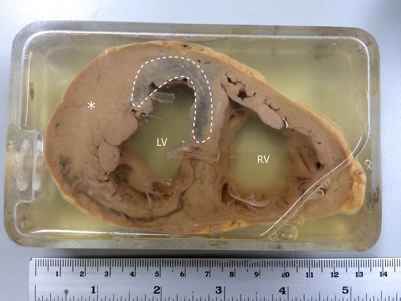
Specimen Showing Myocardial Infarction. MI is observed in the left ventricle and the interventricular septum. The asterisk(*) indicates left ventricular hypertrophy.
Contributed by Wikimedia Commons (CC by 4.0) https://creativecommons.org/licenses/by/4.0/
(Click Image to Enlarge)
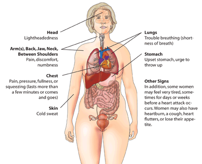
Myocardial Infarction (Heart Attack) Warning Signs in Women.
U.S. Department of Health and Human Services Office on Women's Health
(Click Image to Enlarge)
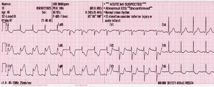
Electrocardiogram With Pardee Waves. Pardee waves indicate acute myocardial infarction in the inferior leads II, III, and augmented voltage foot with reciprocal changes in the anterolateral leads.
Glenlarson, Public Domain, via Wikimedia Commons
(Click Video to Play)
Transesophageal Echocardiography, Pulmonary Embolism. Acute ECG segment elevation mimicking myocardial infarction in a patient with pulmonary embolism.
T Goslar, M Podbregar, Public Domain, via Wikimedia Commons
References
Nascimento BR, Brant LCC, Marino BCA, Passaglia LG, Ribeiro ALP. Implementing myocardial infarction systems of care in low/middle-income countries. Heart (British Cardiac Society). 2019 Jan:105(1):20-26. doi: 10.1136/heartjnl-2018-313398. Epub 2018 Sep 29 [PubMed PMID: 30269080]
Barberi C, van den Hondel KE. The use of cardiac troponin T (cTnT) in the postmortem diagnosis of acute myocardial infarction and sudden cardiac death: A systematic review. Forensic science international. 2018 Nov:292():27-38. doi: 10.1016/j.forsciint.2018.09.002. Epub 2018 Sep 17 [PubMed PMID: 30269044]
Level 1 (high-level) evidenceAlaour B, Liew F, Kaier TE. Cardiac Troponin - diagnostic problems and impact on cardiovascular disease. Annals of medicine. 2018 Dec:50(8):655-665. doi: 10.1080/07853890.2018.1530450. Epub 2018 Nov 21 [PubMed PMID: 30265127]
Massberg S, Polzin A. [Update ESC-Guideline 2017: Dual Antiplatelet Therapy]. Deutsche medizinische Wochenschrift (1946). 2018 Aug:143(15):1090-1093. doi: 10.1055/a-0549-8230. Epub 2018 Jul 30 [PubMed PMID: 30060279]
Scheen AJ. [From atherosclerosis to atherothrombosis : from a silent chronic pathology to an acute critical event]. Revue medicale de Liege. 2018 May:73(5-6):224-228 [PubMed PMID: 29926559]
Berg DD, Wiviott SD, Braunwald E, Guo J, Im K, Kashani A, Gibson CM, Cannon CP, Morrow DA, Bhatt DL, Mega JL, O'Donoghue ML, Antman EM, Newby LK, Sabatine MS, Giugliano RP. Modes and timing of death in 66 252 patients with non-ST-segment elevation acute coronary syndromes enrolled in 14 TIMI trials. European heart journal. 2018 Nov 7:39(42):3810-3820. doi: 10.1093/eurheartj/ehy556. Epub [PubMed PMID: 30239711]
Deng D, Liu L, Xu G, Gan J, Shen Y, Shi Y, Zhu R, Lin Y. Epidemiology and Serum Metabolic Characteristics of Acute Myocardial Infarction Patients in Chest Pain Centers. Iranian journal of public health. 2018 Jul:47(7):1017-1029 [PubMed PMID: 30182001]
Benjamin EJ, Virani SS, Callaway CW, Chamberlain AM, Chang AR, Cheng S, Chiuve SE, Cushman M, Delling FN, Deo R, de Ferranti SD, Ferguson JF, Fornage M, Gillespie C, Isasi CR, Jiménez MC, Jordan LC, Judd SE, Lackland D, Lichtman JH, Lisabeth L, Liu S, Longenecker CT, Lutsey PL, Mackey JS, Matchar DB, Matsushita K, Mussolino ME, Nasir K, O'Flaherty M, Palaniappan LP, Pandey A, Pandey DK, Reeves MJ, Ritchey MD, Rodriguez CJ, Roth GA, Rosamond WD, Sampson UKA, Satou GM, Shah SH, Spartano NL, Tirschwell DL, Tsao CW, Voeks JH, Willey JZ, Wilkins JT, Wu JH, Alger HM, Wong SS, Muntner P, American Heart Association Council on Epidemiology and Prevention Statistics Committee and Stroke Statistics Subcommittee. Heart Disease and Stroke Statistics-2018 Update: A Report From the American Heart Association. Circulation. 2018 Mar 20:137(12):e67-e492. doi: 10.1161/CIR.0000000000000558. Epub 2018 Jan 31 [PubMed PMID: 29386200]
Haig C, Carrick D, Carberry J, Mangion K, Maznyczka A, Wetherall K, McEntegart M, Petrie MC, Eteiba H, Lindsay M, Hood S, Watkins S, Davie A, Mahrous A, Mordi I, Ahmed N, Teng Yue May V, Ford I, Radjenovic A, Welsh P, Sattar N, Oldroyd KG, Berry C. Current Smoking and Prognosis After Acute ST-Segment Elevation Myocardial Infarction: New Pathophysiological Insights. JACC. Cardiovascular imaging. 2019 Jun:12(6):993-1003. doi: 10.1016/j.jcmg.2018.05.022. Epub 2018 Jul 18 [PubMed PMID: 30031700]
Alquézar-Arbé A, Sanchís J, Guillén E, Bardají A, Miró Ò, Ordóñez-Llanos J. Cardiac troponin measurement and interpretation in the diagnosis of acute myocardial infarction in the emergency department: a consensus statement. Emergencias : revista de la Sociedad Espanola de Medicina de Emergencias. 2018 Oct:30(5):336-349 [PubMed PMID: 30260119]
Level 3 (low-level) evidencePerera M, Aggarwal L, Scott IA, Logan B. Received care compared to ADP-guided care of patients admitted to hospital with chest pain of possible cardiac origin. International journal of general medicine. 2018:11():345-351. doi: 10.2147/IJGM.S166570. Epub 2018 Sep 3 [PubMed PMID: 30214268]
Riley RF, Miller CD, Russell GB, Soliman EZ, Hiestand BC, Herrington DM, Mahler SA. Usefulness of Serial 12-Lead Electrocardiograms in Predicting 30-Day Outcomes in Patients With Undifferentiated Chest Pain (the ASAP CATH Study). The American journal of cardiology. 2018 Aug 1:122(3):374-380. doi: 10.1016/j.amjcard.2018.04.031. Epub 2018 May 1 [PubMed PMID: 30196932]
Jneid H, Addison D, Bhatt DL, Fonarow GC, Gokak S, Grady KL, Green LA, Heidenreich PA, Ho PM, Jurgens CY, King ML, Kumbhani DJ, Pancholy S. 2017 AHA/ACC Clinical Performance and Quality Measures for Adults With ST-Elevation and Non-ST-Elevation Myocardial Infarction: A Report of the American College of Cardiology/American Heart Association Task Force on Performance Measures. Journal of the American College of Cardiology. 2017 Oct 17:70(16):2048-2090. doi: 10.1016/j.jacc.2017.06.032. Epub 2017 Sep 21 [PubMed PMID: 28943066]
Level 2 (mid-level) evidenceLarson EA, German DM, Shatzel J, DeLoughery TG. Anticoagulation in the cardiac patient: A concise review. European journal of haematology. 2019 Jan:102(1):3-19. doi: 10.1111/ejh.13171. Epub 2018 Nov 14 [PubMed PMID: 30203452]
Bath PM, Woodhouse LJ, Appleton JP, Beridze M, Christensen H, Dineen RA, Flaherty K, Duley L, England TJ, Havard D, Heptinstall S, James M, Kasonde C, Krishnan K, Markus HS, Montgomery AA, Pocock S, Randall M, Ranta A, Robinson TG, Scutt P, Venables GS, Sprigg N. Triple versus guideline antiplatelet therapy to prevent recurrence after acute ischaemic stroke or transient ischaemic attack: the TARDIS RCT. Health technology assessment (Winchester, England). 2018 Aug:22(48):1-76. doi: 10.3310/hta22480. Epub [PubMed PMID: 30179153]
Adamski P, Adamska U, Ostrowska M, Navarese EP, Kubica J. Evaluating current and emerging antithrombotic therapy currently available for the treatment of acute coronary syndrome in geriatric populations. Expert opinion on pharmacotherapy. 2018 Sep:19(13):1415-1425. doi: 10.1080/14656566.2018.1510487. Epub 2018 Aug 22 [PubMed PMID: 30132731]
Level 3 (low-level) evidenceStone GW, Ellis SG, Gori T, Metzger DC, Stein B, Erickson M, Torzewski J, Williams J Jr, Lawson W, Broderick TM, Kabour A, Piegari G, Cavendish J, Bertolet B, Choi JW, Marx SO, Généreux P, Kereiakes DJ, ABSORB IV Investigators. Blinded outcomes and angina assessment of coronary bioresorbable scaffolds: 30-day and 1-year results from the ABSORB IV randomised trial. Lancet (London, England). 2018 Oct 27:392(10157):1530-1540. doi: 10.1016/S0140-6736(18)32283-9. Epub 2018 Sep 25 [PubMed PMID: 30266412]
Level 1 (high-level) evidenceLopes RD, de Barros E Silva PGM, de Andrade Jesuíno I, Santucci EV, Barbosa LM, Damiani LP, Nakagawa Santos RH, Laranjeira LN, Dall Orto FTC, Beraldo de Andrade P, de Castro Bienert IR, Alexander JH, Granger CB, Berwanger O. Timing of Loading Dose of Atorvastatin in Patients Undergoing Percutaneous Coronary Intervention for Acute Coronary Syndromes: Insights From the SECURE-PCI Randomized Clinical Trial. JAMA cardiology. 2018 Nov 1:3(11):1113-1118. doi: 10.1001/jamacardio.2018.3408. Epub [PubMed PMID: 30264159]
Level 1 (high-level) evidenceChoi AR, Jeong MH, Hong YJ, Sohn SJ, Kook HY, Sim DS, Ahn YK, Lee KH, Cho JY, Kim YJ, Cho MC, Kim CJ, other Korea Acute Myocardial Infarction Registry Investigators. Clinical characteristics and outcomes in acute myocardial infarction patients with versus without any cardiovascular risk factors. The Korean journal of internal medicine. 2019 Sep:34(5):1040-1049. doi: 10.3904/kjim.2018.056. Epub 2018 Sep 1 [PubMed PMID: 30257551]
Piotrowicz R, Wolszakiewicz J. Cardiac rehabilitation following myocardial infarction. Cardiology journal. 2008:15(5):481-7 [PubMed PMID: 18810728]
Ruano-Ravina A, Pena-Gil C, Abu-Assi E, Raposeiras S, van 't Hof A, Meindersma E, Bossano Prescott EI, González-Juanatey JR. Participation and adherence to cardiac rehabilitation programs. A systematic review. International journal of cardiology. 2016 Nov 15:223():436-443. doi: 10.1016/j.ijcard.2016.08.120. Epub 2016 Aug 13 [PubMed PMID: 27557484]
Contractor AS. Cardiac rehabilitation after myocardial infarction. The Journal of the Association of Physicians of India. 2011 Dec:59 Suppl():51-5 [PubMed PMID: 22624283]
Sjölin I, Bäck M, Nilsson L, Schiopu A, Leosdottir M. Association between attending exercise-based cardiac rehabilitation and cardiovascular risk factors at one-year post myocardial infarction. PloS one. 2020:15(5):e0232772. doi: 10.1371/journal.pone.0232772. Epub 2020 May 11 [PubMed PMID: 32392231]
Aeyels D, Seys D, Sinnaeve PR, Claeys MJ, Gevaert S, Schoors D, Sermeus W, Panella M, Bruyneel L, Vanhaecht K. Managing in-hospital quality improvement: An importance-performance analysis to set priorities for ST-elevation myocardial infarction care. European journal of cardiovascular nursing. 2018 Aug:17(6):535-542. doi: 10.1177/1474515118759065. Epub 2018 Feb 16 [PubMed PMID: 29448818]
Level 2 (mid-level) evidenceSchwaab B. [Cardiac Rehabilitation]. Die Rehabilitation. 2018 Apr:57(2):117-126. doi: 10.1055/s-0043-120904. Epub 2017 Dec 7 [PubMed PMID: 29216666]
El Hajj MS, Jaam MJ, Awaisu A. Effect of pharmacist care on medication adherence and cardiovascular outcomes among patients post-acute coronary syndrome: A systematic review. Research in social & administrative pharmacy : RSAP. 2018 Jun:14(6):507-520. doi: 10.1016/j.sapharm.2017.06.004. Epub 2017 Jun 13 [PubMed PMID: 28641999]
Level 1 (high-level) evidence