 Tools
Tools
 Actinomycosis
Introduction
Actinomycosis is a rare subacute-to-chronic infection caused by gram-positive, filamentous, non–acid-fast, anaerobic-to-microaerophilic bacteria of the genus Actinomyces (see Image. Histopathology of Actinomyces Infection). The infection is typically granulomatous and suppurative. Chronic disease is characterized by multiple abscesses that form sinus tracts and contain sulfur granules. Actinomyces israelii has historically been the most frequently isolated species, particularly in cervicofacial disease (see Image. Cervicofacial Actinomycosis). However, more recent molecular studies suggest a broader and evolving species distribution, with A odontolyticus, A meyeri, and A naeslundii increasingly identified as causative agents.[1][2]
Predisposing factors include mucosal disruption, poor dental hygiene, immunosuppression, and prolonged intrauterine device use. Diagnosis is frequently delayed due to the infection’s indolent nature and ability to mimic other granulomatous diseases, including malignancy and tuberculosis. Isolation of the organism also requires prolonged anaerobic culture conditions. Emerging molecular tools, including matrix-assisted laser desorption/ionization time-of-flight mass spectrometry, 16S rRNA gene sequencing, and polymerase chain reaction on formalin-fixed, paraffin-embedded tissue, are improving species identification when conventional cultures fail.[3]
The infection is usually polymicrobial and rarely spreads hematogenously. Other organisms commonly associated with actinomycosis include Aggregatibacter actinomycetemcomitans, Prevotella, Streptococcus, Enterobacteriaceae, Peptostreptococcus, and Staphylococcus. Recent genomic reclassification has transferred several clinically relevant species to novel genera, including Schaalia, Winkia, and Gleimia. Actinomyces species establish infection by evading host defenses and reducing local oxygen tension. Growing evidence also implicates Actinomyces species in medication-related osteonecrosis and osteoradionecrosis of the jaw.[4]
Etiology
Register For Free And Read The Full Article
 Search engine and full access to all medical articles
- 10 free questions in your specialty
- Free CME/CE Activities
- Free daily question in your email
- Save favorite articles to your dashboard
- Emails offering discounts
Learn more about a Subscription to StatPearls Point-of-Care
Etiology
Actinomyces were initially classified as fungi because of their branching morphology. The organisms measure approximately 2 to 30 μm in length and 0.3 to 0.5 μm in width. Actinomyces species often mimic malignancy in intraabdominal infections.[5] Actinomyces normally colonize the human mouth, urogenital tract, and gastrointestinal tract. As commensal organisms, Actinomyces can be difficult to distinguish from true infection.[6][7][8] More than 30 species have been identified, with A israelii being the most frequently reported in human infections.[9][10]
Infection commonly occurs when mucosal or epithelial barriers are disrupted, allowing the organism to invade normally sterile tissue. The most common risk factors include poor dental hygiene, oral surgery, maxillofacial trauma, local tissue inflammation, and diabetes.[11] Thoracic actinomycosis can occur in patients with alcohol use disorder and seizure disorders. Pulmonary infection usually develops as a complication of aspiration. Rarely, disease can result from a human bite. Cervicofacial infection typically occurs after oral cavity surgery in patients with poor oral hygiene. Pelvic actinomycosis is associated with intrauterine device use. Abdominal actinomycosis has been reported after abdominal surgery, most commonly after appendectomy.[12]
Epidemiology
The incidence of actinomycosis is higher in male populations, with an overall male-to-female ratio of approximately 3:1. However, the ratio may be narrower for certain disease sites, such as the central nervous system.[13] The most commonly affected age group is 20 to 60 years, with peak incidence between 40 and 50 years. Prolonged intrauterine device use is associated with increased incidence in female individuals. Actinomycosis has a higher prevalence in populations with low socioeconomic status. No racial predilection has been identified.
Immunosuppression, which may arise from HIV infection, steroid use, treatment with biologic agents like infliximab, organ transplantation, and chemotherapy for hematologic malignancies, also increases the risk of infection. Recent data from cancer center cohorts further highlight radiation therapy and gastrointestinal or genitourinary surgery as significant risk factors for invasive disease. Although uncommon, skin and soft tissue involvement can follow direct skin injury. Hematogenous spread is exceptionally rare.
Pathophysiology
As Actinomyces is a normal component of host flora, the organism has limited virulence and invades deeper tissues primarily after tissue injury and disruption of the normal mucosal barrier. Infection is usually polymicrobial, with up to 5 to 10 additional bacterial species present. Companion bacteria facilitate infection by inhibiting host defenses, reducing oxygen tension, or producing toxins that promote Actinomyces inoculation.[14]
Following mucosal barrier disruption and establishment of infection, the host initiates an intense inflammatory response characterized by suppurative and granulomatous inflammation. The infection does not respect tissue planes and spreads contiguously. This pattern results in the formation of draining sinus tracts, small yellow clumps known as sulfur granules, and potentially extensive tissue fibrosis.[15]
Approximately 60% of infections are cervicofacial and described as “lumpy jaw syndrome.”[16] Predisposing risk factors for cervicofacial infection include infection involving erupting teeth, dental caries, gingivitis, dental extraction, diabetes, alcohol use disorder, malnutrition, and malignancy. Additional risk factors include HIV infection, solid organ transplantation involving the lungs or kidneys, the use of biologic agents like infliximab, and acute lymphoblastic leukemia treated with chemotherapy.
Pulmonary infections primarily result from aspiration of oral and gastrointestinal secretions. Risk factors include the conditions described above and chronic lung diseases that cause tissue destruction and disrupt normal mucosal defenses, such as chronic obstructive pulmonary disease, bronchiectasis, tuberculosis, and silicosis. Pulmonary disease can result from direct or indirect extension of cervicofacial infection. Actinomyces can spread from the lungs to the mediastinum and pleura. Bronchial and laryngeal infections are rare.[17] Vocal cord infection may mimic malignancy.
The genitourinary tract is the second most frequent site of Actinomyces infection. Prolonged intrauterine device use is the primary risk factor.[18] Abdominal actinomycosis most commonly involves the appendix, cecum, and colon and can mimic malignancy. Abdominal surgery is the most significant risk factor.[19] Skin and soft tissue actinomycosis is rare but can occur after skin injury. Hematogenous dissemination is extremely rare.[20]
Growing evidence from recent histopathological studies implicates Actinomyces species in the pathogenesis of medication-related osteonecrosis of the jaw (MRONJ) and osteoradionecrosis (ORN). A 2022 study using targeted histological evaluation identified Actinomyces colonization in a significant proportion of MRONJ bone specimens. However, whether Actinomyces species contribute causally or represent secondary colonization of necrotic tissue remains debated. Histologic staining for Actinomyces species is now considered a common finding in MRONJ lesions.[21]
Histopathology
Gram staining of infected tissue or purulent material is a useful adjunct and, in some cases, more sensitive than culture, which may be negative in approximately 50% of cases. The high rate of negative culture results is attributed to prior antibiotic therapy, polymicrobial infection with competing organisms that inhibit growth, failure to maintain anaerobic conditions during specimen transport and culture, or short incubation periods.[22]
The characteristic finding of actinomycosis is the presence of yellow sulfur granules, which consist primarily of mycelial fragments surrounded by a calcium phosphate matrix (Splendore-Hoeppli phenomenon) that prevents phagocytosis and promotes immune evasion (see Image. Sulfur Granules). The classic microscopic finding is suppurative granulomatous inflammation surrounding sulfur granules, with filamentous gram-positive bacteria visible within the granules. The optimal specimens include tissue biopsy or purulent material from the infected site. Clinicians should communicate suspicion of actinomycosis to microbiologists or pathologists to facilitate prolonged anaerobic incubation and optimize organism recovery.[23] Immunofluorescence has poor sensitivity but high specificity and may confirm the diagnosis.[24]
History and Physical
The clinical presentation depends on the site of infection and varies accordingly. A detailed history, including assessment of risk factors, is essential.[25][26][27]
Cervicofacial Disease
Cervicofacial actinomycosis is the most common form, accounting for approximately 50% of infections. The mandible is the most frequently affected site (53.6%), followed by the cheek (16.4%), chin (13.3%), submaxillary ramus and angle (10.7%), and maxilla (5.7%).[28] The most common presentation is a progressive, painless, indurated mass with or without reddish or bluish discoloration of the overlying skin. The mass eventually progresses to multiple abscesses with sinus tract formation that drain yellow sulfur granules (see Image. Actinomycosis Sinus Tract). Patients may report a history of oral procedures or injury. Difficulty chewing and trismus can occur during chronic stages of infection. Lymphadenopathy is not associated with the disease process.
Genitourinary Presentation
A history of long-term copper intrauterine device (IUD) use is common, with increased duration appearing to confer the greatest risk. IUD-associated pelvic actinomycosis accounts for approximately 3% of all cases. Symptoms often mimic those of gynecologic tumors, including malignant and benign neoplasms. Presentations may include lower abdominal pain, constipation, and vaginal discharge. Fever is uncommon unless infection spreads to the peritoneum.[29]
Gastrointestinal Involvement
Patients may have a history of abdominal surgery, particularly appendectomy, viscus perforation, or fishbone ingestion. Infection may occur in the esophagus, appendix, cecum, or colon, with clinical presentation varying by site. Esophageal disease most commonly presents with dysphagia. Involvement of other gastrointestinal sites is associated with nonspecific symptoms, including low-grade fever, weight loss, fatigue, nausea, vomiting, changes in bowel habits, and abdominal pain with or without a palpable mass. Rarely, Actinomyces infection affects the liver and biliary tract, causing right upper quadrant pain and jaundice (see Image. Liver Actinomycosis).
Pulmonary Localization
Pulmonary actinomycosis can present as an acute-to-subacute infection. However, diagnostic challenges mean most cases are diagnosed during the chronic phase. Initial symptoms are often nonspecific and include low-grade fever and weight loss. Clinical manifestations can mimic those of other chronic pulmonary infections and malignancy. The most common symptoms include chronic productive cough with or without hemoptysis, dyspnea, and chest pain. Pulmonary parenchymal infection can cavitate and form sinus tracts, which are a pathognomonic finding.[30] Extensive disease in immunocompromised individuals can result in severe hypoxemia, respiratory failure, and acute respiratory distress syndrome.
Evaluation
A complete blood count may demonstrate leukocytosis with neutrophilia. Nonspecific inflammatory markers, including C-reactive protein and erythrocyte sedimentation rate, are elevated.[31][32][33] The gold-standard diagnostic test is tissue biopsy culture, with anaerobic pus cultures that demonstrate sulfur granules.[34] Swabs from infected sites should be avoided. Cultures may yield false-negative results.
However, Gram staining can demonstrate branched, beaded, gram-positive filamentous rods with sulfur granules, supporting a preliminary diagnosis. For microscopic examination, granules should be crushed between 2 slides and stained with Gram stain or hematoxylin and eosin to identify the organism. Filamentous bacterial aggregates are readily visualized on hematoxylin and eosin staining and highlighted by Gram (Brown-Hopps or Brown-Brenn) stains. Gram staining may also demonstrate gram-positive filamentous branching bacteria. Serologic tests are not useful for diagnosis. Imaging findings depend on the site of infection and can mimic those of malignancies or other chronic infections, including nocardiosis and tuberculosis.
Cervicofacial Actinomycosis
Imaging findings are nonspecific and do not contribute to the diagnosis, but can help assess the extent of soft-tissue and bone involvement. Dental radiographs are useful for evaluating apical abscesses. Computed tomography (CT) and magnetic resonance imaging (MRI) may demonstrate osteolysis in chronic infection.
Pulmonary Actinomycosis
Imaging findings of actinomycosis are nonspecific. Acute disease can resemble other forms of pneumonia. Chronic disease can present as a pulmonary mass or cavitary lesion. Actinomycosis should be considered in patients with a pulmonary mass suggestive of malignancy. Tuberculosis is an important consideration in the differential diagnosis, particularly in cavitary lesions. CT findings vary, depending on disease duration and may include consolidation, cavitation, pleural effusion, lymph node enlargement, atelectasis, and ground-glass opacification.
The gold-standard diagnostic approach is histological examination and bacterial culture of biopsy specimens obtained via bronchoscopy, CT-guided biopsy, or video-assisted thoracoscopic surgery (VATS). Bronchoscopy is performed by a pulmonologist, CT-guided biopsy by an interventional radiologist, and video-assisted thoracoscopic surgery by a thoracic surgeon. Anaerobic cultures of pleural fluid rarely yield the organism. Sputum cultures are not useful unless cavitary disease is present.
Gastrointestinal Actinomycosis
Imaging findings of actinomycosis are nonspecific and vary with the site of infection and the chronicity of the disease. Chronic disease may demonstrate enterocutaneous fistulas with multiple abdominal abscesses on CT. Culture of infected tissue is the preferred diagnostic test.
Genitourinary Actinomycosis
CT may demonstrate a pelvic mass measuring 6 to 7 cm with a cystic component. Lymphadenopathy occurs in approximately 50% of cases. A tubo-ovarian abscess is highly suggestive of actinomycosis. Bladder actinomycosis can mimic bladder carcinoma, with findings including bladder wall thickening and hematuria.
Pus samples may be obtained after removal of the IUD. Colonization must be distinguished from infection. Tissue biopsy should be obtained in patients presenting with pelvic masses to exclude malignancy.
Treatment / Management
Most cases of actinomycosis may be managed with antibiotic administration, with surgery serving as an adjunctive treatment.[35] Medical management typically involves penicillin G as the first-line agent, administered intravenously at high doses initially, followed by stepdown therapy with oral penicillin V or amoxicillin after 2 to 6 weeks. Long oral amoxicillin treatment courses may be used as stepdown therapy. Amoxicillin may also be used as the primary agent in mild cases. Ceftriaxone has activity against Actinomyces and may be administered as an alternative. Cephalexin has no activity against the organism and should not be used.(A1)
For patients with penicillin allergy, alternative agents include clindamycin and macrolides, such as erythromycin, clarithromycin, or azithromycin. Tetracyclines, such as doxycycline, as well as carbapenems, are also reasonable options. Metronidazole has no activity against Actinomyces and should not be used for actinomycosis. In polymicrobial infections, agents targeting copathogens, such as amoxicillin-clavulanate, may be added. However, the primary regimen should include a β-lactam agent with activity against Actinomyces.
Antimicrobial susceptibility testing is not routinely indicated because of predictable susceptibility patterns, although resistance profiles vary among species.[36] Treatment typically lasts 6 to 12 months, with a minimum of 8 weeks even in favorable cases. Therapy duration may be shortened after surgical resection of the infected site.[37] Surgical management depends on the site and extent of disease and typically involves incision and drainage of abscesses, decompression of closed spaces, and excision of sinus tracts. Treatment response may be monitored with serial imaging.(B3)
Differential Diagnosis
The differential diagnosis depends on the site of infection. Actinomycosis is a slowly progressive condition that can mimic benign and malignant tumors, nocardiosis, and tuberculosis. Differential diagnoses for cervicofacial actinomycosis include brain abscess, dental abscess, and osteomyelitis. Pulmonary actinomycosis should be differentiated from lung abscess, pulmonary tuberculosis, pulmonary sarcoidosis, fungal pneumonia, and aspiration pneumonia. Gastrointestinal actinomycosis should be distinguished from appendicitis, diverticulosis, and Crohn disease. Pelvic inflammatory disease is an important consideration in the differential diagnosis of genitourinary actinomycosis.
Prognosis
Prolonged antibiotic treatment is usually required, and the prognosis is excellent. Improved oral hygiene, widespread antibiotic availability, and advances in surgical techniques have contributed to better clinical outcomes and lower mortality. Extensive or complicated disease often requires both antibiotic therapy and surgery. Morbidity is high in these cases, and death may occur. The addition of surgery to antimicrobial treatment is independently associated with improved outcomes.
Complications
Complications of actinomycosis include abscess formation and local or distant spread of infection. Osteomyelitis may involve the ribs, vertebrae, and mandible.[38] Nervous system complications include meningitis, brain abscess, actinomycetoma, cranial infection, epidural infection, subdural infection, and spinal epidural infection (see Image. Brain Actinomycosis). Dissemination occurs in up to 20% of cases and can involve the lungs, abdomen, and other sites, resulting in a case fatality rate of approximately 11% and persistent disability in about 22% of cases (see Image. Right Leg Actinomycosis). Additional complications include hepatic actinomycosis and endocarditis.[39] Endocarditis can cause perforation of the coronary cusps, resulting in cardiac failure.
Consultations
Optimal management of actinomycosis requires an interprofessional approach. Team members may include an infectious disease specialist, an intensivist, a general surgeon, an endocrinologist, and an internist.
Deterrence and Patient Education
Good oral hygiene and reduced alcohol consumption may lower the risk of cervicofacial, pulmonary, and central nervous system actinomycosis. IUD replacement every 5 years may reduce the risk of pelvic actinomycosis in women.[40] Identification of Actinomyces on cervical Papanicolaou smears does not predict infection, as the organisms often represent normal uterine flora. Patient education and adherence to recommended IUD replacement schedules are important preventive measures.
Pearls and Other Issues
Most patients require placement of a central venous catheter for prolonged outpatient intravenous antibiotic administration. Repeat imaging is necessary to assess treatment response and infection resolution. The prognosis is favorable for most patients, although extensive scarring may occur. Actinomycosis is not contagious and is not transmitted between humans.
Enhancing Healthcare Team Outcomes
Actinomycosis is an uncommon but potentially serious infection that can involve nearly any organ system. Optimal management requires an interprofessional team that may include an infectious disease specialist, an intensivist, a general surgeon, an endocrinologist, an internist, and a specialty-trained infectious diseases pharmacist. Close nursing surveillance is essential in patients with impaired immunity or diabetes because infection can progress rapidly.
Pharmacists play an important role in coordinating antimicrobial therapy, preventing drug-drug interactions, monitoring for adverse drug effects, and educating patients and families about medication adherence. Clinical outcomes depend on the site and severity of infection, immune status, and underlying comorbidities. Morbidity and mortality are low in otherwise healthy patients who receive appropriate treatment.
Media
(Click Image to Enlarge)
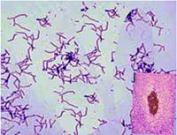
Histopathology of Actinomyces Infection. This microscopic image shows a bacterial colony of Actinomyces. The inset highlights a tissue section with classic sulfur granules surrounded by intense suppurative and granulomatous inflammation characteristic of actinomycosis.
Contributed by S Bhimji, MD
(Click Image to Enlarge)
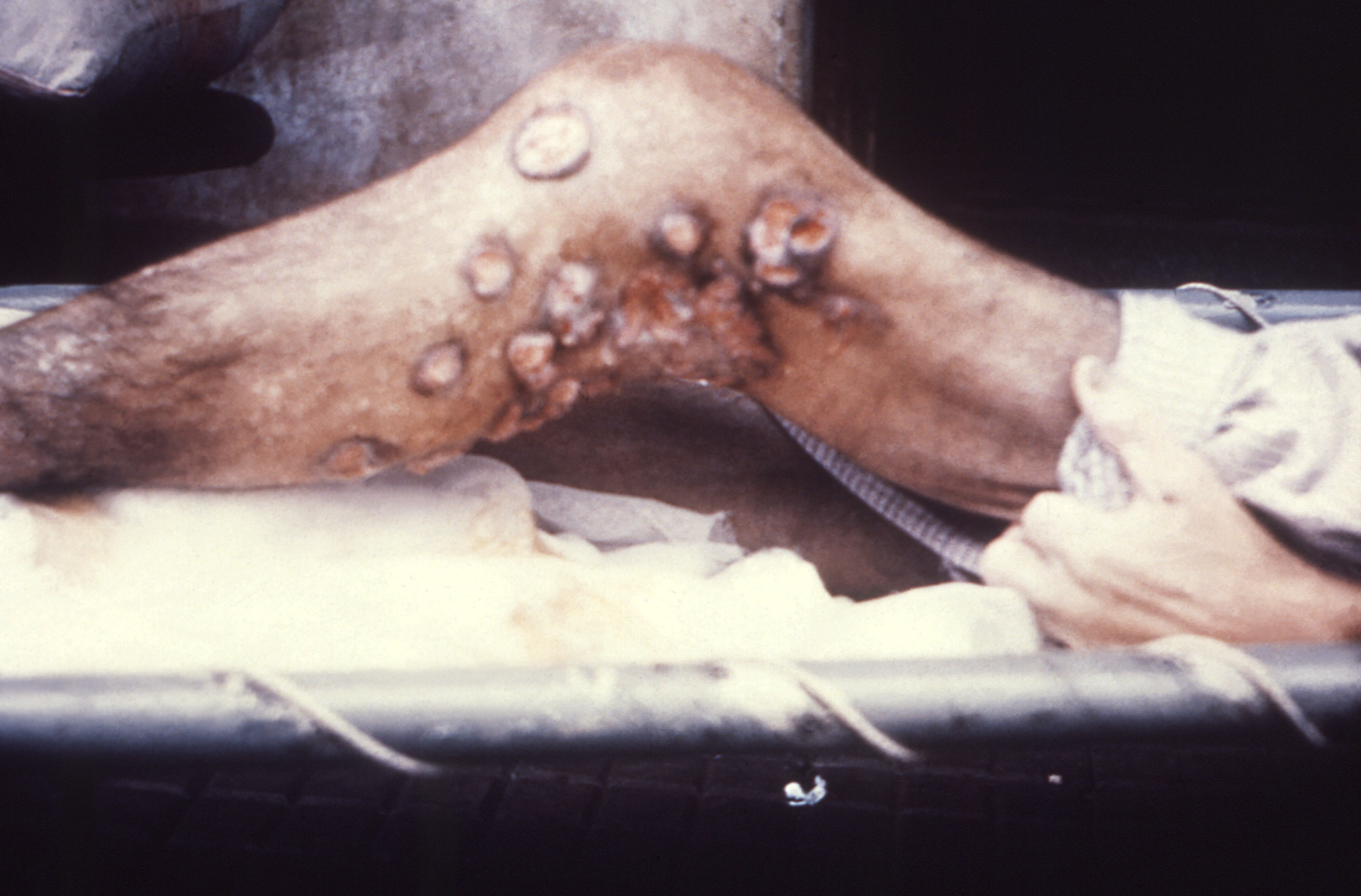
Right Leg Actinomycosis. Seen from a medial view, the right leg of this patient in the region of the knee displays numerous cutaneous lesions diagnosed as actinomycosis (mycetoma), a slowly progressive and destructive infection of the cutaneous and subcutaneous tissues caused by gram-positive aerobic bacteria of the order Actinomycetales. Mycetoma is a slowly progressive, destructive infection of the cutaneous and subcutaneous tissues that can extend to the fascia and eventually involve the underlying bone.
Contributed by the CDC/ Dr. Hardin
(Click Image to Enlarge)
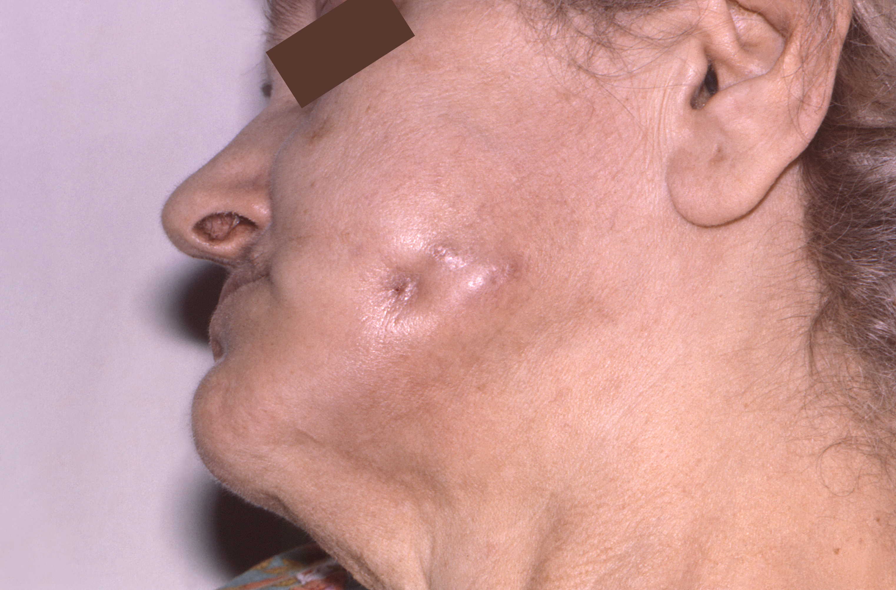
Cervicofacial Actinomycosis. Seen from the left lateral perspective, this female patient had a fistula involving the left cheek caused by actinomycosis, also known as mycetoma, an infection caused by gram-positive, filamentous, aerobic bacteria of the order Actinomycetales. Mycetoma is a slowly progressive, destructive infection of the cutaneous and subcutaneous tissues that can extend to the fascia and eventually involve the underlying bone.
Contributed by the CDC/ Dr. Robert Fass; Ohio State Dept. of Medicine
(Click Image to Enlarge)
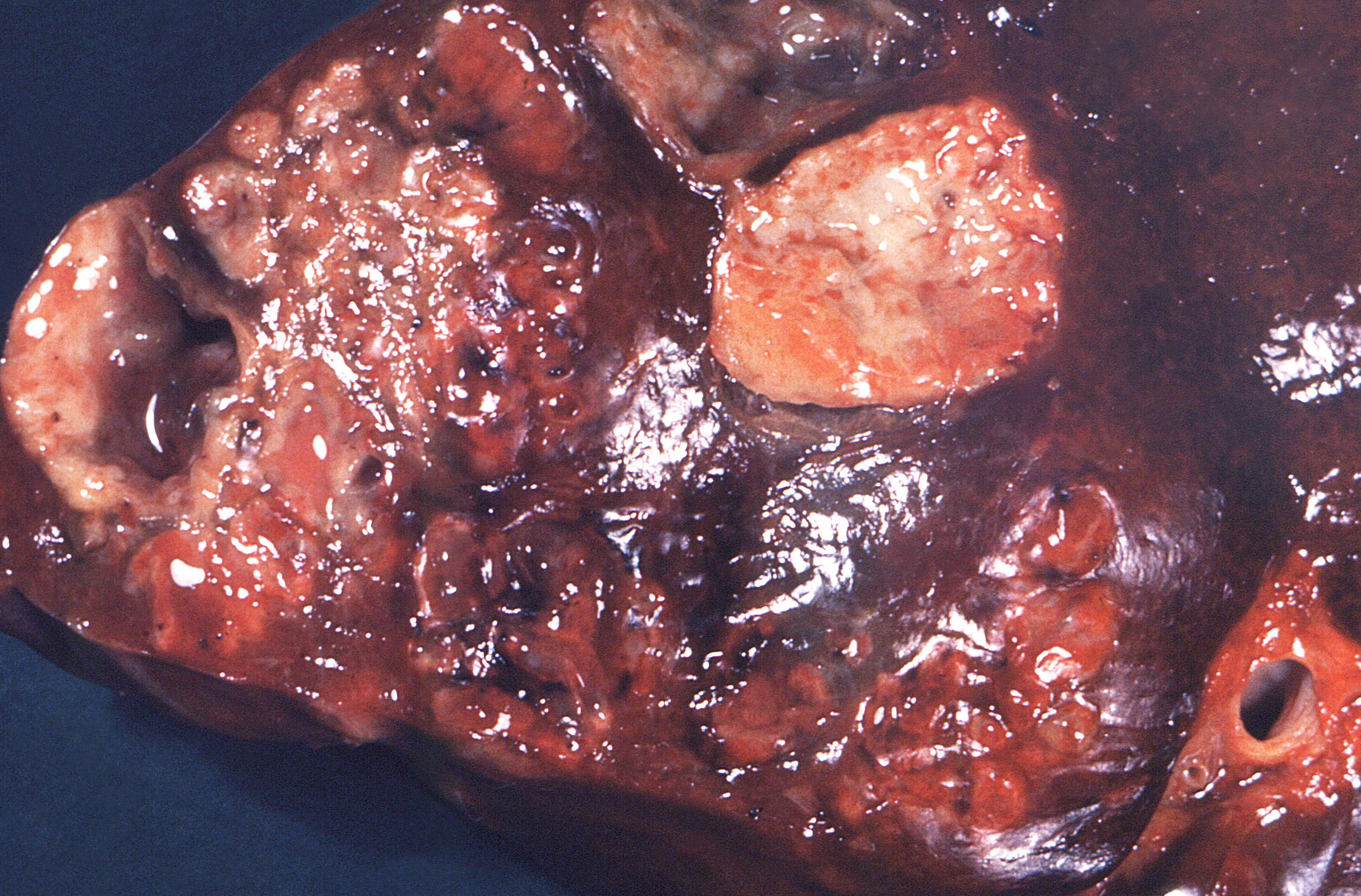
Liver Actinomycosis. This section of a human liver demonstrates numerous suppurative, pus-containing lesions caused by actinomycosis, an infection caused by gram-positive, filamentous, aerobic bacteria of the order Actinomycetales. Actinomycosis is a slowly progressive, destructive infection that primarily involves the cutaneous and subcutaneous tissues, may extend to the fascia and underlying bone, and can rarely disseminate to internal organs, including the liver, as illustrated in this specimen.
Contributed by the CDC/ Dr. Lucille K. Georg
(Click Image to Enlarge)
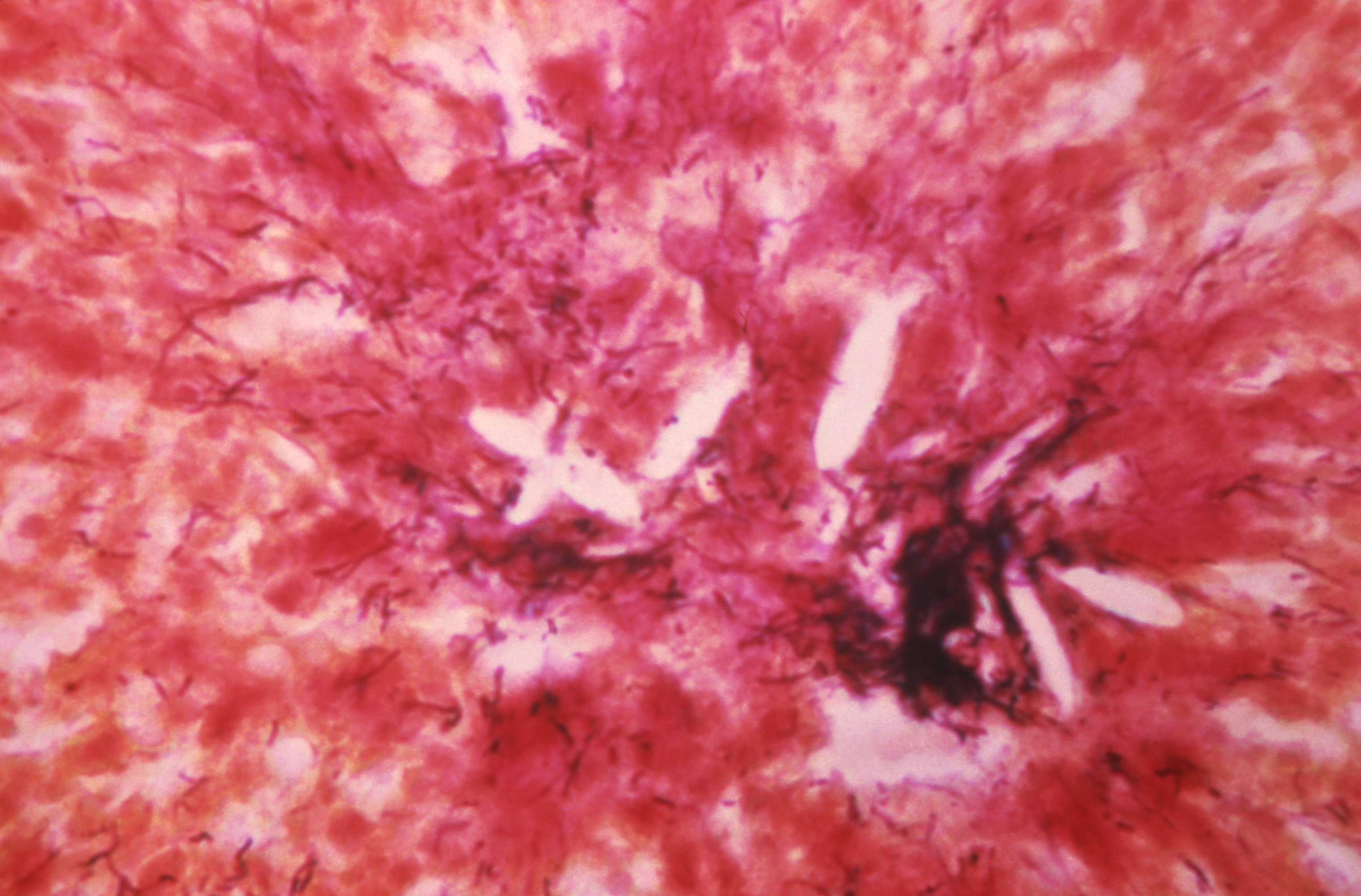
Brain Actinomycosis. This photomicrograph of a gram-stained brain tissue specimen, viewed under 200× magnification, demonstrates a sulfur granule within a chronic inflammatory lesion caused by actinomycosis due to gram-positive, filamentous, aerobic bacteria of the genus Actinomyces.
Contributed by the CDC/ Dr. Richard L. Levin, Greater Southeast Community Hospital, Washington, D.C.
(Click Image to Enlarge)
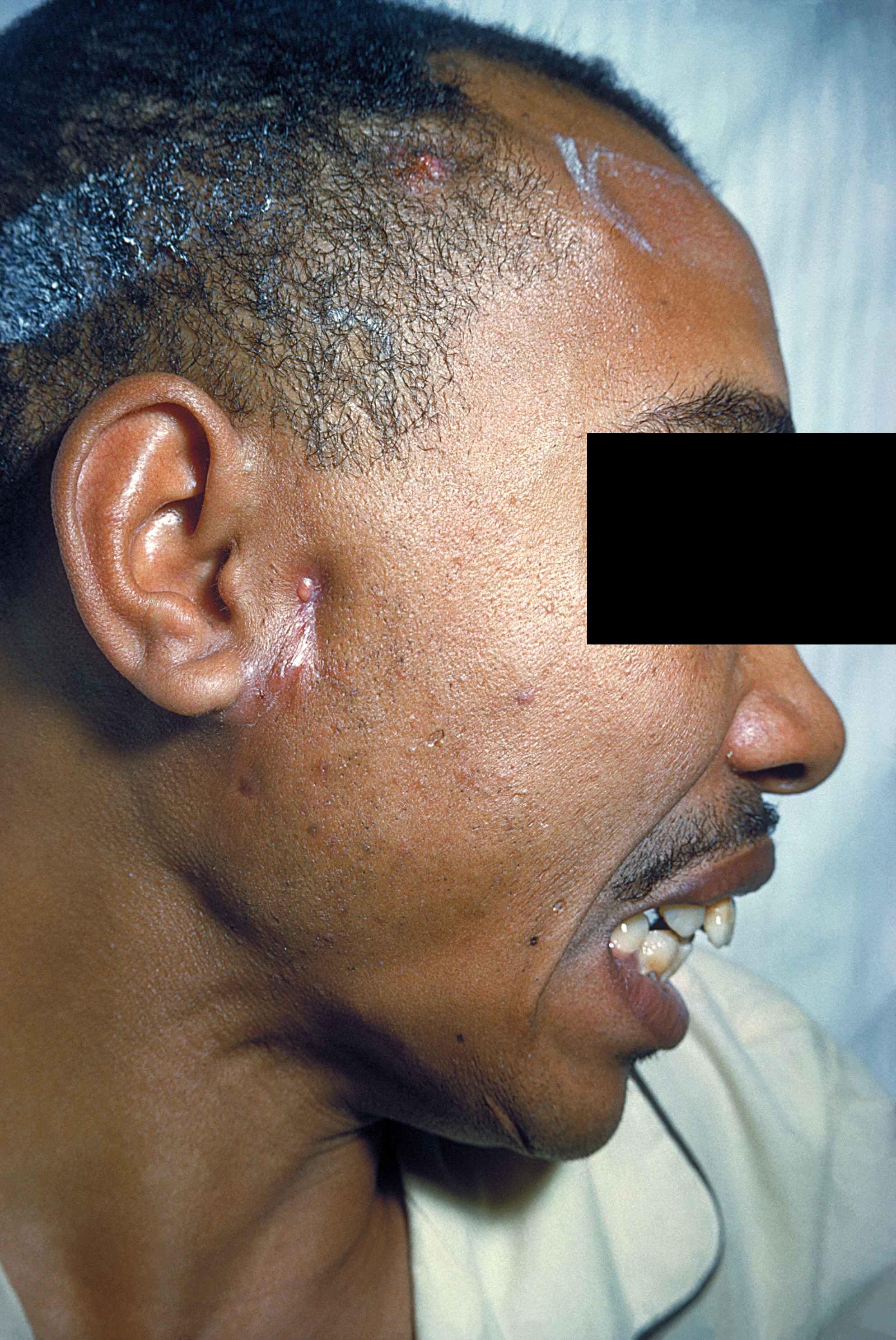
Actinomycosis Sinus Tract. This image depicts a right lateral view of a patient’s head and neck, revealing a cutaneous lesion anterior to the right ear diagnosed as actinomycosis. Actinomyces species, which normally colonize the oral cavity, can become opportunistic pathogens, particularly in immunosuppressed patients. Lesions are characterized by chronic swelling, suppuration, and abscess or granuloma formation.
Contributed by the CDC/ Dr. Thomas F. Sellers; Emory University
(Click Image to Enlarge)
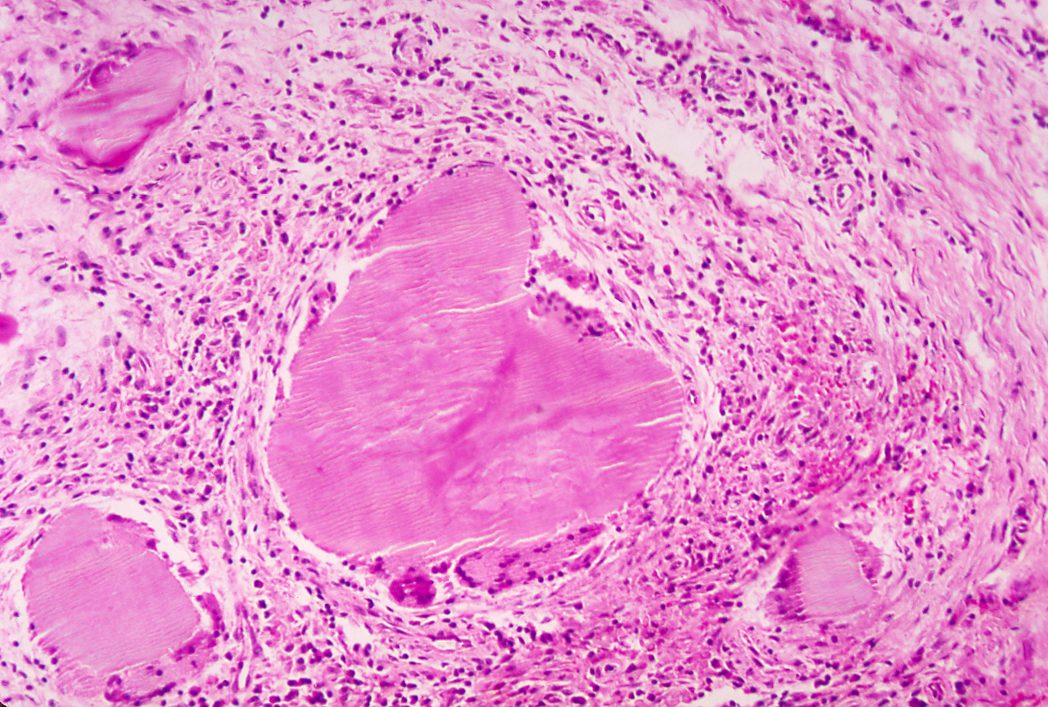
Sulfur Granules. This photomicrograph demonstrates characteristic sulfur granules within a chronic inflammatory lesion associated with actinomycosis. The sulfur granules appear as dense, eosinophilic aggregates of filamentous bacterial colonies surrounded by inflammatory cells.
Contributed from the CDC
References
El-Atoum M, Gailor ME, Segal BH, Bonnewell JP, Almyroudis NG. Microbiology of Actinomyces Species Isolated From Patients With Invasive Disease and Contaminated Samples in a Comprehensive Cancer Center. Open forum infectious diseases. 2024 Oct:11(10):ofae580. doi: 10.1093/ofid/ofae580. Epub 2024 Oct 11 [PubMed PMID: 39450394]
Könönen E, Wade WG. Actinomyces and related organisms in human infections. Clinical microbiology reviews. 2015 Apr:28(2):419-42. doi: 10.1128/CMR.00100-14. Epub [PubMed PMID: 25788515]
Mishra A, Prabhuraj AR, Bhat D, Nandeesh BN, Mhatre R. Intracranial Actinomycosis Manifesting as a Parenchymal Mass Lesion: A Case Report and Review of Literature. World neurosurgery. 2019 Feb:122():190-194. doi: 10.1016/j.wneu.2018.10.134. Epub 2018 Nov 1 [PubMed PMID: 30391617]
Level 3 (low-level) evidenceTippett E, Goyal N, Guy S, Wong J. Actinomyces spp. bloodstream and deep vein thrombus infections in people who inject drugs. Infection. 2019 Jun:47(3):479-482. doi: 10.1007/s15010-018-1246-x. Epub 2018 Nov 8 [PubMed PMID: 30406927]
COPE VZ. Visceral actinomycosis. British medical journal. 1949 Dec 10:2(4640):1311-6 [PubMed PMID: 15407913]
McCann A, Alvi SA, Newman J, Kakarala K, Staecker H, Chiu A, Villwock JA. Atypical Form of Cervicofacial Actinomycosis Involving the Skull Base and Temporal Bone. The Annals of otology, rhinology, and laryngology. 2019 Feb:128(2):152-156. doi: 10.1177/0003489418808541. Epub 2018 Oct 29 [PubMed PMID: 30371104]
Valdés-Peregrina EN, Bonifaz A, Arteaga-Sarmiento JF, Hernández-González M. [Primary intestinal actinomycosis in ilium and colon. A case report and review of the literature]. Revista espanola de patologia : publicacion oficial de la Sociedad Espanola de Anatomia Patologica y de la Sociedad Espanola de Citologia. 2018 Oct-Dec:51(4):253-256. doi: 10.1016/j.patol.2017.10.005. Epub 2017 Dec 6 [PubMed PMID: 30269778]
Level 3 (low-level) evidenceSadeghi S, Azaïs M, Ghannoum J. Actinomycosis Presenting as Macroglossia: Case Report and Review of Literature. Head and neck pathology. 2019 Sep:13(3):327-330. doi: 10.1007/s12105-018-0966-7. Epub 2018 Sep 22 [PubMed PMID: 30244331]
Level 3 (low-level) evidencePulverer G, Schütt-Gerowitt H, Schaal KP. Human cervicofacial actinomycoses: microbiological data for 1997 cases. Clinical infectious diseases : an official publication of the Infectious Diseases Society of America. 2003 Aug 15:37(4):490-7 [PubMed PMID: 12905132]
Level 3 (low-level) evidenceEng RH, Corrado ML, Cleri D, Cherubin C, Goldstein EJ. Infections caused by Actinomyces viscosus. American journal of clinical pathology. 1981 Jan:75(1):113-6 [PubMed PMID: 7457420]
Wasuwanich P, Szymkiewicz D, Kumari J, Vanegas Acosta D. Cervical Facial Actinomycosis Complicated by Bacteremia After 30 Years of Dental Neglect: Successful Treatment With Ceftriaxone and Metronidazole. Cureus. 2024 Jan:16(1):e51734. doi: 10.7759/cureus.51734. Epub 2024 Jan 6 [PubMed PMID: 38318602]
Valour F, Sénéchal A, Dupieux C, Karsenty J, Lustig S, Breton P, Gleizal A, Boussel L, Laurent F, Braun E, Chidiac C, Ader F, Ferry T. Actinomycosis: etiology, clinical features, diagnosis, treatment, and management. Infection and drug resistance. 2014:7():183-97. doi: 10.2147/IDR.S39601. Epub 2014 Jul 5 [PubMed PMID: 25045274]
Meena DS, Kumar D, Sharma M, Malik M, Ravindra A, Santhanam N, Bohra GK, Garg MK. The epidemiology, clinical presentation and treatment outcomes in CNS actinomycosis: a systematic review of reported cases. Orphanet journal of rare diseases. 2023 Jun 2:18(1):133. doi: 10.1186/s13023-023-02744-z. Epub 2023 Jun 2 [PubMed PMID: 37269006]
Level 1 (high-level) evidenceSmego RA Jr, Foglia G. Actinomycosis. Clinical infectious diseases : an official publication of the Infectious Diseases Society of America. 1998 Jun:26(6):1255-61; quiz 1262-3 [PubMed PMID: 9636842]
Kubo M, Osada M, Konno S. A histological and ultrastructural comparison of the sulfur granule of the actinomycosis and actinobacillosis. National Institute of Animal Health quarterly. 1980 Summer:20(2):53-9 [PubMed PMID: 7442815]
Level 3 (low-level) evidenceLerner PI. The lumpy jaw. Cervicofacial actinomycosis. Infectious disease clinics of North America. 1988 Mar:2(1):203-20 [PubMed PMID: 3074108]
Diaz A, Cyberski T, Singh A, Fenton D, Cipriani NA, Lusardi JJ, Corey JP, Blair EA. Laryngeal and pharyngeal actinomycosis: a systematic review and report of 3 cases. American journal of otolaryngology. 2022 Nov-Dec:43(6):103609. doi: 10.1016/j.amjoto.2022.103609. Epub 2022 Aug 18 [PubMed PMID: 36029619]
Level 1 (high-level) evidenceChoi MH, Hong DG, Seong WJ, Lee YS, Park IS. Pelvic actinomycosis confirmed after surgery: single center experience. Archives of gynecology and obstetrics. 2010 Apr:281(4):651-6. doi: 10.1007/s00404-009-1157-2. Epub 2009 Jun 30 [PubMed PMID: 19565255]
Sung HY, Lee IS, Kim SI, Jung SE, Kim SW, Kim SY, Chung MK, Kim WC, Oh ST, Kang WK. Clinical features of abdominal actinomycosis: a 15-year experience of a single institute. Journal of Korean medical science. 2011 Jul:26(7):932-7. doi: 10.3346/jkms.2011.26.7.932. Epub 2011 Jun 20 [PubMed PMID: 21738348]
Felz MW, Smith MR. Disseminated actinomycosis: multisystem mimicry in primary care. Southern medical journal. 2003 Mar:96(3):294-9 [PubMed PMID: 12659364]
Brody A, Scheich B, Dobo-Nagy C. Targeted histological evaluation shows high incidence of actinomyces infection in medication-related osteonecrosis of the jaws. Scientific reports. 2022 Mar 1:12(1):3406. doi: 10.1038/s41598-022-07375-1. Epub 2022 Mar 1 [PubMed PMID: 35233034]
Urs AB, Augustine J, Singh S. Histopathological evaluation of a rare fulminant case of contemporaneous mucormycosis, aspergillosis and actinomycosis. Journal of oral and maxillofacial pathology : JOMFP. 2019 Jan-Apr:23(1):144-146. doi: 10.4103/jomfp.JOMFP_64_19. Epub [PubMed PMID: 31110432]
Level 3 (low-level) evidenceMcHugh KE, Sturgis CD, Procop GW, Rhoads DD. The cytopathology of Actinomyces, Nocardia, and their mimickers. Diagnostic cytopathology. 2017 Dec:45(12):1105-1115. doi: 10.1002/dc.23816. Epub 2017 Sep 9 [PubMed PMID: 28888064]
Leslie DE, Garland SM. Comparison of immunofluorescence and culture for the detection of Actinomyces israelii in wearers of intra-uterine contraceptive devices. Journal of medical microbiology. 1991 Oct:35(4):224-8 [PubMed PMID: 1941993]
Stabrowski T, Chuard C. [Actinomycosis]. Revue medicale suisse. 2019 Oct 9:15(666):1790-1794 [PubMed PMID: 31599519]
Duldulao PM, Ortega AE, Delgadillo X. Mycotic and Bacterial Infections. Clinics in colon and rectal surgery. 2019 Sep:32(5):333-339. doi: 10.1055/s-0039-1687828. Epub 2019 Sep 6 [PubMed PMID: 31507342]
Heo SH, Shin SS, Kim JW, Lim HS, Seon HJ, Jung SI, Jeong YY, Kang HK. Imaging of actinomycosis in various organs: a comprehensive review. Radiographics : a review publication of the Radiological Society of North America, Inc. 2014 Jan-Feb:34(1):19-33. doi: 10.1148/rg.341135077. Epub [PubMed PMID: 24428279]
Sama CB, Mbarga NF, Oben CE, Mbarga JA, Nfor EK, Angwafo Iii FF. Massive paediatric cervicofacial actinomycoses masquerading as an ulcerative malignancy. BMC infectious diseases. 2016 Aug 15:16(1):417. doi: 10.1186/s12879-016-1768-8. Epub 2016 Aug 15 [PubMed PMID: 27526941]
Lippes J. Pelvic actinomycosis: a review and preliminary look at prevalence. American journal of obstetrics and gynecology. 1999 Feb:180(2 Pt 1):265-9 [PubMed PMID: 9988785]
Boot M, Archer J, Ali I. The diagnosis and management of pulmonary actinomycosis. Journal of infection and public health. 2023 Apr:16(4):490-500. doi: 10.1016/j.jiph.2023.02.004. Epub 2023 Feb 8 [PubMed PMID: 36801629]
Ding X, Sun G, Fei G, Zhou X, Zhou L, Wang R. Pulmonary actinomycosis diagnosed by transbronchoscopic lung biopsy: A case report and literature review. Experimental and therapeutic medicine. 2018 Sep:16(3):2554-2558. doi: 10.3892/etm.2018.6483. Epub 2018 Jul 19 [PubMed PMID: 30186488]
Level 3 (low-level) evidenceLim JA, Wong PS, Leong KN, Wong KL, Chow TS. Masking and misleading: concomitant actinomycosis and B-cell lymphoma - a case report and review of literature. Scottish medical journal. 2018 Aug 30:():36933018789312. doi: 10.1177/0036933018789312. Epub 2018 Aug 30 [PubMed PMID: 30165794]
Level 3 (low-level) evidenceWong VK, Turmezei TD, Weston VC. Actinomycosis. BMJ (Clinical research ed.). 2011 Oct 11:343():d6099. doi: 10.1136/bmj.d6099. Epub 2011 Oct 11 [PubMed PMID: 21990282]
Brook I. Actinomycosis: diagnosis and management. Southern medical journal. 2008 Oct:101(10):1019-23. doi: 10.1097/SMJ.0b013e3181864c1f. Epub [PubMed PMID: 18791528]
Nelson AL, Sulak P. IUD patient selection and practice guidelines. Dialogues in contraception. 1998 Spring:5(5):7-12 [PubMed PMID: 12321491]
Level 1 (high-level) evidenceSteininger C, Willinger B. Resistance patterns in clinical isolates of pathogenic Actinomyces species. The Journal of antimicrobial chemotherapy. 2016 Feb:71(2):422-7. doi: 10.1093/jac/dkv347. Epub 2015 Nov 3 [PubMed PMID: 26538502]
Weese WC, Smith IM. A study of 57 cases of actinomycosis over a 36-year period. A diagnostic 'failure' with good prognosis after treatment. Archives of internal medicine. 1975 Dec:135(12):1562-8 [PubMed PMID: 1200725]
Level 3 (low-level) evidenceSezer B, Akdeniz BG, Günbay S, Hilmioğlu-Polat S, Başdemir G. Actinomycosis osteomyelitis of the jaws: Report of four cases and a review of the literature. Journal of dental sciences. 2017 Sep:12(3):301-307. doi: 10.1016/j.jds.2013.02.031. Epub 2013 Sep 20 [PubMed PMID: 30895066]
Level 3 (low-level) evidencePhichaphop C, Apiwattanakul N, Wanitkun S, Boonsathorn S. Bacterial Endocarditis Caused by Actinomyces oris: First Reported Case and Literature Review. Journal of investigative medicine high impact case reports. 2020 Jan-Dec:8():2324709620910645. doi: 10.1177/2324709620910645. Epub [PubMed PMID: 32131639]
Level 3 (low-level) evidenceSehnal B, Beneš J, Kolářová Z, Mojhová M, Zikán M. Pelvic actinomycosis and IUD. Ceska gynekologie. 2018 Winter:83(5):386-390 [PubMed PMID: 30848144]