 Tools
Tools
 Actinic Purpura
Introduction
Actinic purpura is a common benign disorder of the dermal connective tissue caused by chronic sun exposure. This condition was initially described by Bateman in 1818 and is also known as Bateman senile purpura or Bateman disease. Actinic purpura typically affects older adults and is characterized by dark purple blotches on photo-exposed areas, particularly the backs of the hands and the extensor surfaces of the forearms. These lesions normally fade within 3 weeks.[1][2] Some authors have used the term solar purpura as a synonym for actinic purpura, but reserving it for purpuric lesions that occur acutely after sun exposure is preferable (see Image. Actinic Purpura).[3]
Etiology
Register For Free And Read The Full Article
 Search engine and full access to all medical articles
- 10 free questions in your specialty
- Free CME/CE Activities
- Free daily question in your email
- Save favorite articles to your dashboard
- Emails offering discounts
Learn more about a Subscription to StatPearls Point-of-Care
Etiology
Actinic purpura results from skin fragility due to long-term sun exposure, leading to UV-induced dermal atrophy. The dermal connective tissue cannot adequately support the microvasculature, leading to extravasation of blood into the dermis after minor trauma, a finding often neglected in medical history. This is one of the signs of dermatoporosis, which is characterized by skin fragility, skin atrophy, and stellate pseudo-scars.[2][4]
Epidemiology
Actinic purpura is common among older adults, particularly those with fair skin who are more sensitive to sun exposure. The prevalence of actinic purpura increases with age and cumulative UV exposure. The condition is estimated to affect about 12% of individuals older than 50 and 30% of those aged 75 or older; this condition usually affects both sexes equally.[5]
Pathophysiology
Actinic purpura results from extravasation of blood into the dermis. This phenomenon is due to skin atrophy and vascular fragility in older adults, which are exacerbated by chronic sun exposure. The lesions of actinic purpura are commonly located on sun-exposed areas, such as the arms, face, and neck. Skin atrophy in dermatoporosis is due to collagen alteration. These collagen changes may correspond to bone density changes similar to those observed in osteoporosis, suggesting that bone collagen also changes alongside skin collagen, thereby altering bone density. The pronounced skin atrophy caused by photoaging and UV radiation exposure makes the dermal vascular network highly sensitive to even the slightest trauma or shearing force.[6]
The formation of purple patches and macules in actinic purpura results from red blood cell extravasation into the dermis, leading to hemosiderin deposition in the interstitial space. No infiltration of the vessels is observed, and no inflammatory reaction occurs in the dermal tissue. The absence of phagocytosis results in delayed resorption of extravasated blood. This fact results in purple patches and macules of the ecchymotic aspect.[3]
Histopathology
Histopathology of the skin demonstrates epidermal thinning with numerous abnormal keratinocytes arranged in a disorderly pattern. The dermis shows red blood cell extravasation and hemosiderin deposition with no inflammatory cells. The vessel walls of the dermis are normal in structure. Histology of the surrounding skin with solar elastosis showed a blue, homogenized elastotic material present at the base of the epidermis. The amount of collagen in the affected area is markedly decreased.[6][7]
History and Physical
Actinic purpura presents as patches and macules with irregular borders on the forearms and the backs of the hands. However, they can affect other areas, such as the legs, neck, and face. The lesions may appear as dark purple macules and extensive ecchymosis, with average sizes ranging from 1 to 4 cm in diameter.
These macules are asymptomatic and are not associated with pruritus or tenderness. The surrounding skin generally demonstrates altered qualities, appearing thin, pigmented, and inelastic. Besides the typical presentation of actinic purpura, additional signs of photoaging may be evident, including wrinkling, lentigines, a sallow-yellow skin hue, actinic keratoses, and stellate pseudo-scars.[8]
The purpuric lesions persist for about 1 to 3 weeks before resolving spontaneously. However, they do not undergo the phases of inflammation. A residual deposit of hemosiderin in the dermis may leave a brown pigmentation. Actinic purpura can continue to appear due to already established cutaneous and vascular fragility. Although ecchymotic lesions can pose significant aesthetic concerns and psychological effects, they do not carry a risk of serious complications.[3]
Evaluation
The diagnosis of actinic purpura is primarily clinical, based on a detailed patient history and a thorough physical examination, including assessment of lesion distribution on chronically sun-exposed areas, skin quality, and the absence of systemic symptoms. Lesions are painless, noninflammatory, and resolve without scarring within 1 to 3 weeks. Histopathologic evaluation is rarely required but, when performed, demonstrates a thinned epidermis over an atrophic dermis with reduced collagen and abnormal elastic fibers. Dermal vessels maintain normal tensile structure, but red blood cell extravasation and hemosiderin deposition are highlighted by Perl staining. In approximately 10% of cases, neutrophilic infiltration may be observed, mimicking neutrophilic dermatoses or leukocytoclastic vasculitis and potentially leading to misdiagnosis.[9]
Laboratory investigation is generally unnecessary unless atypical features or systemic involvement are suspected. Coagulation studies, complete blood counts, and platelet assessments may be considered when lesions are atypical, widespread, rapidly progressive, or associated with systemic symptoms. Skin biopsy is rarely needed when the diagnosis is uncertain. Histopathology may demonstrate epidermal thinning, dermal atrophy with reduced collagen, solar elastosis, extravasated red blood cells, and hemosiderin deposition, without features of true vasculitis.
Current national and international dermatology guidelines, including those from the American Academy of Dermatology and European Dermatology Forum, recommend a clinical diagnosis without routine laboratory or radiographic evaluation, reserving additional testing for atypical presentations or when alternative diagnoses, such as vasculitis, coagulopathy, or drug-induced purpura, are being considered.[9] Management decisions are guided by the clinical evaluation, emphasizing photoprotection, trauma avoidance, and, when appropriate, topical agents to improve dermal thickness. Clear documentation of lesion appearance, distribution, and patient history supports differentiation from other purpuric disorders and ensures targeted patient education.
Treatment / Management
Actinic purpura is a benign condition that does not require specific treatment, particularly because new lesions continue to develop throughout life. The most effective preventive approach involves protection from excessive sun exposure by applying sunscreen and wearing long-sleeved shirts. Sunscreens with a high protection index (sun protection factor >50) provide sufficient protection against UV-A and UV-B rays and should be applied regularly to all sun-exposed areas.[10][11]
Therapeutic options for actinic purpura lesions that have already formed are limited. Tretinoin or retinoic acid (0.1%) is a vitamin A derivative, which could theoretically reverse UV light–induced severe skin damage. Indeed, topical tretinoin is known for its role in regenerating dermal collagen and reducing the quality of abnormal elastin damaged by solar exposure. However, studies have shown that plaques in actinic purpura do not improve with local tretinoin treatment.[1][3]
In 2002, actinic purpura was successfully treated in a single patient using tissue-engineered skin; however, no similar cases have been reported. More recently, human epidermal growth factor has shown promise as a potential treatment for actinic purpura. When applied twice daily for 6 weeks, it increased the average skin thickness and reduced the number of purpuric lesions. A citrus bioflavonoid blend has also been tested for the treatment of actinic purpura. After 6 weeks, the treated group showed a 50% decrease in purpuric plaques with no reported adverse effects.
Laser therapy is not indicated for actinic purpura, even though it is commonly used to treat other signs of skin aging.[1] Moisturizing creams may be useful to treat associated skin xerosis.[8] Recent studies have shown that a new intense pulsed light protocol is safe and effective in improving the clinical appearance of actinic purpura and preventing future lesions by enhancing skin structure, increasing epidermal thickness, and improving collagen and elastic fiber morphology. The treatment was well tolerated, adverse effects were minimal, and patient satisfaction was high.[12][13][14](A1)
Differential Diagnosis
The differential diagnosis of actinic purpura includes:
Prognosis
Actinic purpura has a favorable prognosis and generally follows a benign course. The lesions typically persist throughout life but resolve within 1 to 3 weeks. Resolution may be accompanied by postinflammatory hyperpigmentation due to hemosiderin deposition. Recurrences are common because the underlying dermal and vascular fragility persists.
Complications
Actinic purpura is a benign condition that typically resolves within 1 to 3 weeks, with residual pigmentation or, in some cases, scarring. Actinic purpura itself is not associated with systemic symptoms. Complications are mainly cosmetic and psychological. The presence of photodamaged, fragile skin may increase the risk of skin tears after minor trauma. These lesions can trigger the patient's emotional distress due to cosmetic disfigurement. Patients may experience distress due to the recurrent patches on the skin.[3]
Deterrence and Patient Education
Deterrence and patient education for actinic purpura focus on minimizing further photodamage and preventing lesion recurrence through lifestyle modification and skin barrier support. Patients should be counseled to consistently use broad-spectrum sunscreens, wear protective clothing, and avoid peak UV exposure. Education should emphasize the role of cumulative sun damage and avoidance of mechanical trauma in such lesions. Gentle skin care routines, including the use of emollients and avoidance of irritants and anticoagulant topical products, typically help reduce vessel fragility. Clinicians should reinforce that actinic purpura is benign yet indicative of photoaged skin, highlighting the importance of ongoing photoprotection and proactive management of cutaneous aging.
Pearls and Other Issues
Actinic purpura is a common, benign disease that primarily affects older adults with fair skin types. The disorder appears to be caused by dermal and vascular fragility induced by chronic sun exposure. This condition does not predispose to serious complications, and the risk of bleeding is minimal. However, it can cause significant aesthetic and cosmetic concerns and can induce a significant psychological impact. Treatment options are limited, mainly relying on vitamin A derivative-based creams and moisturizers. Photoprotection remains the best option and involves wearing protective clothing, regularly applying sunscreen, and using behavioral therapies to avoid excessive sun exposure and trauma.[17]
Enhancing Healthcare Team Outcomes
An interprofessional approach is essential in managing actinic purpura, particularly given its high prevalence among older adults and the fragility of chronically sun-damaged skin. Although actinic purpura is benign, minor trauma can easily lead to purpuric lesions, highlighting the need for coordinated patient education and preventive care. Primary care clinicians collaborate to assess skin integrity, identify patients at higher risk for complications, and provide individualized counseling on photoprotection, trauma avoidance, and skin hydration. Pharmacists support the team by reviewing medications that may increase bleeding risk and advising on topical therapies, such as retinoids or dermal repair agents, to improve skin resilience.
Effective interprofessional communication ensures that all team members are informed of the patient's condition, progress, and any changes in risk status. Care coordination includes reinforcing sun-protective behaviors; monitoring for potential coexisting conditions, such as actinic keratosis; and determining when biopsy or specialist referral is warranted.[10][17] Ethical considerations, including patient autonomy, informed consent, and culturally sensitive sun exposure counseling, are integral to maintaining trust and adherence. By leveraging the unique expertise of each discipline and promoting clear, ongoing communication, the healthcare team enhances patient-centered care, improves outcomes, supports patient safety, and strengthens overall team performance in managing actinic purpura.
Media
(Click Image to Enlarge)
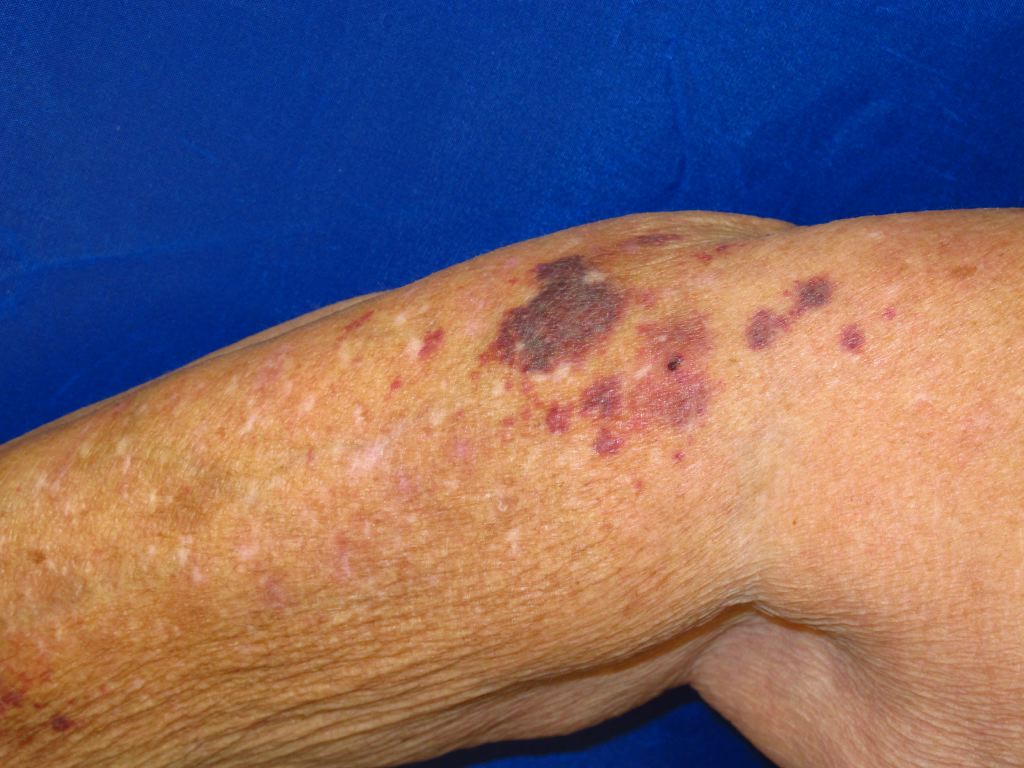
Actinic Purpura. This condition manifests as recurrent, sharply demarcated purpuric macules or patches on sun-exposed areas, most notably the dorsal hands and extensor forearms.
References
Ceilley RI. Treatment of Actinic Purpura. The Journal of clinical and aesthetic dermatology. 2017 Jun:10(6):44-50 [PubMed PMID: 28979656]
Peres GRP, Bandeira da Silva CV, Strazzieri-Pulido KC, de Gouveia Santos VLC. Skin tears in older adult residents of long-term care facilities: prevalence and associated factors. Journal of wound care. 2022 Jun 2:31(6):468-478. doi: 10.12968/jowc.2022.31.6.468. Epub [PubMed PMID: 35678790]
Syed HA, Masood S. Actinic Purpura. StatPearls. 2026 Jan:(): [PubMed PMID: 28846319]
Bortolozo F, Rinaldi M, Souza P, Schütz Paschoal Â, Lemperle G. Dermatoporosis in Upper Limbs Treated With Polymethylmethacrylate Microspheres Using the BioSculpt® Technique. Cureus. 2023 Aug:15(8):e43789. doi: 10.7759/cureus.43789. Epub 2023 Aug 20 [PubMed PMID: 37605716]
Luger A. [The skin in the elderly]. Zeitschrift fur Gerontologie. 1988 Sep-Oct:21(5):264-6 [PubMed PMID: 3070998]
Rayner RL, Carville KJ, Leslie GD, Dhaliwal SS. Clinical purpura and elastosis and their correlation with skin tears in an aged population. Archives of dermatological research. 2019 Apr:311(3):231-247. doi: 10.1007/s00403-019-01899-9. Epub 2019 Feb 20 [PubMed PMID: 30783769]
Borroni RG, Grassi S, Concardi M, Agozzino M, Caspani C, Giordano C, Vignini M, Arbustini E. Involvement of dermal microvascular basement membrane in senile purpura: quantitative immunohistochemical study. Journal of the European Academy of Dermatology and Venereology : JEADV. 2016 Oct:30(10):e63-e65. doi: 10.1111/jdv.13304. Epub 2015 Sep 25 [PubMed PMID: 26404419]
Durso TA, Miletta NR, Ortiz A, Avram M. Laser Therapy for Actinic Purpura. Dermatologic surgery : official publication for American Society for Dermatologic Surgery [et al.]. 2019 Nov:45(11):1435-1436. doi: 10.1097/DSS.0000000000001784. Epub [PubMed PMID: 30640778]
Griffiths CE. The clinical identification and quantification of photodamage. The British journal of dermatology. 1992 Sep:127 Suppl 41():37-42 [PubMed PMID: 1390185]
Lawrence N. New and emerging treatments for photoaging. Dermatologic clinics. 2000 Jan:18(1):99-112 [PubMed PMID: 10626116]
Demetriou C. Reversing precancerous actinic damage by mixing wavelengths (1064 nm, 532 nm). Journal of cosmetic and laser therapy : official publication of the European Society for Laser Dermatology. 2011 Jun:13(3):113-9. doi: 10.3109/14764172.2011.581289. Epub [PubMed PMID: 21609213]
Siperstein R, Wikramanayake TC. Intense Pulsed Light as a Treatment for Senile Purpura: A Pilot Study. Lasers in surgery and medicine. 2021 Sep:53(7):926-934. doi: 10.1002/lsm.23358. Epub 2020 Dec 8 [PubMed PMID: 33615512]
Level 3 (low-level) evidenceKimak A, Żebrowska A. Therapeutic Approach in Pigmented Purpuric Dermatoses-A Scoping Review. International journal of molecular sciences. 2024 Feb 24:25(5):. doi: 10.3390/ijms25052644. Epub 2024 Feb 24 [PubMed PMID: 38473891]
Level 2 (mid-level) evidenceDehghani A, Seddigh MA, Jafarzadeh A, Behrangi E, Lotfi S, Goodarzi A. A systematic review of the safety and effectiveness of laser and light therapies for the treatment of pigmented purpuric dermatoses. Lasers in medical science. 2025 Oct 1:40(1):401. doi: 10.1007/s10103-025-04615-4. Epub 2025 Oct 1 [PubMed PMID: 41032121]
Level 1 (high-level) evidenceJaiswal S, Uprety S, Thapa P, Lamichhane P. Dermoscopic Clues of Scurvy in Clinical Practice. Clinical case reports. 2026 Jun:14(6):e72823. doi: 10.1002/ccr3.72823. Epub 2026 May 27 [PubMed PMID: 42256997]
Level 3 (low-level) evidenceChambers CJ, Liu H, White CR, White KP, Sharon VR. Eruptive purpuric papules on the arms; a case of chemotherapy-induced inflammation of actinic keratoses and review of the literature. Dermatology online journal. 2014 Jan 15:20(1):21246 [PubMed PMID: 24456949]
Level 3 (low-level) evidenceLipozenčić J, Bukvić Mokos Z. Dermatologic lasers in the treatment of aging skin. Acta dermatovenerologica Croatica : ADC. 2010:18(3):176-80 [PubMed PMID: 20887699]