 Tools
Tools
 Achilles Tendinopathy
Introduction
The Achilles tendon, also known as the triceps surae, ranks as the strongest and largest tendon in the human body.[1] This tendon connects the aponeuroses of the gastrocnemius, soleus, and plantaris muscles to the calcaneus bone.[2] A sheath-like structure composed of a single layer of cells surrounds the tendon; this structure is not a true synovial sheath but is known as the paratenon.[2][3][4] The paratenon supplies a significant portion of the blood supply to the tendon.[4]
Results from studies identified a hypovascular area susceptible to injury, located approximately 2 to 6 cm proximal to the calcaneal insertion.[5] The Achilles tendon is essential for transmitting force from the calf muscles to the heel during walking and running.[6] Additionally, various factors can contribute to Achilles tendinopathies, with specific locations more commonly affected. This activity explores insertional and noninsertional tendinitis, paratenonitis, and tendon rupture.
Etiology
Register For Free And Read The Full Article
 Search engine and full access to all medical articles
- 10 free questions in your specialty
- Free CME/CE Activities
- Free daily question in your email
- Save favorite articles to your dashboard
- Emails offering discounts
Learn more about a Subscription to StatPearls Point-of-Care
Etiology
The causes and mechanisms of Achilles tendinopathy include the following:
- Intrinsic factors: These include anatomical factors, age, sex, body weight,[7] metabolic dysfunction,[8] pes cavus, dysmetria, muscle weakness, muscle imbalance, gastrocnemius dysfunction,[9] anatomical variation of the plantaris muscle,[10] tendon vascularity,[11] torsion of the Achilles tendons,[12] slippage of the fascicle,[13] and lateral instability of the ankle.
- Extrinsic factors: These include mechanical overload, constant effort, inadequate equipment,[14] medications (including corticosteroids, anabolic steroids, fluoroquinolones),[15][16] improper footwear, insufficient warming or stretching,[17] hard training surfaces, and direct trauma, among others.[18]
Several factors increase the risk of Achilles tendon rupture, including older age, tendon heterogeneity, fascicle slippage, and excessive exercise in athletes.[19] Athletes tend to present with pathology localized to the Achilles tendon insertion site.[20]
Epidemiology
The Achilles tendon has a cumulative lifetime injury incidence of approximately 24% in athletes. Running-related injuries have a prevalence of 11% to 85% or 2.5 to 59 injuries per 1000 hours of running.[21] Results from one study reported a prevalence of Achilles tendinopathy of 1% to 2% among elite adolescent athletes.[22] Findings from another study reported an injury rate of 9% among recreational athletes.[23] The lifetime injury incidence of 2.35 per 1000 is strongly associated with sporting activities.[24][25][26] This incidence increases in older men.[27] The overall incidence rate of Achilles tendin ruptures is 2.1 per 100,000 person-years, and most Achilles tendon ruptures occur in men, with a 3.5:1 male to female ratio.[28]
Pathophysiology
The etiology of Achilles tendinopathy remains uncertain, and many factors have been implicated, including free radical damage during reperfusion after ischemia, hypoxia, and hyperthermia, as well as impaired tenocyte apoptosis.[29] Macrophages and other phagocytic cells are key regulators of immunobiology, tissue repair, and extracellular matrix remodeling in tendons. Results from current studies have demonstrated heterogeneity among macrophages across tendon compartments, particularly between M1 and M2 macrophages, highlighting the importance of interactions between macrophages and resident tendon cells in specific local niches within this tissue.[30]
Changes in the expression of genes regulating cell-cell and cell-matrix interactions have been reported, with downregulation of MMP3 messenger RNA in tendinopathic Achilles tendon samples.[31] An imbalance in matrix metalloproteinase activity in response to repeated injury or mechanical strain may result in tendon degeneration. The Achilles tendon consists of 2 tendinous portions. The proximal portion gradually merges with the distal portion to form a single, homogeneous tendon. Additionally, the triceps surae muscle group comprises 3 muscular heads; the soleus is monoarticular, and the plantaris and gastrocnemius are biarticular. The mechanical tension of the tendon is concentrated at 2 sites. The sites include the medial or central portion of the paratenon and the middle segment of the tendon, which is the most common site of injury.[32]
As the Achilles tendon descends from its origin, it twists counterclockwise on the right and clockwise on the left, rotating 90°.[33] Smaller soleus fibers insert anteromedially while larger gastrocnemius fibers insert posterolaterally. This configuration is thought to contribute to alterations in tendon biomechanics and influence the pathophysiologic mechanisms of Achilles tendinopathies.[2][13] Tendon damage may even result from stresses within physiological limits because frequent microtrauma may not allow enough time for repair. Microtrauma can also result from nonuniform stress within tendons, producing abnormal load concentrations and frictional forces between fibrils, leading to localized fiber damage.[34] In cases of insertional Achilles tendinopathy, tendon degeneration is characterized by loss of strong parallel type I collagen fibers, fatty infiltration, and capillary proliferation.[2][35] Advanced imaging findings demonstrate tendon thickening, which will be further discussed in the next sections.
Histopathology
The Achilles tendon comprises 95% type I collagen fibers, which are very strong and flexible. A decrease in type I collagen fibers can occur with normal aging or injury. Tendinopathy, encompassing both tendinosis and tendinitis, is characterized by increased proteoglycan and water content and disorganized type III collagen fibers.[35]
The tendon is enveloped by a thin layer of connective tissue called the paratenon, which is rich in elastin and extends into the tendon, binding the collagen bundles together while permitting movement among them.[36] Tendinosis and tendinitis have distinct characteristics. Tendinosis involves degenerative changes in the tendon and its sheath, making the tendon more vulnerable to rupture. At the cellular level, no evidence of acute inflammation is observed. In contrast, tendinitis is marked by an acute inflammatory response resulting from acute trauma, overuse, or improper training.[37]
Paratenonitis is an inflammation affecting the outer layers of the tendon and includes conditions such as tenosynovitis.[38] This pathologic condition results in edema and exudate formation accompanied by inflammatory cells. Subsequently, a fibrinous exudate develops, leading to crepitus and restriction of tendon movement within the sheath. Without treatment, paratenonitis can progress to a chronic state through the development of immature connective tissue that expands into an organized fibrin network known as adhesions.[39]
Results from a study conducted in a rat model showed that the microenvironment of the Achilles tendon promoted the differentiation of stem cells toward chondrogenic and osteogenic lineages, predisposing the tendon to rupture (tendinosis). Following tendon rupture, an inflammatory cascade begins, leading to elevated expression of proinflammatory cytokine messenger RNA, including matrix metalloproteinase 3 (MMP3), cyclooxygenase 2, interleukin 6, and tumor necrosis factor α.[40] Conversely, chronic inflammation triggers neovascularization, which increases the risk of tendon rupture, resulting in greater disability and more complex treatment.[41] Results from a study conducted in rats showed that denervated tendons exhibited morphological alterations, including disorganization of the collagen network, increased production of type III collagen, hypercellularity, altered cell morphology, and elevated expression of the neurokinin 1 receptor. These changes resemble those observed in Achilles tendon injuries.[42]
History and Physical
The term Achilles tendinopathy refers to tendinitis (acute inflammation) and tendinosis (chronic degeneration). Achilles tendinopathy is characterized by pain, inflammation, and stiffness of the Achilles tendon.[43][44][45] The condition can occur in athletes and nonathletes, usually due to an imbalance between muscle power and tendon elasticity.[46] Achilles tendinopathies are divided anatomically into insertional and noninsertional tendinopathies.
Insertional Achilles tendinopathy affects the osseous attachment of the tendon to the posterior calcaneus. Patients with this condition may experience pain in the distal third of the tendon, extending to the insertion site, as well as morning stiffness.[47] Conversely, noninsertional Achilles tendinopathy occurs in the middle to proximal third of the tendon (2 to 6 cm proximal to the insertion). Palpation may reveal nodules and tendon enlargement.[47]
The clinical diagnosis of Achilles tendinopathy is primarily based on the patient history and a detailed physical examination. Characteristic symptoms include Achilles tendon pain that is typically worse in the morning, improves during activity, and often develops after a sudden increase in activity that overloads the tendon. Patients commonly report a gradual onset of pain and stiffness in the posterior heel or lower leg, with chronic recurrent calf or Achilles tendon pain being a hallmark feature. In chronic cases, exercise-induced pain remains the cardinal symptom, whereas crepitations and effusions diminish.
Physical examination begins with inspection of the tendon for bruising, swelling, and visible or palpable tendon enlargement or nodular thickening, particularly 2 to 6 cm proximal to the calcaneal insertion in midportion tendinopathy or directly at the insertion site in insertional tendinopathy. Palpation of the tendon is a key diagnostic component because the technique is simple, reproducible, and reliable for identifying localized tenderness along the affected portion of the Achilles tendon. In some cases, fibrin precipitated from the fibrinogen-rich fluid around the tendon can result in palpable crepitus.
Physical examination should also include a multidimensional assessment of calf muscle capacity with endurance tests such as the standing calf raise test, along with evaluation of the hip and knee to identify biomechanical contributions and muscle imbalances that may predispose patients to tendinopathy. Additional examination findings include pain with active and passive ankle motion, reduced ankle range of motion, and associated conditions, such as retrocalcaneal bursitis or Haglund deformity, in patients with insertional tendinopathy. A complete lower-extremity biomechanical assessment is essential to identify contributing factors, such as pes planus, pes cavus, subtalar joint immobility, or gluteal and foot core muscle weakness, that may require intervention.
Clinical tests include:
- Arc sign: Swelling or nodules within the tendon are palpated while the ankle joint is moved through plantarflexion and dorsiflexion. A positive arc sign is observed when the swelling or nodules move with range of motion.[46][48][49] In cases of paratendinopathy, the area of maximum thickening remains fixed in one position.[48]
- Royal London Hospital test: The point of maximum tenderness is palpated when the ankle is in a neutral position. The patient is then instructed to actively dorsiflex and plantarflex the ankle joint. The previous area of maximum tenderness is again palpated when the ankle joint is in maximum plantarflexion and dorsiflexion. Tendinopathy is present when tenderness significantly decreases or disappears under tension.[46][48]
Evaluation
Imaging Studies
Radiography: Radiography has a limited but specific role in evaluating Achilles tendinopathy because it does not directly visualize soft tissues such as tendons; however, radiography can provide useful complementary information. The primary utility of radiography is to identify bony abnormalities and exclude other conditions that can cause similar symptoms, particularly in insertional Achilles tendinopathy, in which posterior calcaneal exostoses are often present. Lateral ankle radiographs can demonstrate indirect signs of Achilles tendon pathology, including obliteration or alteration of the Kager fat pad (the radiolucent triangle posterior to the ankle and anterior to the Achilles tendon), which becomes obscured by effusion and soft tissue changes associated with tendon injury. In insertional Achilles tendinopathy, radiographs can show multiple parameters, including the calcaneal pitch angle, Fowler-Phillip angle, posterior calcaneal angle, Chauveau-Liet angle, Haglund deformity measurements, and intratendinous calcification length and width, though these parameters have limited diagnostic sensitivity and specificity for the condition. Radiography may also demonstrate increased tendon width when the Achilles tendon is bordered anteriorly by the Kager fat pad and posteriorly by subcutaneous fat, providing an indirect assessment of tendon enlargement. Although radiography can assist in diagnosis by identifying associated bony pathology and excluding differential diagnoses, ultrasonography is considered the first-line imaging modality for Achilles tendinopathy, with MRI reserved for patients who require more detailed soft tissue evaluation.[50][51][52]
Ultrasonography: Ultrasonography is commonly used in the evaluation of tendinopathies because it is readily available, safe, and inexpensive.[53][54] This imaging modality may reveal findings such as increased Achilles tendon thickness, hyperemia associated with hypervascularity, a decreased gastrocnemius-soleus rotation angle, and a reduced Kager fat pad length. Additionally, ultrasonography is useful during interventional treatment.[55] The Achilles tendon's large tissue volume facilitates visualization, and ultrasonography allows dynamic evaluation of the tendon throughout its range of motion.[56]
- Insertional Achilles tendinopathy: Ultrasonography findings include a hypoechoic area and loss of the fibrillar appearance in the distal third of the tendon.[47]
- Noninsertional Achilles tendinopathy: Ultrasonography findings include focal or diffuse thickening of the middle to proximal section of the tendon and hypoechoic areas with loss of the compact linear fibrillar appearance.[47]
Magnetic resonance imaging: This imaging modality provides comprehensive information about joint structures, enabling studies in multiple planes and in both static and dynamic views.[57][58] The normal Achilles tendon is less than 7 mm thick from anterior to posterior on sagittal MRI images. Increased measurements suggest chronic intrasubstance tendinopathy and degeneration.[59] Results from one study showed that MRI had lower sensitivity than ultrasonography in detecting early changes of enthesopathy.[60] However, results from another study demonstrated excellent agreement between tendon thickness measurements obtained from MRI and ultrasonography.[61]
- Insertional Achilles tendinopathy: MRI findings include distal tendon thickening with an intense signal on fat-suppressed images.[47]
- Noninsertional Achilles tendinopathy: MRI findings include thickening of the middle to proximal section of the tendon with heterogeneous signal intensity on T1- and T2-weighted images.[47]
Computed tomography: CT is useful for excluding trabecular structural alterations of the calcaneus in insertional Achilles tendon pathology. However, this imaging modality exposes the patient to radiation.[62][63][64]
Victoria Institute of Sports Assessment-Achilles (VISA-A): VISA-A questionnaire remains the gold standard for assessing pain and function, but additional studies are needed to improve its reliability.[65][66] Nevertheless, this questionnaire is an essential tool for monitoring patients after treatment.
Treatment / Management
Management of Achilles tendinopathy can be divided into conservative and surgical approaches, and further based on whether the condition is acute or chronic. In cases of tendon rupture, surgical treatment is often recommended.
Conservative Management
Conservative treatment strategies are considered first-line in management of Achilles tendinitis and include the following:
- Activity modification or reduction of activity levels [67]
- Nonsteroidal anti-inflammatory drugs (NSAIDs) [68][69]
- Orthotics: Orthotics are particularly beneficial for patients with abnormal foot alignment. Custom orthotics can modify the biomechanics of the foot and ankle, while a 12-15 mm heel lift is typically used as an adjunct for Achilles tendinopathy.
- Cryotherapy: Cryotherapy has an analgesic effect, reduces the metabolic rate of the tendon, and decreases extravasation of blood and protein from the new capillaries found in tendon injuries.
- Lower-tendon compression rehabilitation: This therapy modality consists of a progressive 4-stage tendon-loading program that includes isometric, isotonic, energy-storage-and-release, and sport-specific exercises.[70][71]
- Eccentric stretching exercises: These exercises are an integral component of physical therapy. Moderate-level evidence favors eccentric exercise over concentric exercise for reducing pain.[72][73][74] Eccentric exercises for chronic insertional Achilles tendinopathy are less effective in patients with metabolic syndrome but most effective when combined with extracorporeal shockwave therapy.[75]
- Extracorporeal shock wave therapy (ESWT): Results from studies showed that ESWT reduced pain by 60% and achieved 80% patient satisfaction, improving function and quality of life.[76] ESWT provides effective short-term pain relief and tendon healing, making it the preferred treatment for some healthcare professionals.[63][75] Other findings from studies found that combining ESWT with eccentric exercises yielded the best results.[75]
- Biologic injections:
- Platelet-rich plasma: Platelet-rich plasma is an autologous treatment containing many growth factors and may offer a promising treatment for chronic Achilles tendinopathy, with evidence of pain relief and functional improvement. However, variability in results emphasizes the need for standardized approaches to its use in clinical practice. Results from meta-analyses and recent studies have demonstrated no greater efficacy of platelet-rich plasma than placebo and no reduction in symptom duration.[56][77][78]
- Bone marrow aspirate concentrate: This treatment modality controls inflammation, reduces fibrosis, and recruits tenocytes and mesenchymal cells to damaged tissues. Results from one study showed that 92% of patients with Achilles tendon ruptures who underwent bone marrow aspirate concentrate returned to sports within 6 months, with no reruptures noted.[56][77]
- Peripheral blood mononuclear cells: Peripheral blood mononuclear cells comprise monocytes, macrophages, and lymphocytes. These cells are considered the new generation of regenerative autologous cell concentrates, but studies are lacking in the literature.[56]
- Scaffolds: Scaffolds are composed of a combination of natural and synthetic materials. Among these options, an acellular graft comprising human dermal allograft showed the most promising outcomes in studies. Results from these studies showed an increased return to activity without associated complications.[56]
- Prolotherapy: Prolotherapy (proliferative therapy) is a technique that induces the proliferation of new cells, promoting the regeneration of healthy tissue. Small amounts of hyperosmolar dextrose are injected into the affected area of the Achilles tendon under ultrasonography guidance. Results from one study showed that this procedure improved pain symptoms by 74.2% within 1 year.[72][73][74][76][74][79][80][81][82][83]
- Surgical Management: In 24% to 45.5% of patients with Achilles tendinopathy, conservative treatment is unsuccessful, and a surgical procedure should be considered if symptoms fail to resolve.[84][85] Surgical procedures and approaches can vary and are described in the following sections.
(A1)
Midportion (Noninsertional) Achilles Tendinopathy
- Debridement of diseased tendon: This procedure involves removing the damaged, degenerative portion of the Achilles tendon and repairing the remaining healthy tendon with sutures.
- Tendon transfer (flexor hallucis longus transfer): When extensive tendon damage exists, the flexor hallucis longus tendon from the big toe is transferred to reinforce or replace the damaged Achilles tendon.
- Gastrocnemius recession (calf muscle lengthening): This procedure involves surgical lengthening of tight calf muscles to reduce stress on the Achilles tendon.
- Minimally invasive tendon debridement: This procedure is often performed arthroscopically through smaller incisions, with camera assistance to remove diseased tissue.
Insertional Achilles Tendinopathy
- Achilles tendon detachment with debridement and reattachment: The tendon is detached from the calcaneus, diseased tissue and calcific deposits are removed, and the tendon is reattached using bone anchors or sutures.
- Haglund deformity excision (calcaneal exostectomy): This procedure involves removal of the bony prominence (calcaneal exostosis) at the back of the calcaneus using a chisel or saw.
- Retrocalcaneal bursectomy: This procedure involves removal of the inflamed, fluid-filled bursa between the Achilles tendon and calcaneus.
- Percutaneous Zadek osteotomy: This minimally invasive procedure involves removing a 5-mm wedge of bone from the calcaneus via 2 small incisions, thereby altering tendon fiber orientation and reducing stress.
- Traditional open Zadek osteotomy: This procedure requires an incision of up to 6 cm and a longer recovery time (9 to 12 months vs 6 weeks for the minimally invasive approach).
Additional Procedures
- Tenotomy: Tenotomy involves surgically lengthening a shortened Achilles tendon by making incisions that allow the tendon edges to draw apart and heal in a lengthened position.
- Tendon grafting: Tendon grafting involves replacing part or all of the Achilles tendon with a tendon graft from elsewhere in the foot when severe damage is present.
In cases of insertional Achilles tendinopathy, a combination of surgical interventions, including debridement, resection of the superior calcaneus and bursa, and reattachment of the Achilles tendon, has a success rate of more than 95%. On average, patients can return to activity within approximately 7 months following the surgical procedure.[86] Although calcinosis may be present, it does not interfere with the results. Furthermore, recurrent calcification is common but not associated with inferior surgical outcomes.[86][87]
In patients with noninsertional Achilles tendinopathy, satisfaction rates after surgical treatment range from 69% to 100%.[77] Complication rates are higher in patients with noninsertional tendinopathy. When more than 50% of the tendon is ruptured, reattachment of the Achilles tendon should be considered.[77]
Differential Diagnosis
Posterior heel and ankle pain are common concerns in primary care settings, and the risk of pain is elevated in individuals with higher body mass index and older age. Further evaluation is necessary when patients do not improve with initial treatment approaches.[65] The most common musculoskeletal soft tissue disorders associated with posterior ankle pain include the following:
- Retrocalcaneal bursitis: This condition is easily identified on ultrasonography or MRI.[47]
- Kager fat pad inflammation: Findings include pain on palpation bilaterally at the level of the ankle, anterior to the Achilles tendon.
- Achilles tendon rupture: Thompson test results are positive.[47]
- Achilles paratenonitis: Ultrasonography findings demonstrate fluid and adhesions around the tendon.[47]
- Posterior impingement (os trigonum syndrome): Findings include posterior ankle pain with forced dorsiflexion. Radiography findings demonstrate the presence of an os trigonum.
- Calcaneal stress fracture: Squeeze test results are positive.[47]
- Flexor hallucis longus tendinopathy: Findings include pain during the toe-off phase of gait and increased fluid around the flexor hallucis longus tendon on short tau inversion recovery MRI images.[47]
- Plantar fasciitis: Findings include pain with palpation of the plantar-medial calcaneal tubercle.[66][67]
- Nerve entrapment or neuroma: Findings include pain accompanied by burning, tingling, or numbness. Tinel sign results are positive along the course of the sural nerve.[47]
- Heel pad syndrome: Findings include deep, bruise-like pain in the midheel.[66][67]
- Haglund deformity: This condition causes acute or chronic posterolateral heel pain due to prominence of the calcaneus; the deformity may lead to retrocalcaneal bursitis and often requires surgical resection of the calcaneus.[88]
- Sever disease: This condition is a clinical diagnosis of calcaneal apophysitis in children and adolescents with immature growth plates.[66][67]
- Insertional calcific tendinosis: Mucoid degeneration of the tendon causes gradually worsening pain in middle-aged patients with a high body mass index and often requires surgical detachment and reattachment of the tendon.[88]
- Lumbar S1 radiculopathy: Findings include pain with decreased sensation over the posterolateral ankle.[47]
Erdheim-Chester disease is a rare non–Langerhans cell histiocytosis reported in a 36-year-old man who presented with bilateral xanthogranulomas of the Achilles tendon. Although rare, Erdheim-Chester disease should be considered in the differential diagnosis of intratendinous masses.[68] Additionally, clinicians should evaluate for serious nonmusculoskeletal causes of pain and systemic disease.[69]
Prognosis
The prognosis for patients with Achilles tendinopathy varies significantly between conservative and surgical treatment, with most patients responding well to nonsurgical treatment. Conservative treatment has a favorable long-term prognosis, particularly for acute-to-subacute cases, with eccentric exercise protocols achieving approximately 89% success rates for midportion (noninsertional) Achilles tendinopathy. Most patients with Achilles tendinopathy can recover with nonsurgical treatments, including activity modification, physical therapy, eccentric exercises, and tendon-loading programs, though recovery timelines can be lengthy and require patient commitment to rehabilitation protocols. Achilles tendinopathy has a better prognosis with early, adequate treatment because degenerative tendinopathy represents the final stage of the tendon pathology continuum and carries a poor prognosis due to irreversible changes. However, a small percentage of patients experience persistent symptoms despite conservative treatments lasting several months to more than 1 year, at which point a surgical procedure may be considered.[89][90][91][92]
Results from studies showed good to excellent outcomes after surgical treatment of Achilles tendinopathy in appropriately selected patients, with overall success rates of 80% to 90%. Surgical procedures for insertional Achilles tendinopathy yield good functional outcomes and a satisfactory return to sports when surgical care is tailored to the individual patient. Recovery from a surgical procedure is protracted, typically requiring 9 months to 1 year for full return to all activities, with patients remaining nonweight-bearing for 3 to 4 weeks postoperatively, followed by extensive physical therapy. Results from studies of minimally invasive surgical techniques, such as percutaneous Zadek osteotomy, were promising, with 95% of patients reporting they would choose the procedure again and significantly faster recovery timelines than those associated with traditional open surgical procedures. Final surgical outcomes vary based on the patient's baseline function, degree of tendon degeneration, and adherence to postoperative rehabilitation, with some patients experiencing residual pain or calf weakness despite technically successful procedures. Surgical treatment for Achilles tendinosis carries risks, including infection, nerve damage, wound-healing complications, blood clots, and ongoing pain, which must be weighed against potential benefits when conservative treatment is unsuccessful.[93][94][95][96]
Complications
When considering major and minor complications, the incidence ranges from 3% to 41% after surgical treatment for Achilles tendinopathy.[77] In addition, results from a study by Lohrer et al showed that the success rates of open and minimally invasive surgical procedures were comparable (83.4%), and patient satisfaction rates were comparable.[97] However, complication rates were lower after minimally invasive procedures. Baltes et al classified complications associated with surgical treatment as follows:
- Major complications: Tendon avulsion or rupture, any reoperation, deep vein thrombosis, reflex dystrophy, persistent neuralgia, deep infections, deep suture reactions, and deep wound infections.[77][86]
- Minor complications: Persistent pain, superficial infections, delayed wound healing, scar sensitivity, hypertrophy, mild paresthesia, and prolonged hospitalization.[77][96]
Complication rates also vary depending on whether the surgical procedure is performed for insertional or noninsertional Achilles tendinopathy. For insertional Achilles tendinopathy, the complication rate has been reported as high as 41%; for noninsertional Achilles tendinopathy, it has been reported as high as 85% after surgical procedures.[77][96]
Postoperative and Rehabilitation Care
Following surgical treatment for Achilles tendinopathy, a period of immobilization is required and may involve a cast, walking boot, or posterior splint. The duration of immobilization ranges from 3 to 8 weeks. However, recent articles increasingly advocate accelerated postoperative weight-bearing rehabilitation protocols.
In 2021, Arunakul et al prospectively analyzed conventional and accelerated rehabilitation protocols following surgical treatment for insertional Achilles tendinopathy. Results from the study showed that an accelerated protocol improved short-term functional outcome scores. Templates for both protocols are provided in the following sections.[98]
Conventional Postoperative Protocol
- Weeks 0-2: Nonweight-bearing with immobilization in a cast or splint in the equinus position
- Weeks 2 to 4: Transition to partial weight bearing (25%) with axillary crutches in a controlled ankle motion boot with a 3-layer heel lift (2.4 cm).
- Weeks 4 to 6: Increase partial weight bearing to 50% with axillary crutches and a 1.6-cm heel lift.
- Weeks 6 to 8: Increase partial weight bearing to 75% with axillary crutches in the controlled ankle movement (CAM) boot with a 0.8-cm heel lift.
- Weeks 8 to 10: Full weight bearing with axillary crutches in the CAM boot without a heel lift.
- Week 10: Transition to regular shoes, as tolerated.[98]
Accelerated Postoperative Weight-Bearing Protocol
- Weeks 0 to 2: Nonweight-bearing with immobilization in a cast or splint in equinus position
- Weeks 2 to 3: Transition to full weight bearing as tolerated in a CAM boot with a 3-layer (2.4-cm) step heel lift.
- Weeks 3 to 4: Continue full weight bearing as tolerated in the CAM boot, with the heel lift decreased to 1.6 cm.
- Weeks 4 to 5: Continue full weight bearing as tolerated in the CAM boot, with the heel lift decreased to 0.8 cm.
- Weeks 5 to 6: Full weight bearing in the CAM boot without a heel lift.
- Week 6: Transition to regular shoes, as tolerated.[98]
Consultations
Collaborative care involving multiple specialty groups is necessary to treat Achilles tendinopathy comprehensively. The involvement of various healthcare professionals ensures an interdisciplinary approach, leading to more effective treatment and improved patient outcomes. The 3 specialty groups commonly involved in the treatment of Achilles tendinopathy are as follows:
Podiatry: Podiatrists are crucial in treating Achilles tendinopathy, providing expertise in foot biomechanics, gait analysis, and prescribing orthotics or custom footwear. Podiatrists may also perform corticosteroid injections or extracorporeal shockwave therapy to alleviate pain and promote healing.
Physical therapy: Physical therapists are instrumental in the rehabilitation process for Achilles tendinopathy. They design individualized exercise programs focusing on strengthening the affected tendon, improving flexibility, and addressing any associated biomechanical issues. Physical therapists may use various modalities, such as therapeutic exercises, manual therapy, ultrasonography, and electrical stimulation, to facilitate recovery and restore optimal function.
Orthopedic surgical treatment: Orthopedic surgeons specialize in treating patients for whom conservative treatments do not provide adequate relief, or those with severe tendon damage or rupture; an orthopedic consultation may be necessary. Orthopedic surgeons can assess the need for surgery, perform tendon repairs or reconstructions, and provide postoperative care to facilitate recovery.
Deterrence and Patient Education
Patient education and a more conservative approach can prevent Achilles tendinopathy.[99][100][101][102] Evidence-based strategies include:
- Sport modification: Switching to low-impact activities (eg, swimming, weight training, rowing, and cycling) allows the Achilles tendon time to rest.[102]
- Eccentric exercise therapy: Incorporating eccentric exercises, which involve controlled lengthening of the muscle-tendon unit, has decreased pain and prevented progression to chronic tendinopathy.[57][58][60]
- Nonsteroidal anti-inflammatory drugs: Prior literature supports their use to reduce inflammation and treat pain.[54][55][77]
- Proper footwear: Shoes that provide adequate cushioning and support during heel strike can reduce stress on the Achilles tendon.[84]
- Warm-up and stretching: Stretching the calf muscles before engaging in sports or exercise may be beneficial.[84]
- Gradual progression: Distance and speed should be increased gradually by no more than 10% per week. Avoiding sudden increases in intensity, running on uneven slopes, and performing an appropriate cool-down after exercise are important for preventing overload of the Achilles tendon.[84][85]
- Extracorporeal shock wave therapy: ESWT is a noninvasive treatment that uses shock waves to stimulate healing in the Achilles tendon. This treatment may be beneficial if other interventions have not provided satisfactory results.[63]
Enhancing Healthcare Team Outcomes
Effective treatment of Achilles tendinopathy requires coordinated interprofessional collaboration among orthopedic surgeons, sports medicine clinicians, physical therapists, podiatrists, and nursing staff to optimize patient outcomes and ensure comprehensive care from diagnosis through rehabilitation. Clinicians must clearly communicate diagnostic findings and treatment plans to physical therapists who design and implement evidence-based exercise protocols, particularly the Alfredson eccentric training program, while radiologists provide critical imaging interpretation to guide clinical decision-making and monitor treatment response. Nurses play a vital role in patient education regarding activity modification, proper footwear selection, medication adherence for pain treatment, and monitoring for potential complications, while pharmacists counsel patients on appropriate nonsteroidal anti-inflammatory drug use, potential drug interactions, and the risks associated with fluoroquinolone antibiotics and corticosteroids that can predispose patients to tendinopathy or rupture.
Ethical responsibilities include placing the patient's interests at the center of care, respecting patient autonomy in shared decision-making between conservative and surgical treatment options, and maintaining transparent communication about realistic recovery timelines, treatment risks, and expected functional outcomes. The interprofessional team must use structured communication strategies to facilitate handoffs between clinicians, particularly when transitioning athletes from acute treatment to rehabilitation phases or from unsuccessful conservative treatment to surgical consultation, ensuring continuity of care across treatment modalities. Care coordination is enhanced when team members recognize their professional limitations, actively engage complementary expertise from other disciplines, including podiatry for biomechanical assessment and orthotics, and share accountability for patient outcomes, ensuring that active individuals receive timely diagnosis, appropriate conservative treatment with validated outcome measures, and seamless progression to surgical treatment when indicated after adequate trials of nonsurgical treatment.
Media
(Click Image to Enlarge)
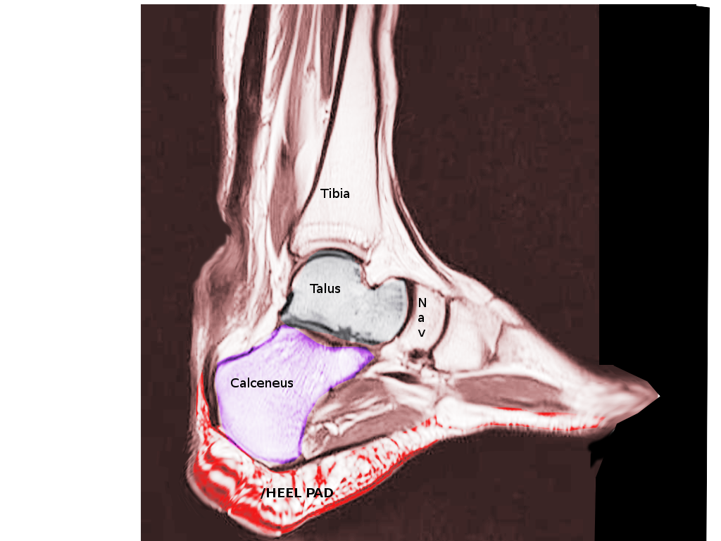
Dorsal Radiograph, Achilles Tendon. Radiograph showing the Achilles tendon, calcaneus, talus, heel pad, and tibia.
Contributed by S Bhimji, MD
(Click Image to Enlarge)
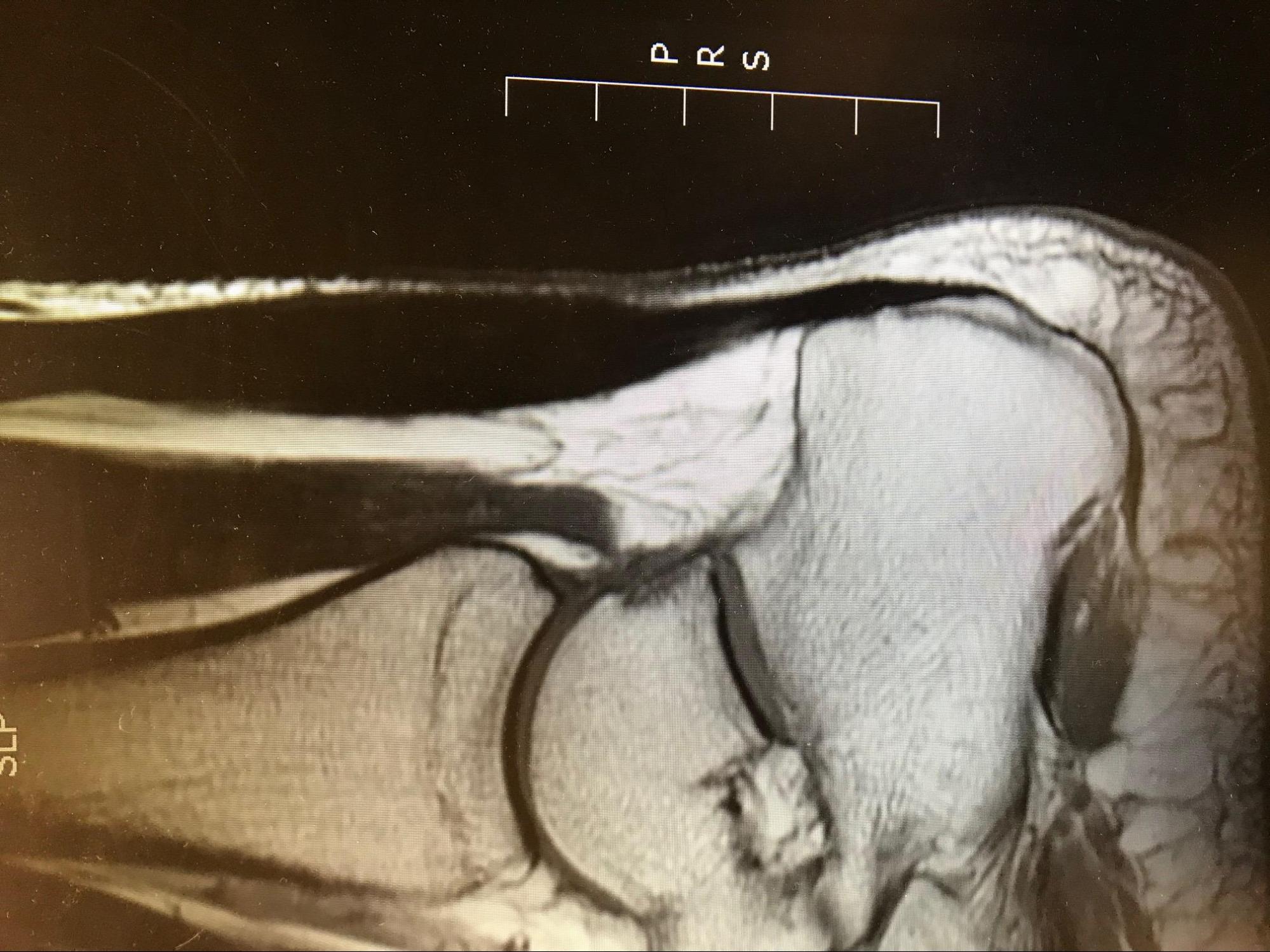
Achilles Tendonitis T1 MRI demonstrating bulbous appearing Achilles tendon with homogenous signal. Contributed by Mark A. Dreyer, DPM, FACFAS
(Click Image to Enlarge)
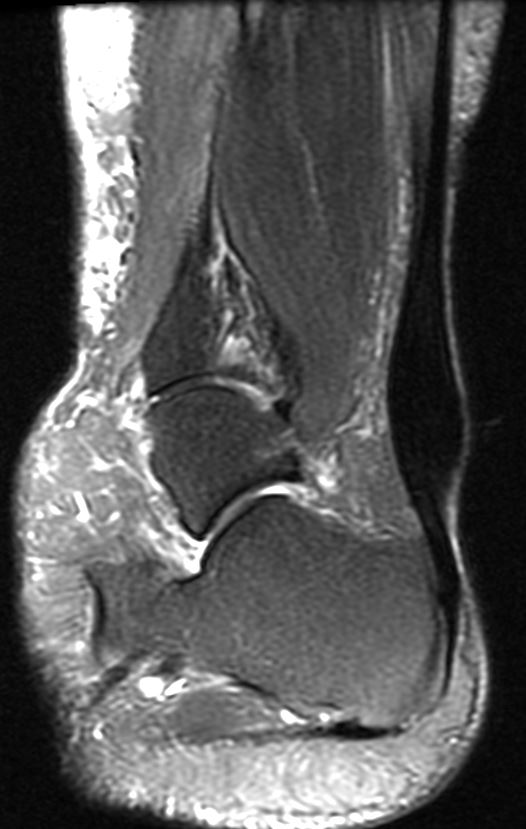
Achilles Tendonitis - MRI demonstrating classic fusiform thickening of the Achilles tendon in the "watershed" area Contributed by Mark A. Dreyer, DPM, FACFAS
(Click Image to Enlarge)
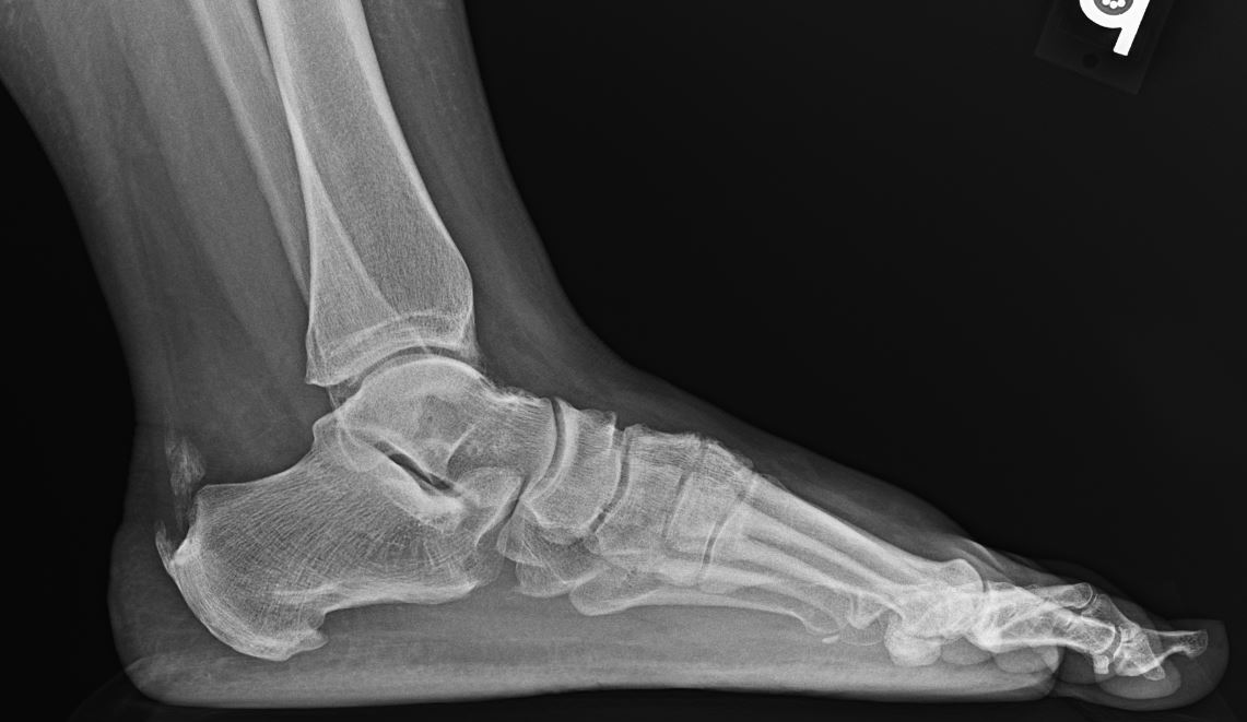
Achilles Tendonitis - lateral weight-bearing radiograph demonstrating a retro-calcaneal bone spur along with intra-tendinous calcification Contributed by Mark A. Dreyer, DPM, FACFAS
References
O'Brien M. The anatomy of the Achilles tendon. Foot and ankle clinics. 2005 Jun:10(2):225-38 [PubMed PMID: 15922915]
Chimenti RL, Cychosz CC, Hall MM, Phisitkul P. Current Concepts Review Update: Insertional Achilles Tendinopathy. Foot & ankle international. 2017 Oct:38(10):1160-1169. doi: 10.1177/1071100717723127. Epub 2017 Aug 8 [PubMed PMID: 28789557]
Weinfeld SB. Achilles tendon disorders. The Medical clinics of North America. 2014 Mar:98(2):331-8. doi: 10.1016/j.mcna.2013.11.005. Epub [PubMed PMID: 24559878]
Ahmed IM, Lagopoulos M, McConnell P, Soames RW, Sefton GK. Blood supply of the Achilles tendon. Journal of orthopaedic research : official publication of the Orthopaedic Research Society. 1998 Sep:16(5):591-6 [PubMed PMID: 9820283]
Mahan J, Damodar D, Trapana E, Barnhill S, Nuno AU, Smyth NA, Aiyer A, Jose J. Achilles tendon complex: The anatomy of its insertional footprint on the calcaneus and clinical implications. Journal of orthopaedics. 2020 Jan-Feb:17():221-227. doi: 10.1016/j.jor.2019.06.008. Epub 2019 Jun 10 [PubMed PMID: 31889745]
Rabusin CL, Menz HB, McClelland JA, Evans AM, Malliaras P, Docking SI, Landorf KB, Gerrard JM, Munteanu SE. Efficacy of heel lifts versus calf muscle eccentric exercise for mid-portion Achilles tendinopathy (HEALTHY): a randomised trial. British journal of sports medicine. 2021 May:55(9):486-492. doi: 10.1136/bjsports-2019-101776. Epub 2020 Sep 28 [PubMed PMID: 32988930]
Level 1 (high-level) evidenceMoon JL, Moon KM, Carlisle DM. Obesity-Related Foot Pain: Diagnosis and Surgical Planning. Clinics in podiatric medicine and surgery. 2019 Jan:36(1):141-151. doi: 10.1016/j.cpm.2018.08.008. Epub 2018 Oct 25 [PubMed PMID: 30446041]
Abate M, Salini V. Mid-portion Achilles tendinopathy in runners with metabolic disorders. European journal of orthopaedic surgery & traumatology : orthopedie traumatologie. 2019 Apr:29(3):697-703. doi: 10.1007/s00590-018-2336-2. Epub 2018 Oct 26 [PubMed PMID: 30367279]
Zhao H, Ren Y, Roth EJ, Harvey RL, Zhang LQ. Concurrent deficits of soleus and gastrocnemius muscle fascicles and Achilles tendon post stroke. Journal of applied physiology (Bethesda, Md. : 1985). 2015 Apr 1:118(7):863-71. doi: 10.1152/japplphysiol.00226.2014. Epub 2015 Feb 5 [PubMed PMID: 25663670]
Olewnik Ł, Wysiadecki G, Podgórski M, Polguj M, Topol M. The Plantaris Muscle Tendon and Its Relationship with the Achilles Tendinopathy. BioMed research international. 2018:2018():9623579. doi: 10.1155/2018/9623579. Epub 2018 May 31 [PubMed PMID: 29955614]
Yang X, Coleman DP, Pugh ND, Nokes LD. The volume of the neovascularity and its clinical implications in achilles tendinopathy. Ultrasound in medicine & biology. 2012 Nov:38(11):1887-95. doi: 10.1016/j.ultrasmedbio.2012.07.002. Epub 2012 Sep 10 [PubMed PMID: 22975033]
Slane LC, Dandois F, Bogaerts S, Vandenneucker H, Scheys L. Non-uniformity in the healthy patellar tendon is greater in males and similar in different age groups. Journal of biomechanics. 2018 Oct 26:80():16-22. doi: 10.1016/j.jbiomech.2018.08.021. Epub 2018 Aug 23 [PubMed PMID: 30224164]
Pękala PA, Henry BM, Ochała A, Kopacz P, Tatoń G, Młyniec A, Walocha JA, Tomaszewski KA. The twisted structure of the Achilles tendon unraveled: A detailed quantitative and qualitative anatomical investigation. Scandinavian journal of medicine & science in sports. 2017 Dec:27(12):1705-1715. doi: 10.1111/sms.12835. Epub 2017 Jan 30 [PubMed PMID: 28139009]
Level 2 (mid-level) evidenceFederer AE, Steele JR, Dekker TJ, Liles JL, Adams SB. Tendonitis and Tendinopathy: What Are They and How Do They Evolve? Foot and ankle clinics. 2017 Dec:22(4):665-676. doi: 10.1016/j.fcl.2017.07.002. Epub 2017 Sep 27 [PubMed PMID: 29078821]
Bolon B. Mini-Review: Toxic Tendinopathy. Toxicologic pathology. 2017 Oct:45(7):834-837. doi: 10.1177/0192623317711614. Epub 2017 May 28 [PubMed PMID: 28553748]
Alušík Š, Paluch Z. [Drug induced tendon injury]. Vnitrni lekarstvi. 2018 Winter:63(12):967-971 [PubMed PMID: 29334747]
Camargo PR, Alburquerque-Sendín F, Salvini TF. Eccentric training as a new approach for rotator cuff tendinopathy: Review and perspectives. World journal of orthopedics. 2014 Nov 18:5(5):634-44. doi: 10.5312/wjo.v5.i5.634. Epub 2014 Nov 18 [PubMed PMID: 25405092]
Level 3 (low-level) evidenceGodoy-Santos AL, Bruschini H, Cury J, Srougi M, de Cesar-Netto C, Fonseca LF, Maffulli N. Fluoroquinolones and the Risk of Achilles Tendon Disorders: Update on a Neglected Complication. Urology. 2018 Mar:113():20-25. doi: 10.1016/j.urology.2017.10.017. Epub 2017 Oct 23 [PubMed PMID: 29074337]
Almekinders LC, Engle CR. Common and Uncommon Injuries in Ultra-endurance Sports. Sports medicine and arthroscopy review. 2019 Mar:27(1):25-30. doi: 10.1097/JSA.0000000000000217. Epub [PubMed PMID: 30601398]
Brund RBK, Rasmussen S, Kersting UG, Arendt-Nielsen L, Palsson TS. Prediction of running-induced Achilles tendinopathy with pain sensitivity - a 1-year prospective study. Scandinavian journal of pain. 2019 Jan 28:19(1):139-146. doi: 10.1515/sjpain-2018-0084. Epub [PubMed PMID: 30407913]
Colbert LH, Hootman JM, Macera CA. Physical activity-related injuries in walkers and runners in the aerobics center longitudinal study. Clinical journal of sport medicine : official journal of the Canadian Academy of Sport Medicine. 2000 Oct:10(4):259-63 [PubMed PMID: 11086751]
Level 2 (mid-level) evidenceCassel M, Risch L, Intziegianni K, Mueller J, Stoll J, Brecht P, Mayer F. Incidence of Achilles and Patellar Tendinopathy in Adolescent Elite Athletes. International journal of sports medicine. 2018 Sep:39(9):726-732. doi: 10.1055/a-0633-9098. Epub 2018 Jun 25 [PubMed PMID: 29940667]
Mulvad B, Nielsen RO, Lind M, Ramskov D. Diagnoses and time to recovery among injured recreational runners in the RUN CLEVER trial. PloS one. 2018:13(10):e0204742. doi: 10.1371/journal.pone.0204742. Epub 2018 Oct 12 [PubMed PMID: 30312310]
Molyneux P, Carroll M, Stewart S, Brenton-Rule A, Rome K. Ultrasound characteristics of the mid-portion of the Achilles tendon in runners: a systematic review protocol. Systematic reviews. 2017 May 30:6(1):108. doi: 10.1186/s13643-017-0501-z. Epub 2017 May 30 [PubMed PMID: 28558847]
Level 1 (high-level) evidencede Jonge S, van den Berg C, de Vos RJ, van der Heide HJ, Weir A, Verhaar JA, Bierma-Zeinstra SM, Tol JL. Incidence of midportion Achilles tendinopathy in the general population. British journal of sports medicine. 2011 Oct:45(13):1026-8. doi: 10.1136/bjsports-2011-090342. Epub [PubMed PMID: 21926076]
Level 2 (mid-level) evidenceWeber J, Buchhorn T. [Midportion Achilles tendinopathy]. Der Unfallchirurg. 2017 Dec:120(12):1038-1043. doi: 10.1007/s00113-017-0411-5. Epub [PubMed PMID: 28921035]
Wezenbeek E, De Clercq D, Mahieu N, Willems T, Witvrouw E. Activity-Induced Increase in Achilles Tendon Blood Flow Is Age and Sex Dependent. The American journal of sports medicine. 2018 Sep:46(11):2678-2686. doi: 10.1177/0363546518786259. Epub 2018 Aug 1 [PubMed PMID: 30067065]
Lemme NJ, Li NY, DeFroda SF, Kleiner J, Owens BD. Epidemiology of Achilles Tendon Ruptures in the United States: Athletic and Nonathletic Injuries From 2012 to 2016. Orthopaedic journal of sports medicine. 2018 Nov:6(11):2325967118808238. doi: 10.1177/2325967118808238. Epub 2018 Nov 26 [PubMed PMID: 30505872]
Lui PPY, Zhang X, Yao S, Sun H, Huang C. Roles of Oxidative Stress in Acute Tendon Injury and Degenerative Tendinopathy-A Target for Intervention. International journal of molecular sciences. 2022 Mar 25:23(7):. doi: 10.3390/ijms23073571. Epub 2022 Mar 25 [PubMed PMID: 35408931]
Majeed Y, Kokozidou M, Gögele C, Traweger A, Lehner C, Tempfer H, Schulze-Tanzil GG. The Role of Phagocytic Cells in the Achilles Tendon. International journal of molecular sciences. 2026 Feb 25:27(5):. doi: 10.3390/ijms27052130. Epub 2026 Feb 25 [PubMed PMID: 41828360]
Ireland D, Harrall R, Curry V, Holloway G, Hackney R, Hazleman B, Riley G. Multiple changes in gene expression in chronic human Achilles tendinopathy. Matrix biology : journal of the International Society for Matrix Biology. 2001 Jun:20(3):159-69 [PubMed PMID: 11420148]
Li HY, Hua YH. Achilles Tendinopathy: Current Concepts about the Basic Science and Clinical Treatments. BioMed research international. 2016:2016():6492597 [PubMed PMID: 27885357]
Edama M, Takabayashi T, Yokota H, Hirabayashi R, Sekine C, Maruyama S, Otani H. Classification by degree of twisted structure of the fetal Achilles tendon. Surgical and radiologic anatomy : SRA. 2021 Oct:43(10):1691-1695. doi: 10.1007/s00276-021-02803-9. Epub 2021 Jul 14 [PubMed PMID: 34263342]
Leadbetter WB. Cell-matrix response in tendon injury. Clinics in sports medicine. 1992 Jul:11(3):533-78 [PubMed PMID: 1638640]
Pirozzi KM. Histophysiology of Fibrocartilage. Clinics in podiatric medicine and surgery. 2022 Jul:39(3):363-370. doi: 10.1016/j.cpm.2022.02.002. Epub 2022 May 21 [PubMed PMID: 35717055]
Kvist M, Józsa L, Järvinen MJ, Kvist H. Chronic Achilles paratenonitis in athletes: a histological and histochemical study. Pathology. 1987 Jan:19(1):1-11 [PubMed PMID: 3588019]
Klatte-Schulz F, Minkwitz S, Schmock A, Bormann N, Kurtoglu A, Tsitsilonis S, Manegold S, Wildemann B. Different Achilles Tendon Pathologies Show Distinct Histological and Molecular Characteristics. International journal of molecular sciences. 2018 Jan 30:19(2):. doi: 10.3390/ijms19020404. Epub 2018 Jan 30 [PubMed PMID: 29385715]
Feilmeier M. Noninsertional Achilles Tendinopathy Pathologic Background and Clinical Examination. Clinics in podiatric medicine and surgery. 2017 Apr:34(2):129-136. doi: 10.1016/j.cpm.2016.10.003. Epub 2016 Dec 24 [PubMed PMID: 28257670]
DeCarbo WT, Bullock MJ. Midsubstance Tendinopathy, Surgical Management. Clinics in podiatric medicine and surgery. 2017 Apr:34(2):175-193. doi: 10.1016/j.cpm.2016.10.006. Epub 2016 Dec 23 [PubMed PMID: 28257673]
Jeong C, Kim SE, Shim KS, Kim HJ, Song MH, Park K, Song HR. Exploring the In Vivo Anti-Inflammatory Actions of Simvastatin-Loaded Porous Microspheres on Inflamed Tenocytes in a Collagenase-Induced Animal Model of Achilles Tendinitis. International journal of molecular sciences. 2018 Mar 12:19(3):. doi: 10.3390/ijms19030820. Epub 2018 Mar 12 [PubMed PMID: 29534523]
Level 3 (low-level) evidenceDakin SG, Newton J, Martinez FO, Hedley R, Gwilym S, Jones N, Reid HAB, Wood S, Wells G, Appleton L, Wheway K, Watkins B, Carr AJ. Chronic inflammation is a feature of Achilles tendinopathy and rupture. British journal of sports medicine. 2018 Mar:52(6):359-367. doi: 10.1136/bjsports-2017-098161. Epub 2017 Nov 8 [PubMed PMID: 29118051]
El-Habta R, Chen J, Pingel J, Backman LJ. Tendinosis-like changes in denervated rat Achilles tendon. BMC musculoskeletal disorders. 2018 Nov 30:19(1):426. doi: 10.1186/s12891-018-2353-7. Epub 2018 Nov 30 [PubMed PMID: 30497469]
Obst SJ, Heales LJ, Schrader BL, Davis SA, Dodd KA, Holzberger CJ, Beavis LB, Barrett RS. Are the Mechanical or Material Properties of the Achilles and Patellar Tendons Altered in Tendinopathy? A Systematic Review with Meta-analysis. Sports medicine (Auckland, N.Z.). 2018 Sep:48(9):2179-2198. doi: 10.1007/s40279-018-0956-7. Epub [PubMed PMID: 29961208]
Level 1 (high-level) evidenceTu P. Heel Pain: Diagnosis and Management. American family physician. 2018 Jan 15:97(2):86-93 [PubMed PMID: 29365222]
Martin RL, Chimenti R, Cuddeford T, Houck J, Matheson JW, McDonough CM, Paulseth S, Wukich DK, Carcia CR. Achilles Pain, Stiffness, and Muscle Power Deficits: Midportion Achilles Tendinopathy Revision 2018. The Journal of orthopaedic and sports physical therapy. 2018 May:48(5):A1-A38. doi: 10.2519/jospt.2018.0302. Epub [PubMed PMID: 29712543]
Maffulli N, Kenward MG, Testa V, Capasso G, Regine R, King JB. Clinical diagnosis of Achilles tendinopathy with tendinosis. Clinical journal of sport medicine : official journal of the Canadian Academy of Sport Medicine. 2003 Jan:13(1):11-5 [PubMed PMID: 12544158]
Vo TP, Ho GWK, Andrea J. Achilles Tendinopathy, A Brief Review and Update of Current Literature. Current sports medicine reports. 2021 Sep 1:20(9):453-461. doi: 10.1249/JSR.0000000000000884. Epub [PubMed PMID: 34524189]
Longo UG, Ronga M, Maffulli N. Achilles Tendinopathy. Sports medicine and arthroscopy review. 2018 Mar:26(1):16-30. doi: 10.1097/JSA.0000000000000185. Epub [PubMed PMID: 29300224]
Matthews W, Ellis R, Furness J, Hing WA. The clinical diagnosis of Achilles tendinopathy: a scoping review. PeerJ. 2021:9():e12166. doi: 10.7717/peerj.12166. Epub 2021 Sep 28 [PubMed PMID: 34692248]
Level 2 (mid-level) evidenceBulstra GH, van Rheenen TA, Scholtes VA. Can We Measure the Heel Bump? Radiographic Evaluation of Haglund's Deformity. The Journal of foot and ankle surgery : official publication of the American College of Foot and Ankle Surgeons. 2015 May-Jun:54(3):338-40. doi: 10.1053/j.jfas.2014.07.006. Epub 2014 Aug 30 [PubMed PMID: 25179453]
Gladman DD, Abufayyah M, Salonen D, Thavaneswaran A, Chandran V. Radiological characteristics of the calcaneal spurs in psoriatic arthritis. Clinical and experimental rheumatology. 2014 May-Jun:32(3):401-3 [PubMed PMID: 24850064]
Level 1 (high-level) evidenceRachel JN, Williams JB, Sawyer JR, Warner WC, Kelly DM. Is radiographic evaluation necessary in children with a clinical diagnosis of calcaneal apophysitis (sever disease)? Journal of pediatric orthopedics. 2011 Jul-Aug:31(5):548-50. doi: 10.1097/BPO.0b013e318219905c. Epub [PubMed PMID: 21654464]
Mahoney JM. Imaging Techniques and Indications. Clinics in podiatric medicine and surgery. 2017 Apr:34(2):115-128. doi: 10.1016/j.cpm.2016.10.014. Epub 2017 Jan 19 [PubMed PMID: 28257669]
Matthews W, Ellis R, Furness J, Hing W. Classification of Tendon Matrix Change Using Ultrasound Imaging: A Systematic Review and Meta-analysis. Ultrasound in medicine & biology. 2018 Oct:44(10):2059-2080. doi: 10.1016/j.ultrasmedbio.2018.05.022. Epub 2018 Jul 12 [PubMed PMID: 30007477]
Level 1 (high-level) evidenceRomero-Morales C, Martín-Llantino PJ, Calvo-Lobo C, Palomo-López P, López-López D, Pareja-Galeano H, Rodríguez-Sanz D. Comparison of the sonographic features of the Achilles Tendon complex in patients with and without achilles tendinopathy: A case-control study. Physical therapy in sport : official journal of the Association of Chartered Physiotherapists in Sports Medicine. 2019 Jan:35():122-126. doi: 10.1016/j.ptsp.2018.12.003. Epub 2018 Dec 5 [PubMed PMID: 30543997]
Level 2 (mid-level) evidenceIndino C, D'Ambrosi R, Usuelli FG. Biologics in the Treatment of Achilles Tendon Pathologies. Foot and ankle clinics. 2019 Sep:24(3):471-493. doi: 10.1016/j.fcl.2019.04.009. Epub 2019 May 21 [PubMed PMID: 31370998]
Bullock MJ, Mourelatos J, Mar A. Achilles Impingement Tendinopathy on Magnetic Resonance Imaging. The Journal of foot and ankle surgery : official publication of the American College of Foot and Ankle Surgeons. 2017 May-Jun:56(3):555-563. doi: 10.1053/j.jfas.2017.01.024. Epub 2017 Feb 28 [PubMed PMID: 28258946]
Tuite MJ. MR imaging of the tendons of the foot and ankle. Seminars in musculoskeletal radiology. 2002 Jun:6(2):119-31 [PubMed PMID: 12077701]
Leslie HD, Edwards WH. Neglected ruptures of the Achilles tendon. Foot and ankle clinics. 2005 Jun:10(2):357-70 [PubMed PMID: 15922924]
Kamel M, Eid H, Mansour R. Ultrasound detection of heel enthesitis: a comparison with magnetic resonance imaging. The Journal of rheumatology. 2003 Apr:30(4):774-8 [PubMed PMID: 12672198]
Albano D, Messina C, Usuelli FG, De Girolamo L, Grassi M, Maccario C, Bignotti B, Tagliafico A, Sconfienza LM. Magnetic resonance and ultrasound in achilles tendinopathy: Predictive role and response assessment to platelet-rich plasma and adipose-derived stromal vascular fraction injection. European journal of radiology. 2017 Oct:95():130-135. doi: 10.1016/j.ejrad.2017.08.006. Epub 2017 Aug 9 [PubMed PMID: 28987658]
Ficek K, Filipek J, Ficek J, Muzalewska M, Humpa F. Calcaneal CT is a useful tool for identifying Achilles tendon disorders: a pilot study. Journal of orthopaedic surgery and research. 2017 Sep 25:12(1):139. doi: 10.1186/s13018-017-0638-4. Epub 2017 Sep 25 [PubMed PMID: 28946912]
Level 3 (low-level) evidenceHuang YB, Zhao YX, Xiao JJ, Li MW, Zhang R, Li SL. [Comparative analysis of the ankle joints in juvenile male soccer players with imaging]. Zhonghua yi xue za zhi. 2016 Jul 5:96(25):1971-5. doi: 10.3760/cma.j.issn.0376-2491.2016.25.003. Epub [PubMed PMID: 27470952]
Level 2 (mid-level) evidenceLang TR, Cook J, Rio E, Gaida JE. What tendon pathology is seen on imaging in people who have taken fluoroquinolones? A systematic review. Fundamental & clinical pharmacology. 2017 Feb:31(1):4-16. doi: 10.1111/fcp.12228. Epub 2016 Oct 5 [PubMed PMID: 27477928]
Level 1 (high-level) evidenceMurphy M, Rio E, Debenham J, Docking S, Travers M, Gibson W. EVALUATING THE PROGRESS OF MID-PORTION ACHILLES TENDINOPATHY DURING REHABILITATION: A REVIEW OF OUTCOME MEASURES FOR SELF- REPORTED PAIN AND FUNCTION. International journal of sports physical therapy. 2018 Apr:13(2):283-292 [PubMed PMID: 30090686]
Palazón-Bru A, Tomás-Rodríguez MI, Mares-García E, Gil-Guillén VF. A Reliability Generalization Meta-Analysis of the Victorian Institute of Sport Assessment Scale for Achilles Tendinopathy (VISA-A). Foot & ankle international. 2019 Apr:40(4):430-438. doi: 10.1177/1071100718816953. Epub 2018 Dec 20 [PubMed PMID: 30569746]
Level 1 (high-level) evidenceMcClinton S, Luedke L, Clewley D. Nonsurgical Management of Midsubstance Achilles Tendinopathy. Clinics in podiatric medicine and surgery. 2017 Apr:34(2):137-160. doi: 10.1016/j.cpm.2016.10.004. Epub 2016 Dec 1 [PubMed PMID: 28257671]
Bussin ER, Cairns B, Bovard J, Scott A. Randomised controlled trial evaluating the short-term analgesic effect of topical diclofenac on chronic Achilles tendon pain: a pilot study. BMJ open. 2017 May 4:7(4):e015126. doi: 10.1136/bmjopen-2016-015126. Epub 2017 May 4 [PubMed PMID: 28473518]
Level 3 (low-level) evidenceHeinemeier KM, Øhlenschlæger TF, Mikkelsen UR, Sønder F, Schjerling P, Svensson RB, Kjaer M. Effects of anti-inflammatory (NSAID) treatment on human tendinopathic tissue. Journal of applied physiology (Bethesda, Md. : 1985). 2017 Nov 1:123(5):1397-1405. doi: 10.1152/japplphysiol.00281.2017. Epub 2017 Aug 31 [PubMed PMID: 28860166]
Pringels L, Capelleman R, Van den Abeele A, Burssens A, Planckaert G, Wezenbeek E, Vanden Bossche L. Effectiveness of reducing tendon compression in the rehabilitation of insertional Achilles tendinopathy: a randomised clinical trial. British journal of sports medicine. 2025 Apr 24:59(9):640-650. doi: 10.1136/bjsports-2024-109138. Epub 2025 Apr 24 [PubMed PMID: 40011018]
Level 1 (high-level) evidenceAli Elsiddig Ahmed E, Muharib R Alruwaili K, Alruwaili AH, Talal M Alruwaili A, Ahmed S Aljudia H, Mohammed N Alhadi N. Efficacy of Platelet-Rich Plasma in Treatment of Achilles Tendinopathy: Systematic Review and Meta-Analysis. Cureus. 2025 Feb:17(2):e79692. doi: 10.7759/cureus.79692. Epub 2025 Feb 26 [PubMed PMID: 40161095]
Level 1 (high-level) evidenceVerrall GM, Dolman BK, Best TM. Applying physical science principles to mid-substance Achilles tendinopathy and the relationship to eccentric lengthening exercises. Scandinavian journal of medicine & science in sports. 2018 Mar:28(3):1159-1165. doi: 10.1111/sms.12978. Epub 2017 Oct 4 [PubMed PMID: 28948634]
McCormack JR, Underwood FB, Slaven EJ, Cappaert TA. Eccentric Exercise Versus Eccentric Exercise and Soft Tissue Treatment (Astym) in the Management of Insertional Achilles Tendinopathy. Sports health. 2016 May/Jun:8(3):230-237. doi: 10.1177/1941738116631498. Epub [PubMed PMID: 26893309]
Wilson F, Walshe M, O'Dwyer T, Bennett K, Mockler D, Bleakley C. Exercise, orthoses and splinting for treating Achilles tendinopathy: a systematic review with meta-analysis. British journal of sports medicine. 2018 Dec:52(24):1564-1574. doi: 10.1136/bjsports-2017-098913. Epub 2018 Aug 31 [PubMed PMID: 30170996]
Level 1 (high-level) evidenceSilbernagel KG, Hanlon S, Sprague A. Current Clinical Concepts: Conservative Management of Achilles Tendinopathy. Journal of athletic training. 2020 May:55(5):438-447. doi: 10.4085/1062-6050-356-19. Epub 2020 Apr 8 [PubMed PMID: 32267723]
Dedes V, Stergioulas A, Kipreos G, Dede AM, Mitseas A, Panoutsopoulos GI. Effectiveness and Safety of Shockwave Therapy in Tendinopathies. Materia socio-medica. 2018 Jun:30(2):131-146. doi: 10.5455/msm.2018.30.141-146. Epub [PubMed PMID: 30061805]
Phisitkul P, Mansur NSB, Netto CC. Failed Surgery for Achilles Tendinopathy. Foot and ankle clinics. 2022 Jun:27(2):431-455. doi: 10.1016/j.fcl.2021.11.027. Epub 2022 May 5 [PubMed PMID: 35680298]
He D, Chen Q, Zhou Y, Zheng C, Li F, Peng Y. Efficacy of platelet-rich plasma as a conservative and surgical adjuvant treatment for chronic midportion Achilles tendinopathy: a systematic review and meta-analysis. The Physician and sportsmedicine. 2026 Apr:54(2):161-171. doi: 10.1080/00913847.2026.2634611. Epub 2026 Feb 23 [PubMed PMID: 41705493]
Level 1 (high-level) evidenceWoitzik E, Jacobs C, Wong JJ, Côté P, Shearer HM, Randhawa K, Sutton D, Southerst D, Varatharajan S, Brison RJ, Yu H, van der Velde G, Stern PJ, Taylor-Vaisey A, Stupar M, Mior S, Carroll LJ. The effectiveness of exercise on recovery and clinical outcomes of soft tissue injuries of the leg, ankle, and foot: A systematic review by the Ontario Protocol for Traffic Injury Management (OPTIMa) Collaboration. Manual therapy. 2015 Oct:20(5):633-45. doi: 10.1016/j.math.2015.03.012. Epub 2015 Mar 28 [PubMed PMID: 25892707]
Level 2 (mid-level) evidenceRabello LM, van den Akker-Scheek I, Brink MS, Maas M, Diercks RL, Zwerver J. Association Between Clinical and Imaging Outcomes After Therapeutic Loading Exercise in Patients Diagnosed With Achilles or Patellar Tendinopathy at Short- and Long-Term Follow-up: A Systematic Review. Clinical journal of sport medicine : official journal of the Canadian Academy of Sport Medicine. 2020 Jul:30(4):390-403. doi: 10.1097/JSM.0000000000000624. Epub [PubMed PMID: 29952842]
Level 1 (high-level) evidenceMurphy M, Travers M, Gibson W, Chivers P, Debenham J, Docking S, Rio E. Rate of Improvement of Pain and Function in Mid-Portion Achilles Tendinopathy with Loading Protocols: A Systematic Review and Longitudinal Meta-Analysis. Sports medicine (Auckland, N.Z.). 2018 Aug:48(8):1875-1891. doi: 10.1007/s40279-018-0932-2. Epub [PubMed PMID: 29766442]
Level 1 (high-level) evidenceLe ADK, Enweze L, DeBaun MR, Dragoo JL. Current Clinical Recommendations for Use of Platelet-Rich Plasma. Current reviews in musculoskeletal medicine. 2018 Dec:11(4):624-634. doi: 10.1007/s12178-018-9527-7. Epub [PubMed PMID: 30353479]
Pham H, Tseng W, Mendeszoon ER, Wong A, Hutchins R, Amin A, Reubens DB. Prolotherapy for Achilles Tendinopathy. Journal of the American Podiatric Medical Association. 2025 Nov-Dec:115(6):. pii: 23-186. doi: 10.7547/23-186. Epub [PubMed PMID: 41468117]
Alfredson H, Masci L, Spang C. Surgical plantaris tendon removal for patients with plantaris tendon-related pain only and a normal Achilles tendon: a case series. BMJ open sport & exercise medicine. 2018:4(1):e000462. doi: 10.1136/bmjsem-2018-000462. Epub 2018 Dec 5 [PubMed PMID: 30588327]
Level 2 (mid-level) evidenceHardy A, Rousseau R, Issa SP, Gerometta A, Pascal-Moussellard H, Granger B, Khiami F. Functional outcomes and return to sports after surgical treatment of insertional Achilles tendinopathy: Surgical approach tailored to the degree of tendon involvement. Orthopaedics & traumatology, surgery & research : OTSR. 2018 Sep:104(5):719-723. doi: 10.1016/j.otsr.2018.05.003. Epub 2018 May 28 [PubMed PMID: 29852319]
Saxena A, Maffulli N, Jin A, Isa E, Arthur WP, Wahl A. Insertional Achilles Tendinopathy: Analysis of 166 Procedures and Return to Activity. The Journal of foot and ankle surgery : official publication of the American College of Foot and Ankle Surgeons. 2021 Nov-Dec:60(6):1117-1123. doi: 10.1053/j.jfas.2021.01.011. Epub 2021 Apr 20 [PubMed PMID: 34024676]
Greiner F, Trnka HJ, Chraim M, Neunteufel E, Bock P. Clinical and Radiological Outcomes of Operative Therapy in Insertional Achilles Tendinopathy With Debridement and Double-Row Refixation. Foot & ankle international. 2021 Sep:42(9):1115-1120. doi: 10.1177/10711007211002814. Epub 2021 Apr 10 [PubMed PMID: 33843294]
Grambart ST, Lechner J, Wentz J. Differentiating Achilles Insertional Calcific Tendinosis and Haglund's Deformity. Clinics in podiatric medicine and surgery. 2021 Apr:38(2):165-181. doi: 10.1016/j.cpm.2020.12.003. Epub 2021 Feb 13 [PubMed PMID: 33745649]
Alfredson H, Cook J. A treatment algorithm for managing Achilles tendinopathy: new treatment options. British journal of sports medicine. 2007 Apr:41(4):211-6 [PubMed PMID: 17311806]
Zhi X, Liu X, Han J, Xiang Y, Wu H, Wei S, Xu F. Nonoperative treatment of insertional Achilles tendinopathy: a systematic review. Journal of orthopaedic surgery and research. 2021 Mar 30:16(1):233. doi: 10.1186/s13018-021-02370-0. Epub 2021 Mar 30 [PubMed PMID: 33785026]
Level 1 (high-level) evidenceJarin IJ, Bäcker HC, Vosseller JT. Functional Outcomes of Insertional Achilles Tendinopathy Treatment: A Systematic Review. JBJS reviews. 2021 Jun 14:9(6):. doi: 10.2106/JBJS.RVW.20.00110. Epub 2021 Jun 14 [PubMed PMID: 34125735]
Level 1 (high-level) evidenceMagnussen RA, Dunn WR, Thomson AB. Nonoperative treatment of midportion Achilles tendinopathy: a systematic review. Clinical journal of sport medicine : official journal of the Canadian Academy of Sport Medicine. 2009 Jan:19(1):54-64. doi: 10.1097/JSM.0b013e31818ef090. Epub [PubMed PMID: 19124985]
Level 1 (high-level) evidenceHall S, Schipper ON, Kaplan JRM, Johnson AH, Gonzalez TA, Vulcano E. Outcomes After Percutaneous Zadek Osteotomy for Insertional Achilles Tendinopathy. Foot & ankle international. 2024 Sep:45(9):931-939. doi: 10.1177/10711007241252803. Epub 2024 Sep 1 [PubMed PMID: 39219246]
Moen R, Hagenbucher JR, Shinabarger AB. Surgical Treatment of Insertional Achilles Tendinopathy: A Systematic Review. Journal of the American Podiatric Medical Association. 2020 Sep 1:110(5):. pii: Article_5. doi: 10.7547/17-052. Epub [PubMed PMID: 33179068]
Level 1 (high-level) evidenceLewis TL, Srirangarajan T, Patel A, Yip GCK, Hussain L, Walker R, Singh S, Latif A, Abbasian A. Clinical outcomes following surgical management of insertional Achilles tendinopathy using a double-row suture bridge technique with mean two-year follow-up. European journal of orthopaedic surgery & traumatology : orthopedie traumatologie. 2023 May:33(4):1179-1184. doi: 10.1007/s00590-022-03270-7. Epub 2022 May 4 [PubMed PMID: 35507118]
Level 2 (mid-level) evidenceBaltes TPA, Zwiers R, Wiegerinck JI, van Dijk CN. Surgical treatment for midportion Achilles tendinopathy: a systematic review. Knee surgery, sports traumatology, arthroscopy : official journal of the ESSKA. 2017 Jun:25(6):1817-1838. doi: 10.1007/s00167-016-4062-9. Epub 2016 Mar 12 [PubMed PMID: 26971111]
Level 1 (high-level) evidenceLohrer H, David S, Nauck T. Surgical treatment for achilles tendinopathy - a systematic review. BMC musculoskeletal disorders. 2016 May 10:17():207. doi: 10.1186/s12891-016-1061-4. Epub 2016 May 10 [PubMed PMID: 27165287]
Level 1 (high-level) evidenceArunakul M, Pholsawatchai W, Arunakul P, Pitakveerakul A. Conventional vs Accelerated Rehabilitation Protocol Following Reattachment of Achilles Tendon for Insertional Achilles Tendinopathy. Foot & ankle international. 2021 Sep:42(9):1121-1129. doi: 10.1177/10711007211003871. Epub 2021 May 24 [PubMed PMID: 34024153]
Barry ME. Patient-education guide. Heel pain. Nursing. 2004 Feb:34(2):43 [PubMed PMID: 14758327]
Chang HJ, Burke AE, Glass RM. JAMA patient page. Achilles tendinopathy. JAMA. 2010 Jan 13:303(2):188. doi: 10.1001/jama.303.2.188. Epub [PubMed PMID: 20068216]
Chinn L, Hertel J. Rehabilitation of ankle and foot injuries in athletes. Clinics in sports medicine. 2010 Jan:29(1):157-67, table of contents. doi: 10.1016/j.csm.2009.09.006. Epub [PubMed PMID: 19945591]
Sartorio F, Zanetta A, Ferriero G, Bravini E, Vercelli S. The EdUReP approach plus manual therapy for the management of insertional Achilles tendinopathy. The Journal of sports medicine and physical fitness. 2018 May:58(5):664-668. doi: 10.23736/S0022-4707.17.06952-3. Epub 2017 Feb 21 [PubMed PMID: 28222571]