 Tools
Tools
 EMS Canine Wound Care
Introduction
Many military and law enforcement agencies now deploy dogs in high-risk and high-fatality operations, such as explosive detection, patrol, tracking, and search and rescue.[1] These canine officers, including military working dogs (MWDs), are highly skilled and invaluable team members who risk their lives to ensure the safety and security of colleagues and the public. A 2024 analysis of MWD mortality confirmed that, as in human combat casualties, the majority of deaths following traumatic injury occur during the immediate and early phases before veterinary evaluation, underscoring the importance of point-of-injury care delivered by nonveterinary personnel.[2] Dog handlers, medics, and other first responders must possess the knowledge and skills required to provide emergency care to an injured canine officer when veterinary services are not available immediately.
Significant anatomic and physiologic differences between canines and humans influence trauma response patterns in working dogs, necessitating adapted approaches to emergency care. Common field emergencies involving deployed canines include hemorrhage, airway obstruction, respiratory compromise, and traumatic brain injury (TBI). This activity outlines essential principles for point-of-injury assessment and intervention. Emphasis is placed on equipping nonveterinary personnel with practical, evidence-informed skills to reduce preventable mortality prior to veterinary care.
Anatomy and Physiology
Register For Free And Read The Full Article
 Search engine and full access to all medical articles
- 10 free questions in your specialty
- Free CME/CE Activities
- Free daily question in your email
- Save favorite articles to your dashboard
- Emails offering discounts
Learn more about a Subscription to StatPearls Point-of-Care
Anatomy and Physiology
Anyone treating a canine officer should understand the anatomic and physiologic differences between canines and humans. Knowledge of normal canine vital signs and perfusion parameters is essential for recognizing physiologic compromise and identifying early signs of shock.
Normal resting body temperature ranges from 100.5 °F to 101.5 °F (38.1 °C–38.6 °C) and may increase to 101.0 °F to 104.0 °F (38.3 °C–40.0 °C) during exercise. Temperatures below 99 °F (37.2 °C) or above 104 °F (40.0 °C) may indicate significant physiologic derangement. Normal resting heart rate ranges from 60 to 75 bpm and may increase to 75 to 130 bpm during exercise. Heart rates greater than 180 bpm or less than 60 bpm may be observed in shock, with marked bradycardia representing a decompensated state.
Normal resting respiratory rate ranges from 10 to 20 breaths/min and may increase to 30 breaths/min or progress to panting during exercise. Respiratory rates greater than 40 breaths/min or less than 8 breaths/min may indicate critical conditions, such as hypoperfusion. Normal mucous membrane color is pink at rest and may appear bright pink during exercise. Pale, white, muddy, or gray mucous membranes are abnormal and may indicate impaired perfusion. Normal capillary refill time is 1 to 2 seconds at rest and approximately 1 second during exercise. A capillary refill time greater than 2 seconds is considered prolonged and may indicate hypoperfusion.[3]
Indications
The most common preventable causes of death in canine trauma–related incidents include hemorrhage from extremity wounds, tension pneumothorax, and airway obstruction. These 3 factors contribute significantly to prehospital fatalities in both civilian trauma and military combat casualties. An estimated 20% to 25% of these preventable deaths occur due to delays in the implementation of early and appropriate basic 1st-aid techniques.[4] A 2024 retrospective analysis of MWD mortality similarly identified hemorrhage, respiratory compromise, and TBI as dominant early causes of death, reinforcing the importance of point-of-injury intervention.
Extremity Wounds
Controlling life-threatening extremity hemorrhage in canines requires recognition that the tapered, conical shape of the canine limb predisposes human windlass-style tourniquets (eg, Combat Application Tourniquet or CAT Special Operations Forces Tactical Tourniquet – Wide or SOFTT-W) to distal slippage and mechanical failure. These devices should not be used as 1st-line therapy. When tourniquet application is indicated, an elastic or stretchable device such as the SWAT-T (Stretch, Wrap, and Tuck Tourniquet), RATS (Rapid Application Tourniquet System), or TK4 (Tactical K9 Tourniquet), or a canine-specific windlass tourniquet such as the TACMED (Tactical Medical) K9 Tourniquet, should be used. The tourniquet should be applied 2 to 3 inches proximal to the wound, with avoidance of articulation sites. A stepwise approach to hemorrhage control is recommended, as follows:
- Direct or circumferential pressure dressings should be applied in combination with hemostatic agents.
- Direct pressure should be maintained over the wound, or wide elastic bandages should be used to create a circumferential pressure dressing.
- Commercial hemostatic dressings or tranexamic acid (TXA) may be applied directly to the wound.
- Wound packing with hemostatic gauze is indicated for noncompressible injuries. A hemostat may be used to clamp a bleeding vessel when it is clearly visualized.
- An elastic or canine-specific tourniquet should be applied proximal to the wound if direct pressure and hemostatic packing fail, with application time recorded. See the related StatPearls chapter “EMS Canine Tourniquet Use” for detailed placement technique.[5]
Junctional tourniquets have not been evaluated in canines. The use of these devices is not recommended at this time.
When canine blood is available for transfusion, the recommended volume is a 10- to 20-mL/kg bolus. Administration may be completed over 4 hours, beginning with 1/4 of the total dose during the first 15 minutes, followed by doubling the infusion rate every 15 minutes until the target rate is achieved. For simplicity, canine Tactical Combat Casualty Care (K9TCCC) recommends a 500-mL initial bolus, as most MWDs weigh over 25 kg.[6] An entire blood unit may be infused rapidly in the setting of ongoing hemorrhage, though expedited administration increases the risk of transfusion reactions, including fever, tachypnea, and facial swelling.
Blood transfusions should be administered via intravenous or intraosseous routes with a filter in place. A single intravenous catheter should be maintained for 0.9% saline administration alone, avoiding concurrent delivery of pharmacologic agents through the same line. A second intravenous catheter may be placed when medications, such as antibiotics and analgesics, must be administered.
A 1:1 ratio of canine plasma to canine packed red blood cells is an alternative option if canine whole blood is unavailable.[7] In veterinary medicine, blood products are commonly supplied in single (125 mL) or double (250 mL) units. When whole blood replacement is approximated using component therapy, each component is typically administered as a corresponding single- or double-unit volume, depending on patient size and transfusion requirements.
Canine freeze-dried plasma (cFDP) is an increasingly available option for austere and prehospital use. Unlike fresh frozen plasma (FFP), cFDP is stored at 1 °C to 25 °C, requires no thawing, and may be reconstituted within minutes, supporting hemostatic resuscitation at the point of injury or at the time of transport to definitive care. In vitro analyses demonstrate that cFDP retains coagulation factor activity and fibrinogen levels comparable to FFP. These findings support the use of cFDP when canine whole blood or FFP is unavailable.[8] Administration of cFDP should occur via the intravenous route at 10 to 20 mL/kg. Human blood products should not be transfused into an MWD under any circumstances due to the high risk of hemolytic reactions.
TXA is readily available in most ambulances. In cases of significant blood loss, a dose of 10 mg/kg may be administered as a slow intravenous push or diluted in 100 mL of normal saline or lactated Ringer solution. TXA should be administered as early as possible when injury occurred within the preceding 3 hours, as evidence demonstrates reduced mortality when the drug is given within this timeframe.[9] Administration beyond 3 hours from injury is generally not recommended, as delayed TXA infusion has been associated with worse outcomes in human trauma data, although this threshold continues to be evaluated in ongoing human trials.[10] Antibiotic therapy is recommended for all open combat wounds.
Chest Wounds
Penetrating chest injuries from ballistic trauma and explosions occur frequently in military and law enforcement canines.[11][12] Breath sounds should be auscultated first. Inspection for an open chest wound may require shaving of hair to improve visualization. Active bleeding should be managed with direct pressure or application of a gloved hand directly over the wound. Occlusive dressings designed for human skin may not adhere effectively to canine hair coats. All open or sucking chest wounds should be managed with application of a vented chest seal as 1st-line therapy. A nonvented chest seal may be used if a vented seal is not available, with close monitoring for development of tension pneumothorax.
The wound should be disinfected prior to chest seal application in stable canines. The surrounding hair should be prepared appropriately prior to application of a chest seal, with trimming performed as needed to improve adhesion. Canine hair coats reduce the ability to achieve an effective occlusive seal. Placement of petroleum-impregnated gauze beneath the seal or application of a loosely placed circumferential chest wrap may improve seal integrity.
Ultrasound may be used to assess lung sliding or the absence of pneumothorax. Lung ultrasound demonstrates higher sensitivity than radiography for the detection of small pneumothoraces.[13] Chest radiography may be used to evaluate for hemothorax or pneumothorax. Absent lung sounds, increased respiratory effort, flail chest, and palpable displaced rib fractures suggest thoracic cavity compromise in the absence of radiographic imaging.
Diagnosis of tension pneumothorax may be supported by needle decompression in canines with chest trauma and worsening respiratory distress, diminished or absent lung sounds, or deteriorating blood pressure. Current K9TCCC guidance recommends using a 14- or 10-gauge, 3.25-inch needle or catheter unit. The needle is inserted at the 7th to 9th intercostal space, either at the midlateral thoracic wall or at the junction of the upper 1/3 and lower 2/3 of the thoracic wall, on the injured side. The needle should be inserted perpendicular to the chest wall and positioned just cranial to the rib to avoid the caudally located neurovascular bundle, with advancement of the catheter to approximately 1/2 its length. Bilateral needle decompression should be considered when a unilateral approach fails to relieve signs of tension pneumothorax due to differences in canine mediastinal anatomy (see Image. Lateral View of Normal Canine Lung Fields).
Muffled heart and lung sounds on auscultation may indicate pleural effusion (ie, hemothorax, pyothorax). Chest radiography may help confirm the diagnosis. Pulmonary contusions may require 24 to 72 hours to become radiographically evident. Intravenous fluids should be administered judiciously in the presence of pulmonary contusion. Most canines require intravenous fluids following trauma. Regardless of fluid status, primary treatment includes supplemental oxygen administration (see Image. Ventrodorsal Radiograph of Normal Canine Costophrenic Margins).
Persistent respiratory distress despite oxygen supplementation may require endotracheal intubation. Hypothermia prevention measures include application of a ready-heat blanket and warming of intravenous fluids or blood products.
Abdominal Wounds
Abdominal wounds or bruising suggest potential associated internal injuries. Bowel evisceration should be managed with irrigation, replacement, and field bandaging. Open thoracic or abdominal wounds require prompt veterinary exploration and definitive treatment as soon as possible.[14]
Contraindications
Initiation of life-saving treatments in an injured canine officer has no absolute contraindications when provider safety is ensured. However, relative contraindications apply to specific interventions. Muzzle placement is contraindicated in canines with respiratory distress, facial trauma, active vomiting, altered mental status, seizures, or suspected heat illness. Human windlass-style limb tourniquets (eg, CAT, SOFTT-W) should not be used as 1st-line hemorrhage control modalities due to conformational mismatch with the canine limb. Human blood products are contraindicated for transfusion into canines due to the high risk of hemolytic transfusion reactions. Junctional tourniquets have not been evaluated in canines and are not currently recommended. TXA is not recommended beyond 3 hours from injury.
Equipment
A canine-capable prehospital kit should include, at minimum, a muzzle constructed of fabric, leather, or improvised materials, such as a belt, triangular bandage, or roller gauze. Personal protective equipment, including gloves, should be available. Hemorrhage control equipment should include an elastic tourniquet (SWAT-T, RATS, or TK4) or a canine-specific windlass tourniquet (TACMED K9), along with hemostatic gauze and compression or pressure bandages. Thoracic injury management supplies should include a vented chest seal as first choice, with a nonvented chest seal as an alternative. Airway and respiratory equipment should include ETTs with internal diameters of 8.0 to 10.0 mm (cuffed) and Miller laryngoscope blades sized #4 or #5. Chest decompression capability should include 14-gauge or 10-gauge, 3.25-inch needle or catheter units.
Intravenous and intraosseous access supplies should be available, including intravenous catheterization equipment and intraosseous access devices such as EZ-IO systems. Resuscitation fluids and pharmacologic agents should include TXA, 0.9% saline or lactated Ringer solution, and canine-specific blood products when available, including canine whole blood and cFDP. Monitoring equipment should include a pulse oximeter suitable for placement on the lip, tongue, toe web, prepuce, or vulva. Thermoregulation equipment should include a ready-heat blanket for the prevention of hypothermia.
Personnel
Optimal prehospital canine care is delivered by a coordinated team. The canine handler should be involved whenever available and typically serves as the primary provider for safe restraint and positioning of the animal. Tactical medics, emergency medicine services (EMS) clinicians, and military medical personnel trained in K9TCCC or K9TECC (Canine Tactical Emergency Casualty Care) can provide airway, breathing, and circulation interventions. Veterinary consultation, when accessible, should be initiated early via telephone or telemedicine to guide care and coordinate evacuation. Veterinary assets are generally concentrated well behind the point of injury. During Operation Iraqi Freedom, fewer than 30 veterinary personnel were available for more than 600 deployed operational canines. Nonveterinary responders frequently serve as the first and sometimes only providers available during the critical early window.
Preparation
Provider safety must be confirmed before initiating care. The canine should be approached from the front or side in a low posture. A properly fitted muzzle should be applied when the handler is absent and the animal is alert, unless muzzle use is contraindicated (see Contraindications). Positioning should be in sternal or lateral recumbency, depending on the injury pattern. Fur at the intended intervention site should be clipped, and the area prepped with surgical disinfectant when time permits. A secondary provider should maintain restraint during all interventions performed by the primary provider. Intravenous access should be established via the cephalic vein on the lateral surface of the forelimbs, the saphenous veins on the lateral surface of the hind limbs, or the external jugular vein within the jugular furrow. Intraosseous access should be placed at the proximomedial tibia using a pediatric intraosseous device or EZ-IO system when peripheral access is unobtainable.
Technique or Treatment
An injured or frightened MWD may act unpredictably during medical care. Gloves and appropriate personal protective equipment should be worn. When available, a trained K9 handler should be involved in managing the canine during the intervention. A properly fitted muzzle may be used in the absence of a handler, provided the animal is not experiencing respiratory distress or other conditions that would make its use unsafe.
Sedation or pain medication should be considered. Human medications should not be given to dogs without checking the safety profile. Many nonsteroidal anti-inflammatory drugs (NSAIDs) intended for human use are toxic to canines. Meloxicam is an exception in that it is approved for veterinary use in dogs when prescribed and administered appropriately. However, most working dogs are German shepherds or Belgian Malinois, typically weighing above 30 kg. Medication doses should be calculated based on the dog's weight. Sedative choice should account for hemodynamic status. Ketamine is commonly preferred in hypovolemic or shock states. Caution is warranted with pure opioids or α2-receptor agonists in unstable patients.
Airway
A dog capable of barking, growling, or whining has a patent airway. Signs of airway obstruction in canines include gagging, noisy breathing, rapid or labored respiration, excessive drooling, stridor, a tripod posture, cyanosis, and pawing at the mouth or neck. Management of airway obstruction may require sedation of the MWD to facilitate visualization of the airway. Direct hand placement within the oral cavity should be avoided. A shoelace may be looped around the jaw and elevated to maintain mouth opening. Alternative techniques include placement of a leash, rope, or rolled gauze between the canine teeth to maintain oral access. Canines have a relatively large tracheal diameter, allowing removal of foreign material by direct extraction or suction when feasible.
Respiratory failure in MWDs weighing more than 25 kg requiring endotracheal intubation necessitates a laryngoscope with Miller blade size #4 or #5, as well as an endotracheal tube (ETT) with an internal diameter of 8.0 to 10.0 mm and cuff inflation capability. ETT depth can be estimated by measuring from the central incisor to the thoracic inlet (point of the shoulder) along the ventral neck prior to intubation. ETT depth should be marked prior to intubation by securing a shoelace over the measured length of the ETT. The shoelace may be secured following intubation by tying it around the mouth (see Image. Landmarks for Estimating Canine Endotracheal Intubation Depth).
Cardiopulmonary resuscitation should be initiated in accordance with the 2024 RECOVER (Reassessment Campaign on Veterinary Resuscitation) guidelines in cases of cardiopulmonary arrest during prehospital care, with high-quality, uninterrupted chest compressions delivered at 100 to 120/min, and endotracheal intubation pursued as soon as feasible. Survival-to-discharge rates for canine cardiac arrest remain low, and resuscitation should not be pursued at the expense of other salvageable casualties.[15]
Hemorrhage interventions in canines include several pharmacologic and blood product options. TXA is administered at 10 mg/kg as a slow intravenous push or diluted in 100 mL of normal saline or lactated Ringer solution. Canine whole blood may be administered as a 10- to 20-mL/kg bolus or as a 500-mL unit bolus. A 1:1 combination of canine plasma and canine packed red blood cells may be used, typically consisting of a 250 mL unit of plasma and a 250 mL unit of packed red blood cells. Reconstituted freeze-dried canine plasma is administered as a 250 mL bolus.
Analgesic options include fentanyl at 4 μg/kg intranasally, 2 to 5 μg/kg intravenously or intraosseously every 20 to 30 minutes, or 10 μg/kg intramuscularly every 20 to 30 minutes. Meloxicam may be administered orally at 0.1 mg/kg once daily. Morphine may be injected intramuscularly at 0.25 to 0.5 mg/kg. Hydromorphone may be administered at 0.1 mg/kg via intravenous, intraosseous, or intramuscular routes. Ketamine may be administered at 2 to 5 mg/kg as a slow push over 1 minute via intravenous, intraosseous, intramuscular, or intranasal routes.
Options for antibiotic therapy include ceftriaxone sodium at 25 mg/kg intravenously or intramuscularly every 12 hours. Cefotaxime sodium may be administered at 25 mg/kg intravenously or intramuscularly every 8 hours. Ertapenem may be given at 15 to 30 mg/kg intravenously or subcutaneously every 8 hours. Moxifloxacin is administered orally at 400 mg once daily. Cefazolin is injected at 15 to 35 mg/kg intravenously, intramuscularly, or subcutaneously every 6 to 8 hours. Antibiotics are recommended for all combat wounds.
Complications
No formal trauma registry or collaborative system has been established for the collection and reporting of prehospital canine care data. Efforts are ongoing to develop a canine-specific Tactical Combat Casualty Care casualty card and standardized prehospital documentation to address this gap.
Clinical Significance
The 3 most common preventable trauma-related causes of death on the battlefield include hemorrhage from extremity wounds, tension pneumothorax, and airway obstruction.[16] Veterinary personnel may not be available for hours to days in a military combat setting, particularly during deployed operations in austere environments. A 2024 retrospective analysis of MWD traumatic mortality demonstrated that the distribution of death following injury mirrors that observed in human combat casualties, with the highest proportion occurring in the immediate phase, ie, within minutes, and driven by hemorrhage, respiratory compromise, and TBI, corresponding to mechanisms targeted by K9TCCC point-of-injury interventions. An earlier cohort study of 11 identified MWD casualties found that most received at least 1 clinical intervention, and 50% survived traumatic injuries.
Management of Concomitant Traumatic Brain Injury
Although hemorrhage is the leading cause of traumatic death in MWDs, head trauma is another major contributor to mortality. A recent retrospective analysis found that TBI accounted for 21.2% of fatal traumatic injuries in MWDs. However, available recommendations for TBI in canine officers are derived from veterinary companion animal literature spanning the last few decades, supplemented by extrapolation from human trauma principles. These sources describe predominantly hospital-based supportive care, including oxygen supplementation, airway and ventilatory support, seizure control, osmotherapy, analgesia, sedation, and intensive neurologic monitoring. Hemorrhage control, airway protection, and respiratory stabilization remain the key prehospital priorities in these injured animals, as such interventions directly address immediately reversible causes of death. However, recognition of neurologic abnormalities at the point of injury may facilitate earlier intervention and expedited transport to veterinary care.[17][18]
Enhancing Healthcare Team Outcomes
The Tactical Combat Casualty Care concept and guidelines have standardized point-of-injury care for human combat casualties and remain among the most significant interventions for reducing combat-related mortality from preventable causes.[19] The K9 Tactical Combat Casualty Care Initiative was developed to provide evidence-based, standardized guidelines for MWDs to reduce preventable deaths. The MARCHE algorithm was modified for canine use as MWD M3ARCH2E, structured as follows:
- M1: Muzzle
- M2: Massive hemorrhage
- M3: Medication
- A: Airway
- R: Respiration
- C: Circulation
- H1: Head injury
- H2: Hypothermia
- E: Everything else
Standardization of canine tactical care strengthens interprofessional response at the point of injury. Using structured algorithms, such as MWD M3ARCH2E, ensures consistent communication and intervention across disciplines.
Nursing, Allied Health, and Interprofessional Team Interventions
Intravenous access in canines can be established via the cephalic vein on the lateral surface of the forelimbs or the saphenous veins on the lateral surface of the hind limbs. The external jugular vein may be accessed on either side of the neck at the jugular furrow. Fur over the selected site should be clipped, and the area should be prepared with surgical disinfectant to maintain sterility and facilitate secure bandaging.
A pulse oximeter may be applied to the lip or tongue. Additional placement sites include the toe web, ear, prepuce, or vulva.
Nursing, Allied Health, and Interprofessional Team Monitoring
Preventive health measures for canines should include routine physical examination, laboratory screening, infectious disease testing, immunization, and parasite prevention. Identification of the canine’s blood type on the collar may also be beneficial.
Media
(Click Image to Enlarge)
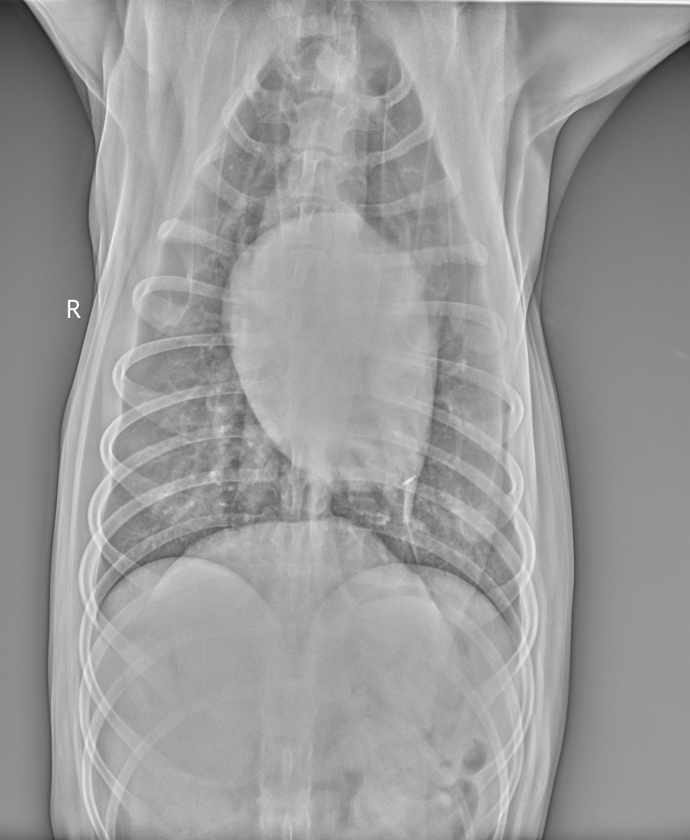
Ventrodorsal Radiograph of Normal Canine Costophrenic Margins. This image demonstrates clear pulmonary vascular markings extending to the bilateral costophrenic margins. The cardiac silhouette is appropriately positioned within the mediastinum, displaying an elongated conformation with the apex pointing toward the left hemithorax, which is typical for deep-chested canine breeds, such as German shepherds and Labrador retrievers.
Contributed by Cecilia Winthrop, DVM
(Click Image to Enlarge)
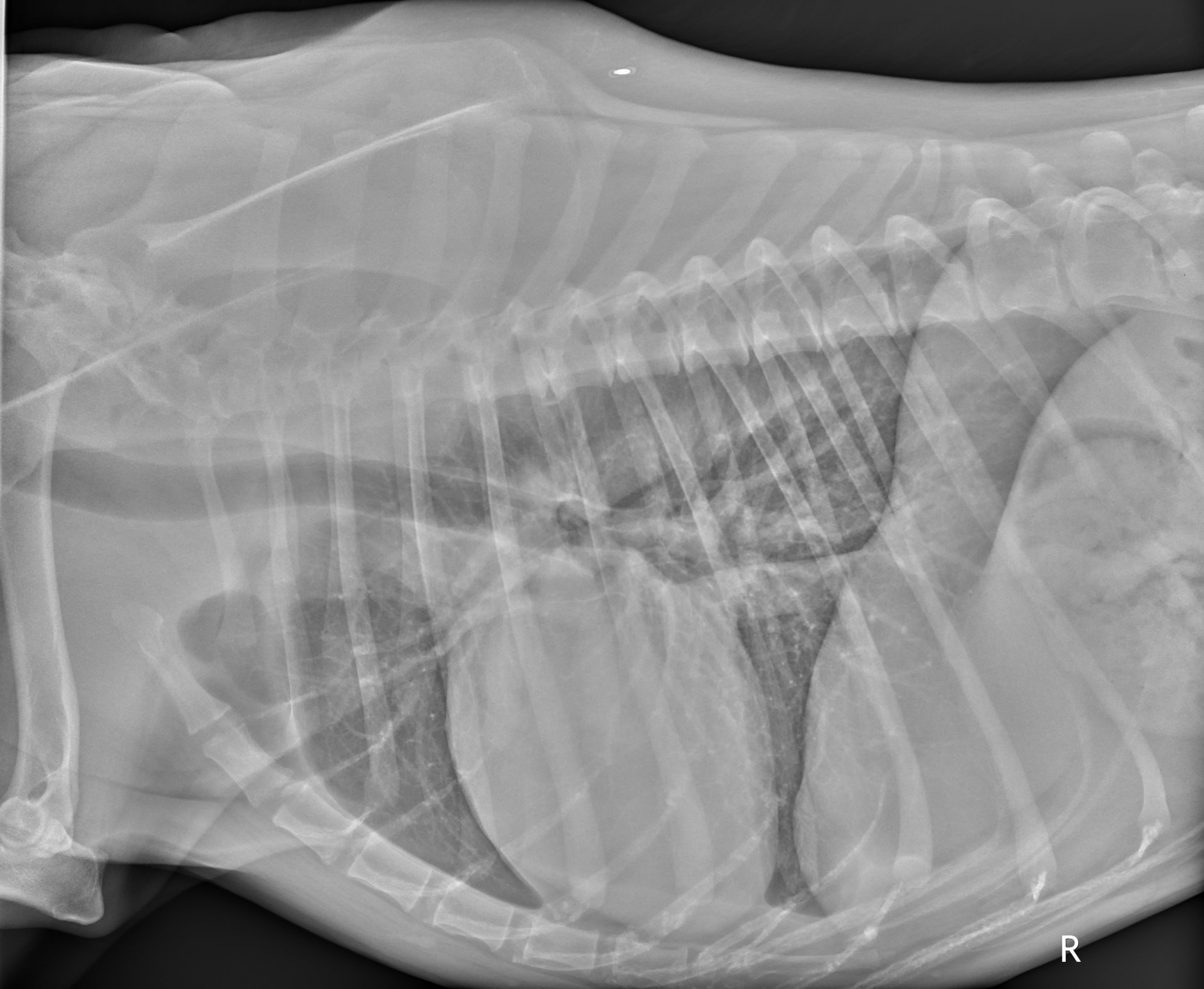
Lateral View of Normal Canine Lung Fields. This x-ray shows a clear, unobstructed view of the canine respiratory system. Normal bronchovascular markings are visible throughout the dorsal and ventral lung fields without evidence of consolidation, nodules, or abnormal fluid accumulation.
Contributed by Cecilia Winthrop, DVM
(Click Image to Enlarge)
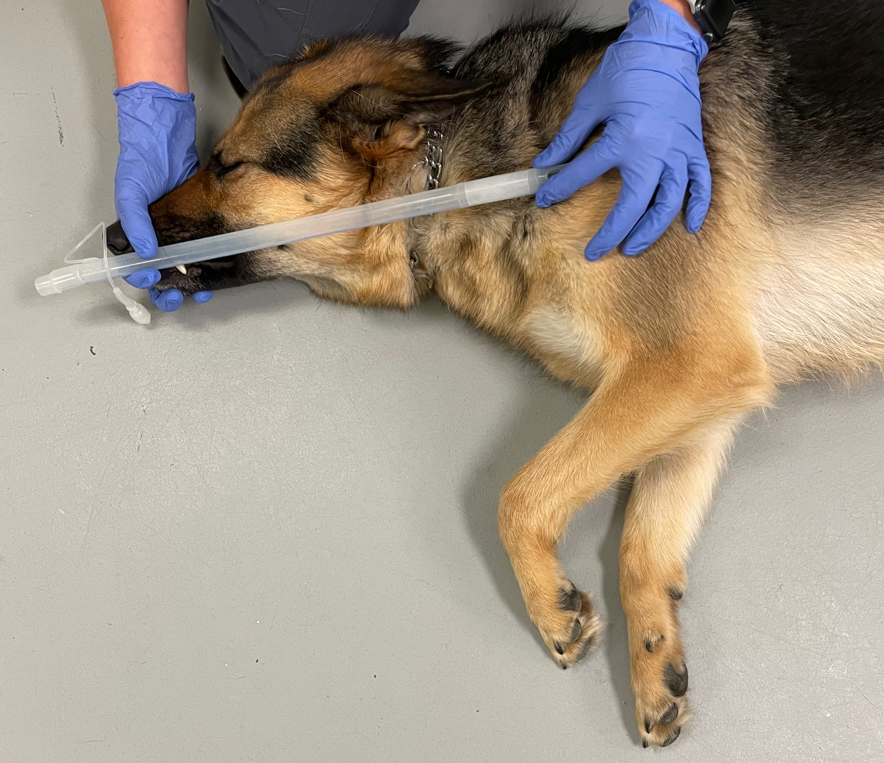
Landmarks for Estimating Canine Endotracheal Intubation Depth. This image shows the proper technique for approximating endotracheal tube insertion depth using external anatomical landmarks on a dog. The tube length is measured from the tip of the incisors to the cranial margin of the thoracic inlet to prevent accidental endobronchial intubation.
Contributed by Cecilia Winthrop, DVM. Obtained with informed consent.
References
Miller L, Pacheco GJ, Janak JC, Grimm RC, Dierschke NA, Baker J, Orman JA. Causes of Death in Military Working Dogs During Operation Iraqi Freedom and Operation Enduring Freedom, 2001-2013. Military medicine. 2018 Sep 1:183(9-10):e467-e474. doi: 10.1093/milmed/usx235. Epub [PubMed PMID: 29547926]
Level 2 (mid-level) evidenceStorer AP, Edwards TH, Rutter CR, Young GE, Mullaney SB. Causes of mortality in military working dog from traumatic injuries. Frontiers in veterinary science. 2024:11():1360233. doi: 10.3389/fvets.2024.1360233. Epub 2024 Jul 8 [PubMed PMID: 39040817]
Taylor WM. Canine tactical field care. Part two--Massive hemorrhage control and physiologic stabilization of the volume depleted, shock-affected, or heatstroke-affected canine. Journal of special operations medicine : a peer reviewed journal for SOF medical professionals. 2009 Spring:9(2):13-21. doi: 10.55460/V7B3-973P. Epub [PubMed PMID: 19813515]
Level 3 (low-level) evidencePalmer LE, Maricle R, Brenner JA. The Operational Canine and K9 Tactical Emergency Casualty Care Initiative. Journal of special operations medicine : a peer reviewed journal for SOF medical professionals. 2015 Fall:15(3):32-38. doi: 10.55460/RMVA-7381. Epub [PubMed PMID: 26360351]
Curry T, Kuhl EA. EMS Canine Tourniquet Use. StatPearls. 2026 Jan:(): [PubMed PMID: 40334034]
Edwards TH, Palmer LE, Baxter RL, Sager TC, Coisman JG, Brown JC, George C, McGraw AC. Canine Tactical Combat Casualty Care (K9TCCC) Guidelines. Journal of special operations medicine : a peer reviewed journal for SOF medical professionals. 2020 Spring:20(1):101-111. doi: 10.55460/YUMR-DBOP. Epub [PubMed PMID: 32203614]
Edwards TH, Pusateri AE, Mays EL, Bynum JA, Cap AP. Lessons Learned From the Battlefield and Applicability to Veterinary Medicine - Part 2: Transfusion Advances. Frontiers in veterinary science. 2021:8():571370. doi: 10.3389/fvets.2021.571370. Epub 2021 May 7 [PubMed PMID: 34026881]
Level 3 (low-level) evidenceEdwards TH, Meledeo MA, Peltier GC, Henderson AF, Pompa LA, Bynum JA. In vitro evaluation of hyperosmotic canine plasma suitable for infusion. Journal of veterinary emergency and critical care (San Antonio, Tex. : 2001). 2024 Jan-Feb:34(1):63-68. doi: 10.1111/vec.13353. Epub 2023 Nov 15 [PubMed PMID: 37966879]
CRASH-3 trial collaborators. Effects of tranexamic acid on death, disability, vascular occlusive events and other morbidities in patients with acute traumatic brain injury (CRASH-3): a randomised, placebo-controlled trial. Lancet (London, England). 2019 Nov 9:394(10210):1713-1723. doi: 10.1016/S0140-6736(19)32233-0. Epub 2019 Oct 14 [PubMed PMID: 31623894]
Level 1 (high-level) evidenceBarrett WJ, Kaucher KA, Orpet RE, Campion EM, Goodloe JM, Fischer PE, Colwell CB, Lyng JW. Tranexamic acid in trauma: A joint position statement and resource document of NAEMSP, ACEP, and ACS-COT. The journal of trauma and acute care surgery. 2025 Sep 1:99(3):357-363. doi: 10.1097/TA.0000000000004727. Epub 2025 Aug 18 [PubMed PMID: 40842057]
Johnson SA, Carr C, Reeves LK, Bean K, Schauer SG. Injuries and Interventions on Transported Military Working Dogs Within the US Central Command. Journal of special operations medicine : a peer reviewed journal for SOF medical professionals. 2022 Spring:22(1):97-101. doi: 10.55460/VTBK-XU21. Epub [PubMed PMID: 35278322]
Reeves LK, Mora AG, Field A, Redman TT. Interventions Performed on Multipurpose Military Working Dogs in the Prehospital Combat Setting: A Comprehensive Case Series Report. Journal of special operations medicine : a peer reviewed journal for SOF medical professionals. 2019 Fall:19(3):90-93. doi: 10.55460/LE5D-P32Y. Epub [PubMed PMID: 31539440]
Level 2 (mid-level) evidenceHwang TS, Yoon YM, Jung DI, Yeon SC, Lee HC. Usefulness of transthoracic lung ultrasound for the diagnosis of mild pneumothorax. Journal of veterinary science. 2018 Sep 30:19(5):660-666. doi: 10.4142/jvs.2018.19.5.660. Epub [PubMed PMID: 30041286]
Taylor WM. Canine tactical field care part three - thoracic and abdominal trauma. Journal of special operations medicine : a peer reviewed journal for SOF medical professionals. 2010 Winter:10(1):50-8 [PubMed PMID: 20306416]
Level 3 (low-level) evidenceBurkitt-Creedon JM, Boller M, Fletcher DJ, Brainard BM, Buckley GJ, Epstein SE, Fausak ED, Hopper K, Lane SL, Rozanski EA, Wolf J. 2024 RECOVER Guidelines: Updated treatment recommendations for CPR in dogs and cats. Journal of veterinary emergency and critical care (San Antonio, Tex. : 2001). 2024 Jul-Aug:34 Suppl 1():104-123. doi: 10.1111/vec.13391. Epub [PubMed PMID: 38924627]
Holcomb JB, Stansbury LG, Champion HR, Wade C, Bellamy RF. Understanding combat casualty care statistics. The Journal of trauma. 2006 Feb:60(2):397-401 [PubMed PMID: 16508502]
Level 3 (low-level) evidenceWart M, Edwards TH, Rizzo JA, Peitz GW, Pigott A, Levine JM, Jeffery ND. Traumatic brain injury in companion animals: Pathophysiology and treatment. Topics in companion animal medicine. 2024 Nov-Dec:63():100927. doi: 10.1016/j.tcam.2024.100927. Epub 2024 Oct 25 [PubMed PMID: 39461414]
Level 3 (low-level) evidencePigott A, Rudloff E. Traumatic Brain Injury-A Review of Intravenous Fluid Therapy. Frontiers in veterinary science. 2021:8():643800. doi: 10.3389/fvets.2021.643800. Epub 2021 Jul 9 [PubMed PMID: 34307515]
Butler FK Jr, Blackbourne LH. Battlefield trauma care then and now: a decade of Tactical Combat Casualty Care. The journal of trauma and acute care surgery. 2012 Dec:73(6 Suppl 5):S395-402. doi: 10.1097/TA.0b013e3182754850. Epub [PubMed PMID: 23192061]