 Tools
Tools
 Laboratory Evaluation of Hereditary Hemochromatosis
Introduction
Hemochromatosis, a disorder caused by excessive iron absorption, was first described in the mid-1800s as bronze diabetes and pigmentary cirrhosis. The term hemochromatosis was believed to have been coined by von Recklinghausen in 1889. A genetic origin of this metabolic problem was first suggested in the 1930s. In 1977, Simon et al reported the association between an HLA class I–like molecule and the presumed hemochromatosis gene on chromosome 6p, establishing the genetic basis of what is now referred to as hereditary hemochromatosis.[1]
Although one of the most common genetic disorders in the United States, affecting over 1 million people, hereditary hemochromatosis is often an incidental finding during routine laboratory iron measurements or the diagnostic workup of other conditions; however, increasing awareness of hereditary hemochromatosis has also contributed to early detection. Early diagnosis allows for intervention before tissue damage occurs due to excessive iron deposition. Iron can deposit in the liver, pancreas, heart, joints, and other endocrine organs if left untreated.[2]
Hereditary hemochromatosis is currently classified into 4 major types, encompassing 5 distinct molecular subtypes. These classifications are based on age of onset, underlying genetic mutation, and mode of inheritance.[3]
- Type I hereditary hemochromatosis is considered the classic form of the disorder, and the onset of symptoms begins in adulthood. Loss-of-function mutations in the hereditary Fe [iron] (HFE) gene are present in approximately 70% of patients diagnosed with hereditary hemochromatosis. This form of hereditary hemochromatosis disproportionately affects males, although females can also be affected.
- Type 2 hereditary hemochromatosis is frequently called juvenile hemochromatosis, as symptoms begin in childhood, and the disease is typically more clinically severe. Unlike type I hereditary hemochromatosis, this form shows no sex preference. Type 2 hereditary hemochromatosis has 2 subtypes: 2a and 2b. Type 2a hereditary hemochromatosis is caused by mutations in the gene initially known as hemojuvelin (HJV) and is now referred to as HFE2. Type 2b hereditary hemochromatosis is due to hepcidin antimicrobial peptide (HAMP) gene mutations.[4]
- Type 3 hereditary hemochromatosis typically has an onset at approximately 30 years of age and is caused by mutations in the transferrin receptor 2 (TFR2) gene.
- Type 4 hereditary hemochromatosis, also known as ferroportin disease, is caused by mutations in the ferroportin/solute carrier family 40 member 1 (SLC40A1) gene. Ferroportin is an iron transmembrane transport protein, and Type 4 hereditary hemochromatosis is the only known form of hemochromatosis that can be inherited in an autosomal dominant fashion. The onset of type 4 hereditary hemochromatosis typically occurs in mid-adulthood.[5][6]
Etiology and Epidemiology
Register For Free And Read The Full Article
 Search engine and full access to all medical articles
- 10 free questions in your specialty
- Free CME/CE Activities
- Free daily question in your email
- Save favorite articles to your dashboard
- Emails offering discounts
Learn more about a Subscription to StatPearls Point-of-Care
Etiology and Epidemiology
Epidemiology
In the United States, about 1 in 300 non-Hispanic White people has hereditary hemochromatosis, with lower rates among people of other races and ethnicities. Approximately 9% to 10% of males with hereditary hemochromatosis develop liver disease. Types 1, 2, and 3 hereditary hemochromatosis are transmitted via an autosomal recessive pattern, whereas type 4 hereditary hemochromatosis is predominantly an autosomal dominant disease. [7]
Hereditary hemochromatosis is the most common inherited disorder among people of northern European ancestry. The United States, Europe, and Australia have a similar disease prevalence of 1 case per 200 to 400 people, with the highest prevalence in those of Irish and Scandinavian ancestry. Hereditary hemochromatosis–associated mutations are rare among people of Asian, African, Hispanic, and Pacific Islander ancestry. Despite the high prevalence of the gene mutation, clinical penetrance is low and variable. Up to 25% of people with C282Y homozygosity are clinically asymptomatic.[8][9]
Etiology
Over 100 known mutations in the HFE gene can cause type 1 hereditary hemochromatosis. The HFE protein regulates the production of hepcidin, a protein produced by the liver that regulates iron homeostasis. The p.Cys282Tyr or C282Y variant is the most common genetic mutation in type 1 hereditary hemochromatosis, which occurs due to a single nucleotide change (845 G-A), substituting cysteine with tyrosine at amino acid 282.[10] This mutation prevents the altered HFE protein from inserting into the cell's surface membrane. The second most common mutation in Type 1 hereditary hemochromatosis replaces histidine with aspartic acid at position 63 (written as p.His63Asp or H63D).[11]
Patients with type 1 hereditary hemochromatosis have 2 mutated copies of the HFE gene. Homozygous C282Y and heterozygous C282Y/H63D mutations of the HFE gene on chromosome 6 are responsible for up to 95% of type 1 hereditary hemochromatosis cases.[8]
Interestingly, the fact that the C282Y homozygosity was mainly detected in patients of Northern European ancestry and is not observed in other regions of the world, including Asia and Africa, led to the recognition of a heterogenous genetic basis of hereditary hemochromatosis and the discovery of other variants involving the non-HFE genes, particularly the HAMP and HJV genes associated with severe early-onset juvenile forms of hereditary hemochromatosis.[1]
Although the C282Y variant was initially described as the primary mutation responsible for hereditary hemochromatosis, several other non-HFE genes, including variants of HAMP, HJV, SLC40A1, and TFR2 genes, have been identified in other types of hereditary hemochromatosis.[12][13][14]
Pathophysiology
The clinical symptoms of hereditary hemochromatosis result from genetic defects in iron metabolism proteins, leading to iron overload and deposition in the liver, pancreas, myocardium, joints, and multiple endocrine organs, including the pituitary, thyroid, parathyroid, and adrenal glands. Humans cannot actively excrete iron; regulation of intestinal iron absorption is the primary mechanism for maintaining iron homeostasis. The total body iron pool in healthy adults ranges from 3 to 4 g; approximately 0.5 g is stored in the liver.[15] In severe, untreated hereditary hemochromatosis, total body iron stores may exceed 50 g, of which one-third is stored in hepatocytes. Excessive iron appears directly toxic to tissues, although cells that are not irreversibly injured recover function as the iron is removed. The characteristic findings of severe, untreated hereditary hemochromatosis are micronodular cirrhosis, diabetes mellitus, and abnormal skin pigmentation.
Normal iron homeostasis is a complex mechanism that is incompletely understood. Humans must absorb iron from their diet; iron is used in multiple physiological processes, including DNA biosynthesis, oxygen transport, and cellular energy generation.[15] Hepcidin, the protein product of HAMP, is a circulating peptide hormone that is a negative regulator of iron absorption by enterocytes. When circulating hepcidin levels are high, iron absorption in the gut is reduced.[16] Ferroportin is an iron transmembrane transport protein encoded by SLC4OA1 that transports iron from the enterocyte into the vasculature. Ferroportin also transports iron out of reticuloendothelial cells. The proteolytic degradation of ferroportin is facilitated by hepcidin.[17]
The HFE, HJV, and TFR2 gene products are hepatocyte membrane proteins that sense iron. In the presence of normal or elevated iron levels, these gene products activate hepcidin production, reducing enterocyte absorption and promoting ferroportin degradation.[8][15]
Mutations in HFE, HFE2, HAMP, HJV, and TFR2 significantly impair hepcidin synthesis, ultimately increasing intestinal iron absorption and iron deposition in tissues. The gain-of-function mutation of SLC40A1 impairs hepcidin-ferroportin binding. HFE-related mutations include C282Y, H63D, and S65C; together, these account for over 90% of cases of hereditary hemochromatosis, most of which are due to C282Y homozygosity.[18][19][20]
The liver is most severely affected by untreated hereditary hemochromatosis. Whatever the underlying genetic defect, excess iron deposition within hepatocytes results in lipid peroxidation via iron-catalyzed free radical reactions, stimulation of collagen formation by stellate cells, and DNA damage by reactive oxygen species. Ferroptosis is a recently discovered form of nonapoptotic cell death mediated by reactive oxygen species and lipid peroxidation induction.[15] The risk of hepatocellular carcinoma in patients with hereditary hemochromatosis is increased 200-fold.
Iron uptake into pancreatic beta-cells leads to impaired insulin synthesis and release, whereas liver fibrosis leads to high levels of circulating insulin and insulin resistance.[21] Iron deposition in the pancreas can result in hyperglycemia; hereditary hemochromatosis should be suspected if there is skin hyperpigmentation, joint pain, hypogonadism, or features of liver disease in addition to persistent hyperglycemia.[22] The pathogenesis of diabetes mellitus in hereditary hemochromatosis is considered multifactorial; both insulin deficiency and resistance may contribute. Iron overload can impair insulin secretion and glucose tolerance early in hereditary hemochromatosis before cirrhosis occurs.[23]
The myocardium is also affected by iron deposition. In hereditary hemochromatosis, iron deposition initially occurs within subepicardial cardiac myocytes; as the disease progresses, it spreads throughout the myocardium. Myocardial hypertrophy leads to diastolic dysfunction. If iron deposition continues, cardiomyopathy, systolic dysfunction, and arrhythmias ensue.[24]
Deposition of iron in the cells of the anterior pituitary gland may result in reduced production of luteinizing hormone and follicle-stimulating hormone.[25] In non-HFE forms of hereditary hemochromatosis, iron deposition starts early, is more severe, and occurs in the pituitary, especially the gonadotropes, although other lineages are affected.[26]
Joint pain affects up to 75% of patients with hereditary hemochromatosis even before the diagnosis is made. Overt clinical symptoms related to joint involvement typically appear in the fifth decade of life but may appear as early as the third decade.[27] Hemochromatosis-associated pseudogout (chondrocalcinosis) is not common, but in patients with severe chondrocalcinosis, the frequency of association may justify screening for hemochromatosis, especially in younger males.[28] The inhibition of pyrophosphatases and synovial iron sequestration are likely mechanisms causing damage to the articular cartilage.[29]
Specimen Requirements and Procedure
Evaluation of hereditary hemochromatosis begins with biochemical iron studies, which require strict adherence to pre-analytical conditions. Serum iron and total iron-binding capacity, when interpreted alongside ferritin, are used to assess iron status. Serum and plasma yield comparable results for iron measurement; however, due to significant diurnal variation, an early-morning fasting sample is preferred, as iron concentrations may decrease by up to 30% later in the day.[30] Peak levels typically occur in the early- to late-morning hours. Specimens should be centrifuged promptly after collection, as storage of whole blood without separation renders samples unsuitable for analysis.[31] Serum iron remains stable for up to 1 week at 4 to 8 °C and up to 1 year when frozen at −20 °C.[32] Hemolyzed samples must be rejected because erythrocyte iron causes falsely elevated results, and the effect of hemolysis is unpredictable.[33]
Ferritin testing should be performed on serum collected in plain red-top or serum separator tubes. No specific patient preparation is required.[34] Ferritin specimens are stable for at least 1 week at 4 °C and up to 6 months at −20 °C. Frozen samples should not be thawed at 37 °C, and repeated freeze-thaw cycles or vigorous mixing should be avoided to prevent protein denaturation.[32] Because ferritin is an acute-phase reactant, concurrent measurement of C-reactive protein using a serum separator tube sample is recommended to exclude inflammation-related hyperferritinemia.[35]
If biochemical findings suggest iron overload, confirmatory HFE genetic testing is performed. This test requires whole blood collected in an EDTA (lavender-top) tube; ACD (yellow-top) tubes may be used according to laboratory protocol. Whole blood specimens for molecular analysis should not be frozen before DNA extraction, as freezing may compromise cellular integrity and DNA quality.[36]
Assessment of iron-related organ involvement requires additional specimens. Liver injury is evaluated using serum liver function tests, including alanine aminotransferase, aspartate aminotransferase, and gamma-glutamyl transferase.[37] Metabolic complications are assessed using HbA1c measured in EDTA whole blood or fasting plasma glucose collected in a fluoride oxalate tube.[38] When screening for hypogonadism, total testosterone should be measured in a morning sample collected between 7:00 and 10:00 AM to account for diurnal variation and capture peak physiological concentrations.[39]
Diagnostic Tests
Serum ferritin concentration and serum transferrin saturation (TSAT) are increased in patients with hereditary hemochromatosis. Further testing should be considered if the ferritin levels exceed 200 µg/L in menstruating females or 300 µg/L in males or nonmenstruating females, or if the TSAT is >45%.[1] Total iron-binding capacity values >450 mcg/dL (80.55 mmol/L) can also help diagnose pathological iron accumulation.[40] The negative predictive value for iron overload is 97% when serum ferritin is normal and TSAT <45%.[41]
Ferritin is an acute-phase reactant that is often elevated in conditions of inflammation or malignancy; therefore, hyperferritinemia is a nonspecific finding that must be interpreted within the clinical context.[34] A persistently elevated TSAT level (>45%) is a more reliable indicator of hereditary hemochromatosis. Other findings that raise suspicion of hereditary hemochromatosis include MRI evidence of hepatic iron overload or iron deposits in hepatocytes on liver biopsy.[42]
The absence of acquired risk factors for hepcidin deficiency, such as alcohol misuse or end-stage liver disease, favors the possibility of hereditary hemochromatosis.[43] Secondary causes of iron overload must be excluded; these include iatrogenic iron overload in the setting of regular blood transfusions, reduced hepcidin production due to hepatic dysfunction, chronic renal failure, adult-onset Still disease, hemophagocytic lymphohistiocytosis, hereditary hyperferritinemia, and disorders of ineffective erythropoiesis such as the thalassemias, other hemolytic anemias, and myelodysplastic syndromes.[44]
Genetic Testing
If serum ferritin levels are persistently elevated and the TSAT is >45%, the next diagnostic step is genetic testing; HFE genotyping is recommended.[45][46] Homozygous C282Y and compound heterozygous C282Y/H63D mutations of the HFE gene are responsible for up to 95% of all hereditary hemochromatosis cases; types 2 through 4 hereditary hemochromatosis comprise a small percentage of remaining cases and are caused by non-HFE mutations.[46] The incidence of C282Y or H63D compound heterozygosity is less than 5%.[47]
Since mutations in HJV, HAMP, TFR2, or the ferroportin gene are far less common, genetic testing for non-HFE hemochromatosis may be considered in cases where other causes of hyperferritinemia have been ruled out, there is a family history of iron overload, or hepatic iron overload can be demonstrated on MRI or liver biopsy. HJV, HAMP, and TFR2 mutations are usually observed in early-onset disease with severe clinical manifestations and are recessive, whereas the ferroportin mutations are dominant.[48][49][50]
Interestingly, although C282Y homozygosity is strongly associated with an increased risk of cirrhosis, phenotypic variability remains significant due to incomplete penetrance. However, a significant positive statistical association exists between abdominal pain and cirrhosis in hemochromatosis probands with this homozygous mutation (see Image. Flow Chart for the Investigation of Hereditary Hemochromatosis).[9][51]
Testing for Liver Involvement
Liver biopsy with histochemical, semi-quantitative assessment, or chemical determination of iron content was previously considered the gold standard of diagnosis in hereditary hemochromatosis. However, in current clinical practice, the diagnosis is made via genetic testing for specific mutations, with elevated serum ferritin and TSAT.[51] A diagnostic liver biopsy is indicated when the diagnosis is unclear, as in certain types of non-HFE hemochromatosis, dysmetabolic iron overload syndrome, nonalcoholic fatty liver disease, and some types of alcoholic liver disease presenting with elevated ferritin and moderate iron overload. A liver biopsy can also assess the degree of fibrosis and detect hepatocellular carcinoma.[52]
Histopathological evaluation of the liver in hereditary hemochromatosis reveals characteristic intracytoplasmic deposits of golden-yellow hemosiderin granules within hepatocytes. These deposits are initially located in the periportal region (see Image. Micrograph of Hepatic Iron Deposition in Hereditary Hemochromatosis) but eventually are found throughout the lobule, including within Kupffer cells and biliary epithelium. These hemosiderin granules appear blue with the Prussian blue stain (see Image. High-Magnification Micrograph of Hepatic Iron Deposition in Hereditary Hemochromatosis). Parenchymal architecture is preserved in early-stage hereditary hemochromatosis; the onset of cirrhosis distorts normal architecture.
Noninvasive modalities such as magnetic resonance imaging (MRI) are now preferred over invasive liver biopsy for quantifying hepatic iron deposition.[53][54] MRI for noninvasive hepatic iron quantification also improves the diagnostic yield of next-generation sequencing in patients with hyperferritinemia.[55]
Other noninvasive techniques for detecting liver fibrosis in patients with hereditary hemochromatosis include the aspartate aminotransferase-to-platelet ratio index, fibrosis-4 index, Hepascore, and hepatic transient elastography (FibroScan).[56][57][58] The aspartate aminotransferase-to-platelet ratio index cut-off value >0.44 and the fibrosis-4 index cut-off value >1.1 have shown good diagnostic utility, correctly identifying liver biopsy-diagnosed advanced hepatic fibrosis in 85% and 80% of cases, respectively.[59] Hepascore incorporates the clinical variables of sex and age with blood-based markers, including bilirubin, gamma-glutamyl transferase, hyaluronic acid, and alpha2-macroglobulin, to detect hepatic fibrosis. Transient elastography (FibroScan) uses ultrasound to assess hepatic fibrosis and can be performed in an outpatient setting. Hepascore and transient elastography are likely most reliable when serum ferritin levels are >1000 μg/L.[60]
Testing for Cardiac Involvement
Suddies suggest that serial assessment of serum non–transferrin-bound iron levels may guide the initiation of treatment to avoid cardiac and other organ damage. However, standardization of this protocol has proven difficult.[61]
Electrocardiographic changes in hereditary hemochromatosis are typically nonspecific and recognized late in the course of the disease. However, standard echocardiography can reveal cardiac abnormalities even in the early stages of hereditary hemochromatosis. Two-dimensional speckle tracking echocardiography and three-dimensional real-time echocardiography are novel techniques that appear more precise and distinctive in detecting subtle cardiac abnormalities in early-stage hereditary hemochromatosis.[40]
Cardiac MRI plays a pivotal role in detecting cardiac abnormalities associated with hereditary hemochromatosis. A T2 relaxation time of less than 20 ms is regarded as a reliable marker of myocardial iron overload. Despite its diagnostic value, the routine use of cardiac MRI remains constrained by cost and limited availability.[62][63] Cardiac biopsy is rarely performed in the diagnosis and evaluation of hereditary hemochromatosis. A cardiac biopsy may be indicated in cases where the diagnosis is unclear, and other causes of heart failure or infiltrative diseases need to be ruled out.[40]
Testing for Pancreatic Involvement
Pancreatic iron deposition may lead to hyperglycemia, and hereditary hemochromatosis is associated with an increased risk of type 2 diabetes. Screening and diagnosis can be achieved through fasting serum glucose measurement, glucose tolerance testing, or hemoglobin A1c assessment. Less commonly, hereditary hemochromatosis may result in chronic pancreatitis; in such cases, MRI provides an effective means of evaluating pancreatic iron deposition.[64]
Testing Hormone Levels to Evaluate Hypogonadism
Hypogonadism is the second most common endocrine abnormality in hereditary hemochromatosis, with a frequency ranging from 10% to 100%.[65] Assessment of serum follicle-stimulating hormone, luteinizing hormone, testosterone, and, occasionally, gonadotropin-releasing hormone is advised for coexisting infertility or reproductive system disorders. Brain MRI can detect pituitary iron deposits.[66] Evidence suggests that screening for common endocrinopathies is beneficial in patients with hereditary hemochromatosis and pituitary iron deposition, even in the absence of symptoms.[67] Additionally, if the serum ferritin is ≥300 μg/L, screening is beneficial regardless of clinical manifestations.[68] In premenopausal females, hypogonadism is evidenced by secondary amenorrhea and decreased FSH and LH serum levels.[69]
Testing for Musculoskeletal Involvement
Joint pain precedes a hereditary hemochromatosis diagnosis in up to 75% of patients. Plain radiography of the affected joints can be used initially to determine the extent of arthritis. A validated rheumatological scoring system based on joint radiographs has been used in hemochromatosis-related arthritis. Osteopenia and osteoporosis can be assessed using dual-energy x-ray absorptiometry (DEXA) based on bone density measurements.[70] The trabecular bone score, based on the spatial grayscale analysis of DEXA images, enables the evaluation of bone microarchitecture.[29]
Testing Procedures
Iron profiles are performed using routine clinical chemistry and immunological assays.[71] Serum iron is typically measured by spectrophotometric methods, while ferritin and transferrin are quantified using automated immunoassays.[72]
Ferritin testing is used to measure stored iron in the body and is performed using highly sensitive automated immunoassay platforms, most commonly enzyme-linked immunosorbent assays, chemiluminescent microparticle immunoassays, or immunoturbidimetric assays. These methods rely on specific antibodies that bind serum ferritin, allowing accurate quantification of ferritin concentration through colorimetric, luminescent, or turbidimetric signal detection.[73]
Transferrin testing measures the principal iron-transport protein in circulation and is primarily performed using turbidimetric or nephelometric immunoassays. In these assays, antibodies directed against human transferrin form immune complexes with the analyte in the patient's serum. The resulting change in turbidity or light scatter is measured, providing a value directly proportional to the transferrin concentration. Transferrin saturation is subsequently calculated using serum iron and transferrin values.[74]
Molecular testing is conducted using real-time polymerase chain reaction (PCR) with fluorescence resonance energy transfer hybridization probes to differentiate wild-type from mutant HFE alleles.[75] Identification of homozygosity for the C282Y mutation confirms the diagnosis of classic hereditary hemochromatosis. Samples found to be heterozygous for C282Y are reflexed for H63D mutation analysis.[76]
Histopathological assessment, supported by immunohistochemistry, is used to detect and localize tissue iron deposition. Radiological techniques are additionally utilized to assess iron accumulation and structural involvement of the liver, heart, musculoskeletal, endocrine, and other organ systems, as well as to evaluate disease progression and the extent of tissue damage.[77]
Interfering Factors
Iron profile measurements are subject to several preanalytical and analytical interferences. Hemolysis can falsely elevate serum iron levels by releasing intracellular iron, whereas lipemia and icterus may interfere with spectrophotometric measurements. Diurnal variation and recent oral or parenteral iron supplementation can also affect serum iron results.[78] Transferrin saturation calculations may be impacted by inaccuracies in either serum iron or transferrin measurements. Transferrin measurement by turbidimetric or nephelometric immunoassays may also be affected by hyperlipidemia, paraproteinemia, and severe hemolysis, which can alter light-scattering and turbidity readings.[79][80] Inflammatory states may reduce transferrin concentrations, influencing calculated transferrin saturation without reflecting true iron status.[81]
Ferritin immunoassays are susceptible to multiple biological and analytical interferences. As an acute-phase reactant, ferritin levels may be elevated in the presence of inflammation, infection, liver disease, malignancy, or metabolic syndrome, independent of iron stores.[48] Secondary causes of iron overload, including ineffective erythropoiesis; repeated blood transfusions; reduced hepcidin production from acute or chronic liver injury; chronic kidney disease; adult-onset Still disease; hemophagocytic lymphohistiocytosis; aceruloplasminemia; atransferrinemia; hereditary hyperferritinemia; dietary iron overload syndromes, such as Bantu siderosis; and oral or injectable iron excess, must also be considered when interpreting ferritin results.[82]
Ferritin immunoassay procedures can be further influenced by heterophilic antibodies or human anti-animal antibodies, leading to discordant values that may require alternative testing methods or confirmatory assays.[83] In vivo factors such as ethanol intake, iron salts, and oral contraceptives can elevate ferritin levels, whereas exogenous erythropoietin administration can lower them.[84] Immunonephelometric analytical methods are additionally affected by increased turbidity or lipemia, which can impair accurate signal measurement.[85] High biotin intake may interfere with chemiluminescent immunoassays, leading to falsely increased or decreased ferritin readings.[86]
Molecular testing using real-time PCR with fluorescence resonance energy transfer hybridization probes may be affected by poor DNA quality, inadequate sample quantity, or the presence of PCR inhibitors such as heme or anticoagulants.[87] Rare HFE variants outside routinely tested regions can result in false-negative findings, whereas contamination and allele dropout, though uncommon, represent additional technical limitations.[88]
Histopathological and immunohistochemical evaluation may be influenced by sampling error, uneven tissue iron distribution, prior phlebotomy or chelation therapy, and variability in staining techniques. Interpretation may also be affected by coexisting liver pathology, including steatosis or fibrosis.[89]
Radiological assessment of iron deposition, particularly MRI-based techniques, may be limited by motion artifacts, severe fibrosis, coexisting hepatic steatosis, or technical variability between scanners and protocols.[90] Accurate cardiac and hepatic iron quantification requires standardized acquisition and interpretation to minimize inter-observer and inter-institutional variability.[91]
Clinical Significance
Hereditary hemochromatosis is a prevalent genetic disorder with low and variable penetrance; up to 25% of patients with type 1 hereditary hemochromatosis are clinically asymptomatic. The characteristic laboratory findings of elevated serum ferritin and transferrin saturation or elevated liver transaminases are frequently found incidentally during routine laboratory screening or evaluation for other common disorders. In these circumstances, further diagnostic testing is required to arrive at a specific diagnosis. Primary hereditary hemochromatosis must be differentiated from secondary causes of iron overload.
Primary hereditary hemochromatosis is frequently managed via serial phlebotomy. Secondary hemochromatosis is managed with iron chelation and treatment of the underlying cause of iron overload.
Type 1 hereditary hemochromatosis is the most common form of the disorder and is inherited in an autosomal recessive pattern. Once a diagnosis of hereditary hemochromatosis is established, screening and genetic testing should be offered to the proband's first- and second-degree relatives.[92] Additionally, if the proband plans to conceive, their partner should be offered screening and genetic testing. Antenatal counseling should be offered to asymptomatic carriers in addition to patients with symptomatic hereditary hemochromatosis; the risks of transmitting the mutation to potential offspring should be explained.[93]
Quality Control and Lab Safety
To ensure optimal precision and accuracy of test results, timely calibration and quality checks, conducted through internal and external quality control (QC) systems, should be performed in accordance with the laboratory's quality policy. Strict adherence to standard operating procedures and international guidelines is essential to enhance diagnostic accuracy.[94]
The analytical examination process's QC monitors a measurement procedure to verify that it meets performance specifications appropriate for patient care or that an error condition must be corrected.[95] For non-waived tests, laboratory regulations require, at a minimum, the analysis of at least 2 levels of QC materials once every 24 hours. If necessary, laboratories can assay QC samples more frequently to ensure accurate results. QC samples should be assayed after calibration or maintenance of an analyzer to verify the correct method performance.[96] To address situations in which manufacturers' QC recommendations fall short of regulatory requirements, laboratories may implement an individualized QC plan. This approach involves conducting a risk assessment across all testing phases to identify potential sources of error and establishing a QC strategy to minimize their likelihood.[97]
The design of a QC plan must consider the analytical performance capability of a measurement procedure and the risk of harm to patients if an erroneous laboratory test result is used for a clinical care decision. An erroneous laboratory test result is a hazardous condition that may or may not cause harm to a patient, depending on what action or inaction a clinical care provider takes based on the erroneous result.[98]
The acceptable range and rules for interpreting QC results are based on the probability of detecting a significant analytical error condition with an acceptably low false-alert rate.[97] The desired process control performance characteristics must be established for each measurement before selecting the appropriate QC rules.[99] Westgard multi-rules are typically used to evaluate the QC runs. If a run is declared out of control, the system, including instruments, standards, and controls, should be investigated to determine the cause of the problem. No analysis should be performed until the issue has been resolved.[100]
Changing reagent lots can unexpectedly affect QC results. Careful evaluation of QC target values across reagent lots is necessary. The matrix-related interaction between a QC material and a reagent can vary across reagent lots; QC results may not reliably reflect a measurement procedure's performance for patient samples after a reagent lot change.[101] Clinical patient samples should be used to verify the consistency of results between old and new reagent lots, given the unpredictability of matrix-related bias in QC materials.[102]
Laboratories are required to participate in an external QC or proficiency testing program, as mandated by the Centers for Medicare & Medicaid Services under the Clinical Laboratory Improvement Amendments regulations.[94] Participation helps ensure the laboratory's accuracy and reliability compared with other laboratories performing the same or comparable assays. Results are monitored by the Centers for Medicare & Medicaid Services through required participation and scoring, as well as by voluntary accreditation organizations. The proficiency testing plan should be incorporated into the laboratory's quality assessment plan and overall quality program.[103]
All specimens, control materials, and calibrator materials should be considered potentially infectious. The usual precautions required for handling all laboratory reagents should be exercised. Disposal of all waste material should be in accordance with local guidelines. Gloves, a lab coat, and safety glasses should be worn when handling human blood specimens. All plastic tips, sample cups, and gloves that come into contact with blood should be placed in a biohazard waste container. All disposable glassware should be discarded into the sharps waste containers.[104] All work surfaces should be protected with disposable absorbent benchtop paper, which should be discarded into biohazard waste containers weekly or whenever blood contamination occurs. All work surfaces should be wiped weekly.[105]
Enhancing Healthcare Team Outcomes
Improving outcomes for patients with hereditary hemochromatosis requires a coordinated, interprofessional approach that integrates early recognition, accurate diagnosis, longitudinal monitoring, and patient-centered management. Clinicians, advanced practitioners, nurses, laboratory professionals, genetic counselors, pharmacists, and other healthcare professionals each play a critical role across the care continuum. Core clinical skills include recognizing characteristic laboratory patterns of iron overload, appropriately utilizing genetic testing, and assessing organ involvement to guide timely intervention and prevent irreversible complications.
Strategic management of hereditary hemochromatosis relies on evidence-based protocols for diagnostic evaluation, therapeutic phlebotomy, and long-term surveillance. Ethical considerations are central to care, particularly regarding informed consent for genetic testing, confidentiality, and counseling of affected family members. Clearly defined professional responsibilities support shared decision-making and ensure accountability, while flattening traditional hierarchies to value each team member's contributions.
Effective interprofessional communication facilitates accurate interpretation of laboratory results, coordination of specialty referrals, and consistent patient education. Nurses and allied health professionals reinforce adherence to treatment and monitoring plans, whereas pharmacists support medication safety and the management of comorbidities. Through structured care coordination, the interprofessional team enhances patient safety, improves clinical outcomes, and delivers comprehensive, high-quality care for individuals with hereditary hemochromatosis.
Media
(Click Image to Enlarge)
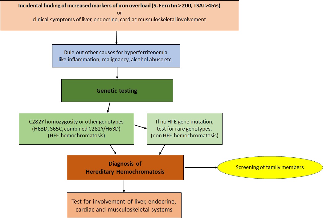
Flow Chart for the Investigation of Hereditary Hemochromatosis.
Contributed by J Divakaran, MD
(Click Image to Enlarge)
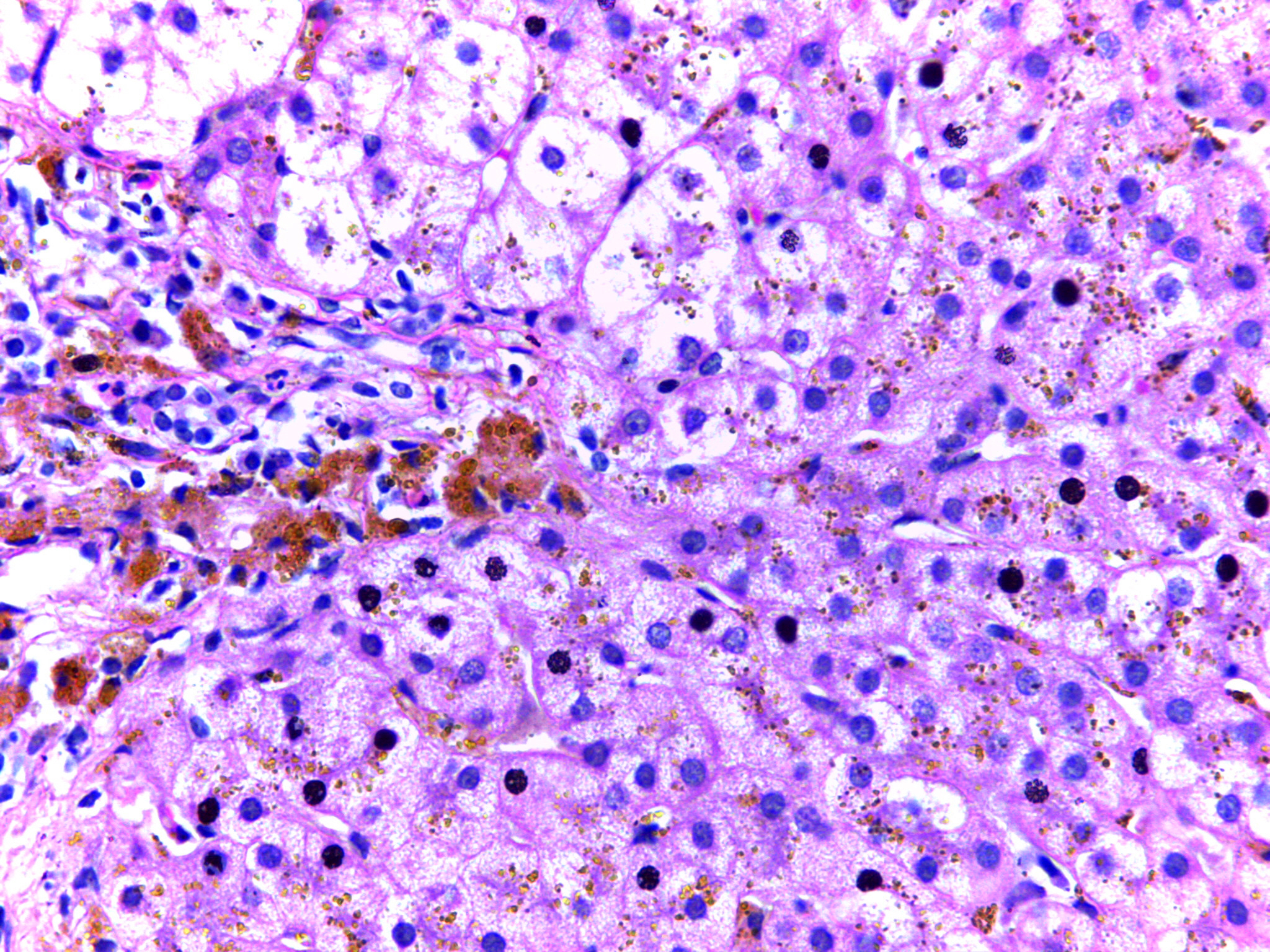
Micrograph of Hepatic Iron Deposition in Hereditary Hemochromatosis. The image shows hepatocytes with coarse, golden-yellow hemosiderin granules in the cytoplasm. These granules stain with Prussian blue.
Calicut Medical College, Public Domain, via Wikimedia Commons
(Click Image to Enlarge)
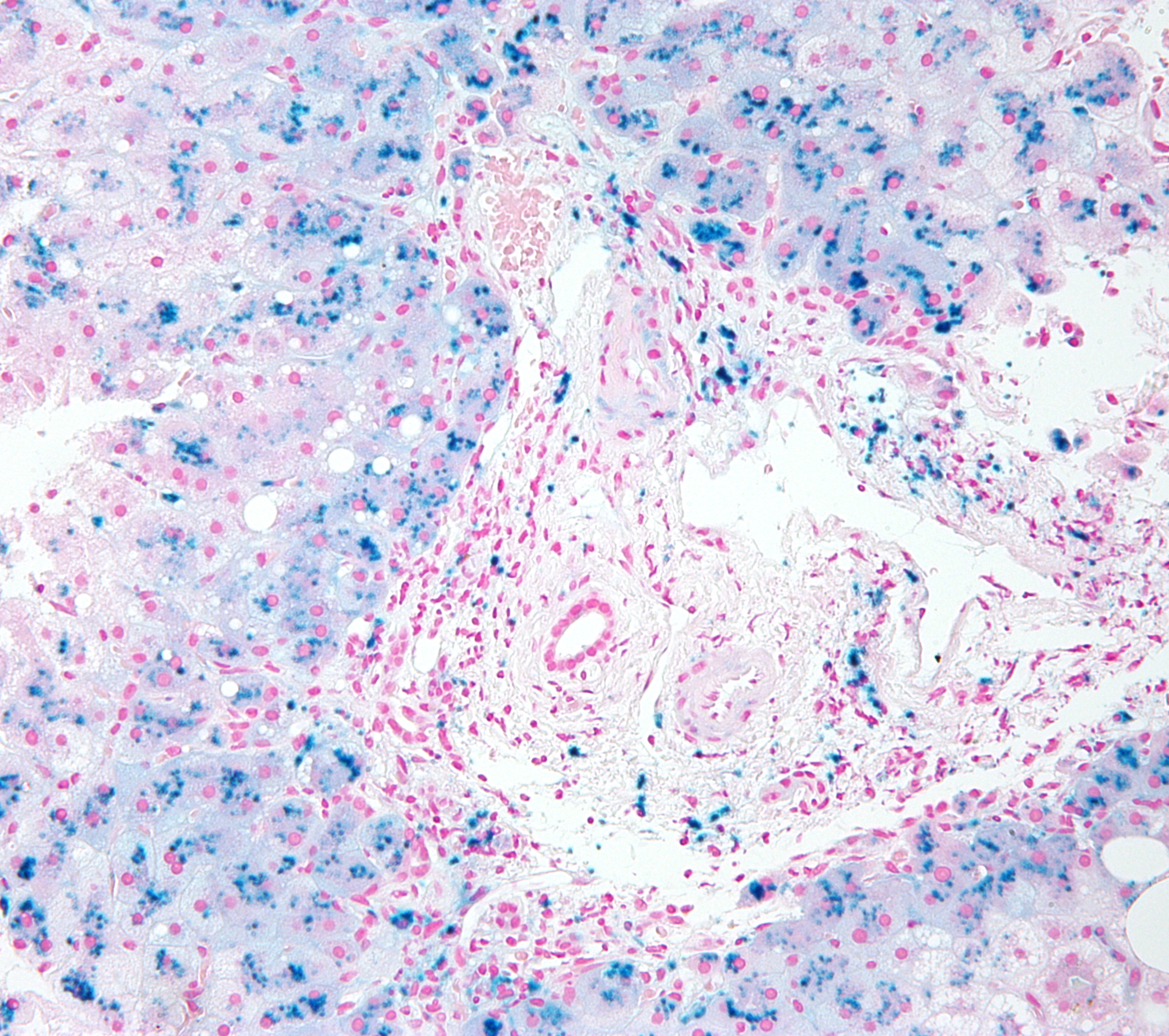
High-Magnification Micrograph of Hepatic Iron Deposition in Hereditary Hemochromatosis. Liver micrograph showing hemosiderin deposits within hepatocytes, which stain blue with Prussian blue, confirming iron accumulation characteristic of hereditary hemochromatosis.
Nephron, Public Domain, via Wikimedia Commons
References
Girelli D, Busti F, Brissot P, Cabantchik I, Muckenthaler MU, Porto G. Hemochromatosis classification: update and recommendations by the BIOIRON Society. Blood. 2022 May 19:139(20):3018-3029. doi: 10.1182/blood.2021011338. Epub [PubMed PMID: 34601591]
Killeen RB, Vadakekut ES. Hemochromatosis. StatPearls. 2026 Jan:(): [PubMed PMID: 28613612]
Gerhard GS, Paynton BV, DiStefano JK. Identification of Genes for Hereditary Hemochromatosis. Methods in molecular biology (Clifton, N.J.). 2018:1706():353-365. doi: 10.1007/978-1-4939-7471-9_19. Epub [PubMed PMID: 29423808]
Crawford DHG. Hereditary hemochromatosis types 1, 2, and 3. Clinical liver disease. 2014 May:3(5):96-97. doi: 10.1002/cld.339. Epub 2014 May 27 [PubMed PMID: 30992896]
Zhang W, Lv T, Huang J, Ou X. Type 4B hereditary hemochromatosis associated with a novel mutation in the SLC40A1 gene: A case report and a review of the literature. Medicine. 2017 Sep:96(38):e8064. doi: 10.1097/MD.0000000000008064. Epub [PubMed PMID: 28930842]
Level 3 (low-level) evidenceSpeletas M, Kioumi A, Loules G, Hytiroglou P, Tsitouridis J, Christakis J, Germenis AE. Analysis of SLC40A1 gene at the mRNA level reveals rapidly the causative mutations in patients with hereditary hemochromatosis type IV. Blood cells, molecules & diseases. 2008 May-Jun:40(3):353-9 [PubMed PMID: 17997113]
Girelli D, Marchi G, Busti F. Diagnosis and management of hereditary hemochromatosis: lifestyle modification, phlebotomy, and blood donation. Hematology. American Society of Hematology. Education Program. 2024 Dec 6:2024(1):434-442. doi: 10.1182/hematology.2024000568. Epub [PubMed PMID: 39644049]
Kane SF, Roberts C, Paulus R. Hereditary Hemochromatosis: Rapid Evidence Review. American family physician. 2021 Sep 1:104(3):263-270 [PubMed PMID: 34523883]
Sandnes M, Vorland M, Ulvik RJ, Reikvam H. HFE Genotype, Ferritin Levels and Transferrin Saturation in Patients with Suspected Hereditary Hemochromatosis. Genes. 2021 Jul 28:12(8):. doi: 10.3390/genes12081162. Epub 2021 Jul 28 [PubMed PMID: 34440336]
Milman N, Pedersen P. Evidence that the Cys282Tyr mutation of the HFE gene originated from a population in Southern Scandinavia and spread with the Vikings. Clinical genetics. 2003 Jul:64(1):36-47 [PubMed PMID: 12791037]
Barton JC, Edwards CQ, Acton RT. HFE gene: Structure, function, mutations, and associated iron abnormalities. Gene. 2015 Dec 15:574(2):179-92. doi: 10.1016/j.gene.2015.10.009. Epub 2015 Oct 9 [PubMed PMID: 26456104]
Lanzara C, Roetto A, Daraio F, Rivard S, Ficarella R, Simard H, Cox TM, Cazzola M, Piperno A, Gimenez-Roqueplo AP, Grammatico P, Volinia S, Gasparini P, Camaschella C. Spectrum of hemojuvelin gene mutations in 1q-linked juvenile hemochromatosis. Blood. 2004 Jun 1:103(11):4317-21 [PubMed PMID: 14982873]
Level 2 (mid-level) evidenceMontosi G, Donovan A, Totaro A, Garuti C, Pignatti E, Cassanelli S, Trenor CC, Gasparini P, Andrews NC, Pietrangelo A. Autosomal-dominant hemochromatosis is associated with a mutation in the ferroportin (SLC11A3) gene. The Journal of clinical investigation. 2001 Aug:108(4):619-23 [PubMed PMID: 11518736]
Level 3 (low-level) evidenceTang S, Bai L, Gao Y, Hou W, Song W, Liu H, Hu Z, Duan Z, Zhang L, Zheng S. A Novel Mutation of Transferrin Receptor 2 in a Chinese Pedigree With Type 3 Hemochromatosis: A Case Report. Frontiers in genetics. 2022:13():836431. doi: 10.3389/fgene.2022.836431. Epub 2022 Apr 8 [PubMed PMID: 35464850]
Level 3 (low-level) evidenceRoemhild K, von Maltzahn F, Weiskirchen R, Knüchel R, von Stillfried S, Lammers T. Iron metabolism: pathophysiology and pharmacology. Trends in pharmacological sciences. 2021 Aug:42(8):640-656. doi: 10.1016/j.tips.2021.05.001. Epub 2021 Jun 2 [PubMed PMID: 34090703]
Rishi G, Wallace DF, Subramaniam VN. Hepcidin: regulation of the master iron regulator. Bioscience reports. 2015 Mar 31:35(3):. doi: 10.1042/BSR20150014. Epub 2015 Mar 31 [PubMed PMID: 26182354]
Thomas C, Oates PS. Ferroportin/IREG-1/MTP-1/SLC40A1 modulates the uptake of iron at the apical membrane of enterocytes. Gut. 2004 Jan:53(1):44-9 [PubMed PMID: 14684575]
Bardou-Jacquet E, Ben Ali Z, Beaumont-Epinette MP, Loreal O, Jouanolle AM, Brissot P. Non-HFE hemochromatosis: pathophysiological and diagnostic aspects. Clinics and research in hepatology and gastroenterology. 2014 Apr:38(2):143-54. doi: 10.1016/j.clinre.2013.11.003. Epub 2013 Dec 8 [PubMed PMID: 24321703]
Golfeyz S, Lewis S, Weisberg IS. Hemochromatosis: pathophysiology, evaluation, and management of hepatic iron overload with a focus on MRI. Expert review of gastroenterology & hepatology. 2018 Aug:12(8):767-778. doi: 10.1080/17474124.2018.1496016. Epub 2018 Jul 19 [PubMed PMID: 29966105]
Piperno A, Pelucchi S, Mariani R. Inherited iron overload disorders. Translational gastroenterology and hepatology. 2020:5():25. doi: 10.21037/tgh.2019.11.15. Epub 2020 Apr 5 [PubMed PMID: 32258529]
Barton JC, Acton RT. Diabetes in HFE Hemochromatosis. Journal of diabetes research. 2017:2017():9826930. doi: 10.1155/2017/9826930. Epub 2017 Feb 26 [PubMed PMID: 28331855]
Lee YN, Huda MS. Uncommon forms of diabetes. Clinical medicine (London, England). 2021 Jul:21(4):e337-e341. doi: 10.7861/clinmed.2021-0369. Epub [PubMed PMID: 35192474]
Hramiak IM, Finegood DT, Adams PC. Factors affecting glucose tolerance in hereditary hemochromatosis. Clinical and investigative medicine. Medecine clinique et experimentale. 1997 Apr:20(2):110-8 [PubMed PMID: 9088667]
Gujja P, Rosing DR, Tripodi DJ, Shizukuda Y. Iron overload cardiomyopathy: better understanding of an increasing disorder. Journal of the American College of Cardiology. 2010 Sep 21:56(13):1001-12. doi: 10.1016/j.jacc.2010.03.083. Epub [PubMed PMID: 20846597]
Level 3 (low-level) evidenceBergeron C, Kovacs K. Pituitary siderosis. A histologic, immunocytologic, and ultrastructural study. The American journal of pathology. 1978 Nov:93(2):295-309 [PubMed PMID: 362939]
Level 3 (low-level) evidenceSantiago de Sousa Azulay R, Magalhães M, Tavares MDG, Dualibe R, Barbosa L, Sá Gaspar S, Faria AM, Nascimento GC, Damianse SDSP, Rocha VCC, Gomes MB, Dos Santos Faria M. Novel Mutation in the Hemojuvelin Gene (HJV) in a Patient with Juvenile Hemochromatosis Presenting with Insulin-dependent Diabetes Mellitus, Secondary Hypothyroidism and Hypogonadism. The American journal of case reports. 2020 Apr 24:21():e923108. doi: 10.12659/AJCR.923108. Epub 2020 Apr 24 [PubMed PMID: 32327622]
Level 3 (low-level) evidenceBanaszkiewicz K, Sikorska K, Panas D, Sworczak K. The Role of the Trabecular Bone Score in the Assessment of Osteoarticular Disorders in Patients with HFE-Hemochromatosis: A Single-Center Study from Poland. Genes. 2021 Aug 25:12(9):. doi: 10.3390/genes12091304. Epub 2021 Aug 25 [PubMed PMID: 34573286]
Timms AE, Sathananthan R, Bradbury L, Athanasou NA, Wordsworth BP, Brown MA. Genetic testing for haemochromatosis in patients with chondrocalcinosis. Annals of the rheumatic diseases. 2002 Aug:61(8):745-7 [PubMed PMID: 12117686]
Dallos T, Sahinbegovic E, Aigner E, Axmann R, Schöniger-Hekele M, Karonitsch T, Stamm T, Farkas M, Karger T, Cavallaro A, Stölzel U, Keysser G, Datz C, Schett G, Manger B, Zwerina J. Validation of a radiographic scoring system for haemochromatosis arthropathy. Annals of the rheumatic diseases. 2010 Dec:69(12):2145-51. doi: 10.1136/ard.2009.122119. Epub 2010 Aug 2 [PubMed PMID: 20679473]
Level 2 (mid-level) evidencePfeiffer CM, Looker AC. Laboratory methodologies for indicators of iron status: strengths, limitations, and analytical challenges. The American journal of clinical nutrition. 2017 Dec:106(Suppl 6):1606S-1614S. doi: 10.3945/ajcn.117.155887. Epub 2017 Oct 25 [PubMed PMID: 29070545]
Dale JC, Burritt MF, Zinsmeister AR. Diurnal variation of serum iron, iron-binding capacity, transferrin saturation, and ferritin levels. American journal of clinical pathology. 2002 May:117(5):802-8 [PubMed PMID: 12090432]
Monden S, Tanaka M, Takahashi H. [Pre-test blood samples at clinical laboratories--effects of temperature and duration of blood sample storage on measurements]. Rinsho byori. The Japanese journal of clinical pathology. 2008 Mar:56(3):239-42 [PubMed PMID: 18411808]
Lippi G, Salvagno GL, Montagnana M, Brocco G, Guidi GC. Influence of hemolysis on routine clinical chemistry testing. Clinical chemistry and laboratory medicine. 2006:44(3):311-6 [PubMed PMID: 16519604]
Daru J, Colman K, Stanworth SJ, De La Salle B, Wood EM, Pasricha SR. Serum ferritin as an indicator of iron status: what do we need to know? The American journal of clinical nutrition. 2017 Dec:106(Suppl 6):1634S-1639S. doi: 10.3945/ajcn.117.155960. Epub 2017 Oct 25 [PubMed PMID: 29070560]
Urbanski G, Chabrun F, Lavigne C, Lacout C, Delattre E, Reynier P, Requin J. Serum ferritin/C-reactive protein ratio is a simple and effective biomarker for diagnosing iron deficiency in the context of systemic inflammation. QJM : monthly journal of the Association of Physicians. 2024 Feb 7:117(1):9-15. doi: 10.1093/qjmed/hcad218. Epub [PubMed PMID: 37758245]
Lanktree MB, Lanktree BB, Paré G, Waye JS, Sadikovic B, Crowther MA. Examining the clinical use of hemochromatosis genetic testing. Canadian journal of gastroenterology & hepatology. 2015 Jan-Feb:29(1):41-5 [PubMed PMID: 25706573]
Lala V, Zubair M, Minter DA. Liver Function Tests. StatPearls. 2026 Jan:(): [PubMed PMID: 29494096]
Raut S, Naik K, Parab V, Choudhary N, Ghosh K. Utility of Estimating Glycated Hemoglobin in Sodium Fluoride Tube and by Turbidimetric Immunoinhibition Method: Our Experience in a Tertiary Care Cancer Centre. Cureus. 2025 Mar:17(3):e81164. doi: 10.7759/cureus.81164. Epub 2025 Mar 25 [PubMed PMID: 40276456]
Crawford ED, Poage W, Nyhuis A, Price DA, Dowsett SA, Gelwicks S, Muram D. Measurement of testosterone: how important is a morning blood draw? Current medical research and opinion. 2015:31(10):1911-4. doi: 10.1185/03007995.2015.1082994. Epub 2015 Sep 11 [PubMed PMID: 26360789]
Level 3 (low-level) evidenceDaniłowicz-Szymanowicz L, Świątczak M, Sikorska K, Starzyński RR, Raczak A, Lipiński P. Pathogenesis, Diagnosis, and Clinical Implications of Hereditary Hemochromatosis-The Cardiological Point of View. Diagnostics (Basel, Switzerland). 2021 Jul 16:11(7):. doi: 10.3390/diagnostics11071279. Epub 2021 Jul 16 [PubMed PMID: 34359361]
Bassett ML, Halliday JW, Ferris RA, Powell LW. Diagnosis of hemochromatosis in young subjects: predictive accuracy of biochemical screening tests. Gastroenterology. 1984 Sep:87(3):628-33 [PubMed PMID: 6745616]
Moyer TP, Highsmith WE, Smyrk TC, Gross JB Jr. Hereditary hemochromatosis: laboratory evaluation. Clinica chimica acta; international journal of clinical chemistry. 2011 Aug 17:412(17-18):1485-92. doi: 10.1016/j.cca.2011.04.007. Epub 2011 Apr 13 [PubMed PMID: 21510925]
Hagve TA, Asberg A, Ulvik R, Borch-Iohnsen B, Thorstensen K. [Hemochromatosis--from an underdiagnosed curiosity to a common disease]. Tidsskrift for den Norske laegeforening : tidsskrift for praktisk medicin, ny raekke. 2009 Apr 30:129(9):863-6. doi: 10.4045/tidsskr.08.0084. Epub [PubMed PMID: 19415085]
Brissot P, Troadec MB, Loréal O, Brissot E. Pathophysiology and classification of iron overload diseases; update 2018. Transfusion clinique et biologique : journal de la Societe francaise de transfusion sanguine. 2019 Feb:26(1):80-88. doi: 10.1016/j.tracli.2018.08.006. Epub 2018 Aug 15 [PubMed PMID: 30173950]
Bacon BR, Adams PC, Kowdley KV, Powell LW, Tavill AS, American Association for the Study of Liver Diseases. Diagnosis and management of hemochromatosis: 2011 practice guideline by the American Association for the Study of Liver Diseases. Hepatology (Baltimore, Md.). 2011 Jul:54(1):328-43. doi: 10.1002/hep.24330. Epub [PubMed PMID: 21452290]
Level 1 (high-level) evidenceEuropean Association for the Study of the Liver. EASL Clinical Practice Guidelines on haemochromatosis. Journal of hepatology. 2022 Aug:77(2):479-502. doi: 10.1016/j.jhep.2022.03.033. Epub 2022 Jun 1 [PubMed PMID: 35662478]
Level 1 (high-level) evidenceWalsh A, Dixon JL, Ramm GA, Hewett DG, Lincoln DJ, Anderson GJ, Subramaniam VN, Dodemaide J, Cavanaugh JA, Bassett ML, Powell LW. The clinical relevance of compound heterozygosity for the C282Y and H63D substitutions in hemochromatosis. Clinical gastroenterology and hepatology : the official clinical practice journal of the American Gastroenterological Association. 2006 Nov:4(11):1403-10 [PubMed PMID: 16979952]
Hsu CC, Senussi NH, Fertrin KY, Kowdley KV. Iron overload disorders. Hepatology communications. 2022 Aug:6(8):1842-1854. doi: 10.1002/hep4.2012. Epub 2022 Jun 14 [PubMed PMID: 35699322]
Barton JC, Lafreniere SA, Leiendecker-Foster C, Li H, Acton RT, Press RD, Eckfeldt JH. HFE, SLC40A1, HAMP, HJV, TFR2, and FTL mutations detected by denaturing high-performance liquid chromatography after iron phenotyping and HFE C282Y and H63D genotyping in 785 HEIRS Study participants. American journal of hematology. 2009 Nov:84(11):710-4. doi: 10.1002/ajh.21524. Epub [PubMed PMID: 19787796]
Level 2 (mid-level) evidenceWang Y, Du Y, Liu G, Guo S, Hou B, Jiang X, Han B, Chang Y, Nie G. Identification of novel mutations in HFE, HFE2, TfR2, and SLC40A1 genes in Chinese patients affected by hereditary hemochromatosis. International journal of hematology. 2017 Apr:105(4):521-525. doi: 10.1007/s12185-016-2150-8. Epub 2016 Nov 28 [PubMed PMID: 27896572]
Barton JC, Barton JC, Patel N, McLaren GD. Abdominal pain and cirrhosis at diagnosis of hemochromatosis: Analysis of 219 referred probands with HFE p.C282Y homozygosity and a literature review. PloS one. 2021:16(12):e0261690. doi: 10.1371/journal.pone.0261690. Epub 2021 Dec 21 [PubMed PMID: 34932603]
Milman NT, Schioedt FV, Junker AE, Magnussen K. Diagnosis and Treatment of Genetic HFE-Hemochromatosis: The Danish Aspect. Gastroenterology research. 2019 Oct:12(5):221-232. doi: 10.14740/gr1206. Epub 2019 Oct 4 [PubMed PMID: 31636772]
Henninger B, Alustiza J, Garbowski M, Gandon Y. Practical guide to quantification of hepatic iron with MRI. European radiology. 2020 Jan:30(1):383-393. doi: 10.1007/s00330-019-06380-9. Epub 2019 Aug 7 [PubMed PMID: 31392478]
França M, Carvalho JG. MR imaging assessment and quantification of liver iron. Abdominal radiology (New York). 2020 Nov:45(11):3400-3412. doi: 10.1007/s00261-020-02574-8. Epub [PubMed PMID: 32435848]
Viveiros A, Schaefer B, Panzer M, Henninger B, Plaikner M, Kremser C, Franke A, Franzenburg S, Hoeppner MP, Stauder R, Janecke A, Tilg H, Zoller H. MRI-Based Iron Phenotyping and Patient Selection for Next-Generation Sequencing of Non-Homeostatic Iron Regulator Hemochromatosis Genes. Hepatology (Baltimore, Md.). 2021 Nov:74(5):2424-2435. doi: 10.1002/hep.31982. Epub 2021 Jul 13 [PubMed PMID: 34048062]
Lin ZH, Xin YN, Dong QJ, Wang Q, Jiang XJ, Zhan SH, Sun Y, Xuan SY. Performance of the aspartate aminotransferase-to-platelet ratio index for the staging of hepatitis C-related fibrosis: an updated meta-analysis. Hepatology (Baltimore, Md.). 2011 Mar:53(3):726-36. doi: 10.1002/hep.24105. Epub 2011 Feb 11 [PubMed PMID: 21319189]
Level 1 (high-level) evidenceVallet-Pichard A, Mallet V, Nalpas B, Verkarre V, Nalpas A, Dhalluin-Venier V, Fontaine H, Pol S. FIB-4: an inexpensive and accurate marker of fibrosis in HCV infection. comparison with liver biopsy and fibrotest. Hepatology (Baltimore, Md.). 2007 Jul:46(1):32-6 [PubMed PMID: 17567829]
Adams LA, Bulsara M, Rossi E, DeBoer B, Speers D, George J, Kench J, Farrell G, McCaughan GW, Jeffrey GP. Hepascore: an accurate validated predictor of liver fibrosis in chronic hepatitis C infection. Clinical chemistry. 2005 Oct:51(10):1867-73 [PubMed PMID: 16055434]
Level 2 (mid-level) evidenceChin J, Powell LW, Ramm LE, Hartel GF, Olynyk JK, Ramm GA. Utility of Serum Biomarker Indices for Staging of Hepatic Fibrosis Before and After Venesection in Patients With Hemochromatosis Caused by Variants in HFE. Clinical gastroenterology and hepatology : the official clinical practice journal of the American Gastroenterological Association. 2021 Jul:19(7):1459-1468.e5. doi: 10.1016/j.cgh.2020.07.052. Epub 2020 Aug 1 [PubMed PMID: 32745684]
Ong SY, Khoo T, Nicoll AJ, Gurrin L, Worland T, Pateria P, Ramm LE, Testro A, Anderson GJ, Skoien R, Powell LW, Ramm GA, Olynyk JK, Delatycki MB. Utility and limitations of Hepascore and transient elastography to detect advanced hepatic fibrosis in HFE hemochromatosis. Scientific reports. 2021 Jul 19:11(1):14654. doi: 10.1038/s41598-021-94083-x. Epub 2021 Jul 19 [PubMed PMID: 34282177]
Jacobs EM, Hendriks JC, van Tits BL, Evans PJ, Breuer W, Liu DY, Jansen EH, Jauhiainen K, Sturm B, Porter JB, Scheiber-Mojdehkar B, von Bonsdorff L, Cabantchik ZI, Hider RC, Swinkels DW. Results of an international round robin for the quantification of serum non-transferrin-bound iron: Need for defining standardization and a clinically relevant isoform. Analytical biochemistry. 2005 Jun 15:341(2):241-50 [PubMed PMID: 15907869]
Shizukuda Y, Tripodi DJ, Zalos G, Bolan CD, Yau YY, Leitman SF, Waclawiw MA, Rosing DR. Incidence of cardiac arrhythmias in asymptomatic hereditary hemochromatosis subjects with C282Y homozygosity. The American journal of cardiology. 2012 Mar 15:109(6):856-60. doi: 10.1016/j.amjcard.2011.11.011. Epub 2011 Dec 22 [PubMed PMID: 22196777]
Level 2 (mid-level) evidenceAnderson LJ. Assessment of iron overload with T2* magnetic resonance imaging. Progress in cardiovascular diseases. 2011 Nov-Dec:54(3):287-94. doi: 10.1016/j.pcad.2011.07.004. Epub [PubMed PMID: 22014495]
Andersson R, Tingstedt B, Xia J. Pathogenesis of chronic pancreatitis: a comprehensive update and a look into the future. Scandinavian journal of gastroenterology. 2009:44(6):661-3. doi: 10.1080/00365520902718739. Epub [PubMed PMID: 19199163]
McDermott JH, Walsh CH. Hypogonadism in hereditary hemochromatosis. The Journal of clinical endocrinology and metabolism. 2005 Apr:90(4):2451-5 [PubMed PMID: 15657376]
Zafar AM, Zuberi L, Khan AH, Ahsan H. Utility of MRI in assessment of pituitary iron overload. JPMA. The Journal of the Pakistan Medical Association. 2007 Sep:57(9):475-7 [PubMed PMID: 18072648]
Level 3 (low-level) evidenceSmit SL, Peters TMA, Gisbertz IAM, Moolenaar W, Hendriks Y, Vincent HH, Houtsma D, Loosveld OJL, van Herwaarden AE, Rennings AJM, Swinkels DW. Variable workup calls for guideline development for type 2A hereditary haemochromatosis. The Netherlands journal of medicine. 2018 Oct:76(8):365-373 [PubMed PMID: 30362946]
El Osta R, Grandpre N, Monnin N, Hubert J, Koscinski I. Hypogonadotropic hypogonadism in men with hereditary hemochromatosis. Basic and clinical andrology. 2017:27():13. doi: 10.1186/s12610-017-0057-8. Epub 2017 Jul 8 [PubMed PMID: 28694969]
Kelly TM, Edwards CQ, Meikle AW, Kushner JP. Hypogonadism in hemochromatosis: reversal with iron depletion. Annals of internal medicine. 1984 Nov:101(5):629-32 [PubMed PMID: 6435491]
Bazzocchi A, Ponti F, Albisinni U, Battista G, Guglielmi G. DXA: Technical aspects and application. European journal of radiology. 2016 Aug:85(8):1481-92. doi: 10.1016/j.ejrad.2016.04.004. Epub 2016 Apr 14 [PubMed PMID: 27157852]
Worwood M. The laboratory assessment of iron status--an update. Clinica chimica acta; international journal of clinical chemistry. 1997 Mar 18:259(1-2):3-23 [PubMed PMID: 9086290]
Garcia-Casal MN, Peña-Rosas JP, Urrechaga E, Escanero JF, Huo J, Martinez RX, Lopez-Perez L. Performance and comparability of laboratory methods for measuring ferritin concentrations in human serum or plasma: A systematic review and meta-analysis. PloS one. 2018:13(5):e0196576. doi: 10.1371/journal.pone.0196576. Epub 2018 May 3 [PubMed PMID: 29723227]
Level 1 (high-level) evidenceDahman LSB, Sumaily KM, Sabi EM, Hassan MA, Thalab AMB, Sayad AS, Kolaib SMB, Alhadhrmi FM. A Comparative Study for Measuring Serum Ferritin Levels with Three Different Laboratory Methods: Enzyme-Linked Immunosorbent Assay versus Cobas e411 and Cobas Integra 400 Methods. Diagnostics (Basel, Switzerland). 2022 Jan 27:12(2):. doi: 10.3390/diagnostics12020320. Epub 2022 Jan 27 [PubMed PMID: 35204412]
Level 2 (mid-level) evidenceCotton F, Thiry P, Boeynaems J. Measurement of soluble transferrin receptor by immunoturbidimetry and immunonephelometry. Clinical biochemistry. 2000 Jun:33(4):263-7 [PubMed PMID: 10936583]
Parks SB, Popovich BW, Press RD. Real-time polymerase chain reaction with fluorescent hybridization probes for the detection of prevalent mutations causing common thrombophilic and iron overload phenotypes. American journal of clinical pathology. 2001 Mar:115(3):439-47 [PubMed PMID: 11242801]
Singh P, Millson C, Huang C, Driver RJ. Identifying haemochromatosis patients with C282Y homozygosity from inpatient electronic patient records in England using a novel algorithm: a retrospective observational study. BMJ open. 2025 Feb 18:15(2):e089369. doi: 10.1136/bmjopen-2024-089369. Epub 2025 Feb 18 [PubMed PMID: 39965948]
Level 2 (mid-level) evidenceAlvarenga AM, Brissot P, Santos PCJL. Haemochromatosis revisited. World journal of hepatology. 2022 Nov 27:14(11):1931-1939. doi: 10.4254/wjh.v14.i11.1931. Epub [PubMed PMID: 36483608]
Krasowski MD. Educational Case: Hemolysis and Lipemia Interference With Laboratory Testing. Academic pathology. 2019 Jan-Dec:6():2374289519888754. doi: 10.1177/2374289519888754. Epub 2019 Nov 22 [PubMed PMID: 31803827]
Level 3 (low-level) evidenceFernández Prendes C, Castro Castro MJ, Sánchez Navarro L, Rapún Mas L, Morales Indiano C, Arrobas Velilla T. Handling of lipemic samples in the clinical laboratory. Advances in laboratory medicine. 2023 Apr:4(1):5-27. doi: 10.1515/almed-2023-0003. Epub 2023 Feb 20 [PubMed PMID: 37359904]
Level 3 (low-level) evidenceMali B, Armbruster D, Serediak E, Ottenbreit T. Comparison of immunoturbidimetric and immunonephelometric assays for specific proteins. Clinical biochemistry. 2009 Oct:42(15):1568-71. doi: 10.1016/j.clinbiochem.2009.06.016. Epub 2009 Jun 26 [PubMed PMID: 19560450]
Gonzales GF, Aguilar J, Yucra S, Vásquez-Velásquez C. A novel diagnostic approach to differentiate iron overload from inflammation in children using transferrin saturation (TSAT) and ferritin-based indices: A cross-sectional study. Science progress. 2025 Oct-Dec:108(4):368504251385071. doi: 10.1177/00368504251385071. Epub 2025 Oct 9 [PubMed PMID: 41066209]
Level 2 (mid-level) evidenceGattermann N, Muckenthaler MU, Kulozik AE, Metzgeroth G, Hastka J. The Evaluation of Iron Deficiency and Iron Overload. Deutsches Arzteblatt international. 2021 Dec 10:118(49):847-856. doi: 10.3238/arztebl.m2021.0290. Epub [PubMed PMID: 34755596]
Grouzmann E, Dayer N, Bain C, Neville CM, McCusker MD, Wang RY. Eliminating heterophilic antibody interference for ferritin detection using Olympus F(ab')2 based reagent. Clinical laboratory. 2008:54(9-10):355-7 [PubMed PMID: 19097493]
Level 3 (low-level) evidenceTate J, Ward G. Interferences in immunoassay. The Clinical biochemist. Reviews. 2004 May:25(2):105-20 [PubMed PMID: 18458713]
Dignass A, Farrag K, Stein J. Limitations of Serum Ferritin in Diagnosing Iron Deficiency in Inflammatory Conditions. International journal of chronic diseases. 2018:2018():9394060. doi: 10.1155/2018/9394060. Epub 2018 Mar 18 [PubMed PMID: 29744352]
Kabiri P, Weiskirchen R, van Helden J. The biotin interference within interference suppressed immunoassays. Journal of clinical laboratory analysis. 2021 Sep:35(9):e23940. doi: 10.1002/jcla.23940. Epub 2021 Aug 4 [PubMed PMID: 34346119]
García ME, Blanco JL, Caballero J, Gargallo-Viola D. Anticoagulants interfere with PCR used to diagnose invasive aspergillosis. Journal of clinical microbiology. 2002 Apr:40(4):1567-8 [PubMed PMID: 11923400]
Hamdi-Rozé H, Beaumont-Epinette MP, Ben Ali Z, Le Lan C, Loustaud-Ratti V, Causse X, Loreal O, Deugnier Y, Brissot P, Jouanolle AM, Bardou-Jacquet E. Rare HFE variants are the most frequent cause of hemochromatosis in non-c282y homozygous patients with hemochromatosis. American journal of hematology. 2016 Dec:91(12):1202-1205. doi: 10.1002/ajh.24535. Epub 2016 Aug 22 [PubMed PMID: 27518069]
Brunt EM, Olynyk JK, Britton RS, Janney CG, Di Bisceglie AM, Bacon BR. Histological evaluation of iron in liver biopsies: relationship to HFE mutations. The American journal of gastroenterology. 2000 Jul:95(7):1788-93 [PubMed PMID: 10925986]
Narayanasamy S, Franca M, Idilman IS, Yin M, Venkatesh SK. Advanced Imaging Techniques for Assessing Fat, Iron, and Fibrosis in Chronic Liver Disease. Gut and liver. 2025 Jan 15:19(1):31-42. doi: 10.5009/gnl240302. Epub 2025 Jan 8 [PubMed PMID: 39774121]
Wood JC, Enriquez C, Ghugre N, Tyzka JM, Carson S, Nelson MD, Coates TD. MRI R2 and R2* mapping accurately estimates hepatic iron concentration in transfusion-dependent thalassemia and sickle cell disease patients. Blood. 2005 Aug 15:106(4):1460-5 [PubMed PMID: 15860670]
Santos PCJL, Krieger JE, Pereira AC. Molecular diagnostic and pathogenesis of hereditary hemochromatosis. International journal of molecular sciences. 2012:13(2):1497-1511. doi: 10.3390/ijms13021497. Epub 2012 Feb 1 [PubMed PMID: 22408404]
Guggenbuhl P, Brissot P, Loréal O. Miscellaneous non-inflammatory musculoskeletal conditions. Haemochromatosis: the bone and the joint. Best practice & research. Clinical rheumatology. 2011 Oct:25(5):649-64. doi: 10.1016/j.berh.2011.10.014. Epub [PubMed PMID: 22142745]
Badrick T. Integrating quality control and external quality assurance. Clinical biochemistry. 2021 Sep:95():15-27. doi: 10.1016/j.clinbiochem.2021.05.003. Epub 2021 May 7 [PubMed PMID: 33965412]
Level 2 (mid-level) evidenceHowanitz PJ, Howanitz JH. Quality control for the clinical laboratory. Clinics in laboratory medicine. 1983 Sep:3(3):541-51 [PubMed PMID: 6357609]
Level 2 (mid-level) evidenceKinns H, Pitkin S, Housley D, Freedman DB. Internal quality control: best practice. Journal of clinical pathology. 2013 Dec:66(12):1027-32. doi: 10.1136/jclinpath-2013-201661. Epub 2013 Sep 26 [PubMed PMID: 24072731]
Level 2 (mid-level) evidenceKearney E. Internal quality control. Methods in molecular biology (Clifton, N.J.). 2013:1065():277-89. doi: 10.1007/978-1-62703-616-0_18. Epub [PubMed PMID: 23996371]
Level 2 (mid-level) evidenceWestgard JO. A Total Quality-Control Plan with Right-Sized Statistical Quality-Control. Clinics in laboratory medicine. 2017 Mar:37(1):137-150. doi: 10.1016/j.cll.2016.09.011. Epub 2016 Dec 20 [PubMed PMID: 28153361]
Level 2 (mid-level) evidenceWestgard JO. Internal quality control: planning and implementation strategies. Annals of clinical biochemistry. 2003 Nov:40(Pt 6):593-611 [PubMed PMID: 14629798]
Level 2 (mid-level) evidenceBayat H. Selecting multi-rule quality control procedures based on patient risk. Clinical chemistry and laboratory medicine. 2017 Oct 26:55(11):1702-1708. doi: 10.1515/cclm-2016-1077. Epub [PubMed PMID: 28236626]
Level 2 (mid-level) evidenceTopic E, Nikolac N, Panteghini M, Theodorsson E, Salvagno GL, Miler M, Simundic AM, Infusino I, Nordin G, Westgard S. How to assess the quality of your analytical method? Clinical chemistry and laboratory medicine. 2015 Oct:53(11):1707-18. doi: 10.1515/cclm-2015-0869. Epub [PubMed PMID: 26408611]
Level 2 (mid-level) evidenceDalenberg DA, Schryver PG, Klee GG. Analytical performance specifications: relating laboratory performance to quality required for intended clinical use. Clinics in laboratory medicine. 2013 Mar:33(1):55-73. doi: 10.1016/j.cll.2012.11.005. Epub 2012 Dec 20 [PubMed PMID: 23331729]
Level 2 (mid-level) evidenceCenters for Disease Control and Prevention (CDC) (2) Centers for Medicare & Medicaid Services (CMS), HHS. Medicare, Medicaid, and CLIA programs; laboratory requirements relating to quality systems and certain personnel qualifications. Final rule. Federal register. 2003 Jan 24:68(16):3639-714 [PubMed PMID: 12545998]
Level 2 (mid-level) evidenceRojo-Molinero E, Alados JC, de la Pedrosa EG, Leiva J, Pérez JL. [Safety in the Microbiology laboratory]. Enfermedades infecciosas y microbiologia clinica. 2015 Jun-Jul:33(6):404-10. doi: 10.1016/j.eimc.2014.06.014. Epub 2014 Nov 8 [PubMed PMID: 25444041]
Level 3 (low-level) evidenceLo J. Biological safety in the medical laboratory. Hong Kong medical journal = Xianggang yi xue za zhi. 2015 Jun:21(3):200. doi: 10.12809/hkmj154581. Epub [PubMed PMID: 26045068]