 Tools
Tools
 Harlequin Syndrome
Introduction
Harlequin syndrome is a rare dysautonomic disorder first described by Lance et al in 1988 (see Image. Harlequin Syndrome).[1] The condition is characterized by unilateral facial flushing and sweating with sharply demarcated midline separation. Unilateral interruption of sympathetic fibers responsible for facial vasomotor and sudomotor control produces the clinical manifestations.
Most cases are idiopathic, but Harlequin syndrome may be triggered by exercise, emotional stress, heat exposure, or ingestion of spicy foods. In some patients, underlying cervical or upper thoracic pathology causes compression or disruption of sympathetic fibers, and surgical or anesthetic injury in the neck or upper thoracic region can also produce the syndrome.
Despite its striking and sometimes alarming appearance, Harlequin syndrome is generally benign. Sudden onset of hemifacial discoloration frequently generates anxiety in patients and caregivers. Clinician familiarity with the syndrome ensures accurate diagnosis, appropriate reassurance, and identification of secondary causes when present.
Harlequin syndrome should be distinguished from neonatal harlequin color change, which is a separate transient vasomotor phenomenon in newborns rather than the typical acquired dysautonomic disorder.
Etiology
Register For Free And Read The Full Article
 Search engine and full access to all medical articles
- 10 free questions in your specialty
- Free CME/CE Activities
- Free daily question in your email
- Save favorite articles to your dashboard
- Emails offering discounts
Learn more about a Subscription to StatPearls Point-of-Care
Etiology
Harlequin syndrome may be idiopathic or occur secondary to structural lesions, medical procedures, or surgical interventions that disrupt sympathetic innervation to the face. Idiopathic cases account for more than half of reported instances, with no identifiable cause. Rare congenital presentations have been described, but these are uncommon and should be distinguished from neonatal harlequin color change. No consistent genetic association has been established. Limited reports have described SCN10A variants in congenital Harlequin syndrome, but this association remains preliminary and should be interpreted cautiously.[2]
Secondary Harlequin syndrome arises from the disruption or impairment of sympathetic pathways. Structural lesions represent a well-recognized cause, including Pancoast tumors, cervical sympathetic chain tumors, cervical carotid artery dissection, birth-related trauma, thoracic syringomyelia, medullary infarction, and thalamic hemorrhage.
A significant proportion of cases are iatrogenic, most commonly following regional or neuraxial anesthesia. Transient sympathetic blockade produces temporary hemifacial flushing and sweating, with implicated procedures including thoracic epidural anesthesia, targeted thoracic and upper limb regional blocks, including paravertebral, intercostal, interscalene, and erector spinae plane techniques, internal jugular venous catheterization, and neck cannulation for venoarterial extracorporeal membrane oxygenation.
Surgical interventions involving the neck and mediastinum may also cause secondary Harlequin syndrome through compression, traction injury, or accidental resection of sympathetic fibers. Reported procedures include carotid endarterectomy, thyroidectomy, anterior cervical discectomy, neck schwannoma excision, rotational spine surgeries, thoracic sympathectomy, and excision of cervical or mediastinal masses.
Other reported causes of secondary Harlequin syndrome include occlusion of the 3rd thoracic radicular artery following exercise. Viral illnesses and neurologic or systemic disorders, including human herpesvirus 6, cytomegalovirus, parvovirus B19, varicella-zoster virus, Guillain-Barré syndrome, multiple sclerosis, hyperthyroidism, and diabetic autonomic neuropathy, have been reported in association with Harlequin syndrome. However, these associations do not establish direct causation in every case.[3][4]
Neonatal harlequin color change is a distinct, benign, transient vasomotor phenomenon characterized by erythema of one half of the body with contralateral pallor, usually with a sharp midline demarcation.[5][6][7] It usually occurs in the first days of life, may recur with lateral positioning, and typically resolves spontaneously within minutes. [8] The condition is thought to reflect hypothalamic immaturity, resulting in transient autonomic dysregulation and vasomotor instability, rather than focal sympathetic pathway injury. It has been reported in both preterm and term infants and may be accentuated by factors that influence vascular tone, including prostaglandin E1. Because it is benign and self-limited, it should be clearly distinguished from classic Harlequin syndrome.
Epidemiology
Harlequin syndrome is reported more frequently in women. True incidence remains unknown because of reliance on sporadic case reports. Most cases are acquired, whereas congenital Harlequin syndrome is uncommon. Importantly, neonatal harlequin color change is a distinct and relatively common transient neonatal vasomotor phenomenon and should not be considered equivalent to classic Harlequin syndrome.
Pathophysiology
The sympathetic nervous system regulates facial vasomotor tone and sweating. First-order neurons originate in the hypothalamus, descend through the posterolateral brainstem, and synapse at spinal cord levels C8 to T2. Second-order neurons exit the spinal cord at T1 to T3 and ascend via the cervical sympathetic chain to synapse at the superior cervical ganglion (C3–C4). Postganglionic fibers travel along the internal and external carotid arteries to supply facial sudomotor and vasodilator function, primarily from T2 to T3. Oculosympathetic fibers responsible for pupillary dilation originate at T1.
Unilateral injury, compression, or blockade of this pathway produces ipsilateral loss of sweating and impaired vasomotor regulation, resulting in a pale, cool, and anhidrotic side. In contrast, the contralateral intact side demonstrates compensatory flushing and hyperhidrosis during exertion, heat exposure, or emotional stress. Involvement of T4 fibers may extend discoloration to the upper chest and arm, whereas T1 involvement results in Horner syndrome. Horner syndrome, characterized by miosis, ptosis, and anhidrosis, may occur with or without the Harlequin sign. The extent of facial and upper thoracic discoloration correlates with the degree and level of sympathetic disruption.[9]
History and Physical
Harlequin syndrome typically presents acutely and is often triggered by heat, exercise, or emotional stress. The affected side of the face appears pale, cool, and anhidrotic, whereas the contralateral side demonstrates compensatory flushing, hyperemia, and sweating. The midline demarcation is sharp, resembling the mask of the Italian theatrical character Harlequin.
Episodes may last from seconds to hours and generally resolve completely. Recurrent episodes can occur with repeated exposure to triggers. Discoloration may extend to the neck, chest, or arm, depending on the level of sympathetic involvement.
Associated features may include cluster headaches, unilateral forehead sweating, nasal discharge, lacrimation, pupillary constriction, or mild ptosis. Sensory or motor deficits are generally not observed. A subclinical form may present solely as facial temperature asymmetry without visible flushing. Harlequin syndrome has also been reported in association with hemicrania continua.[10]
Secondary causes may produce additional signs related to the underlying pathology. In these cases, discoloration may be prolonged or permanent and exacerbated by stress. A thorough neurological, ocular, and systemic examination is essential. This is particularly important in patients with persistent symptoms, atypical features, or suspected secondary causes.
Evaluation
The diagnosis of Harlequin syndrome is primarily clinical, based on characteristic unilateral facial flushing in the appropriate context. Episodes occurring after anesthetic or surgical procedures are typically transient and self-resolving, and additional investigation is frequently unnecessary. Provocative stress testing can reproduce symptoms. Further evaluation is warranted in patients with new-onset idiopathic symptoms, persistent or progressive findings, associated pain, Horner syndrome, neurologic deficits, or concern for structural or vascular pathology. Quantitative sudomotor axon reflex testing (QSART) and thermoregulatory sweat testing (TST) provide objective assessment of sympathetic sudomotor function when indicated. These studies may be useful in selected cases but are not required routinely.[11] Chest radiography, computed tomography, or magnetic resonance imaging of the lung apex, carotid arteries, spinal cord, and brain may be necessary to exclude secondary causes before classifying a case as idiopathic.
Treatment / Management
Idiopathic Harlequin syndrome is benign and self-limiting, resolving once the triggering stimulus subsides. Reassurance and patient education constitute the primary management approach.
Harlequin syndrome following regional anesthesia results from sympathetic blockade and typically resolves with regression of the block. Head-up positioning and reduction or cessation of local anesthetic infusion may facilitate recovery. The occurrence of Harlequin syndrome in this setting may indicate unintended cephalad spread of anesthesia and warrants clinical vigilance.
Postsurgical cases resulting from compressive or traction injury may persist for hours to days. Complete transection of T2 to T4 fibers can produce permanent symptoms, which may be socially distressing. For persistent and socially distressing symptoms, interventional therapies such as sympathetic block, botulinum toxin injection, or sympathectomy have been reported; however, the evidence supporting these measures is limited to case reports and small series, and they should therefore be reserved for selected patients.[12][13][14](B3)
Identification of secondary causes requires targeted evaluation to determine the appropriate therapeutic approach. Management of the underlying pathology is essential when secondary causes are identified.
Differential Diagnosis
In infants, neonatal harlequin color change may enter the differential diagnosis but should be distinguished from classic Harlequin syndrome. Other dysautonomic disorders with overlapping features include Horner syndrome, Ross syndrome, Holmes–Adie syndrome, and other segmental autonomic disorders. Horner syndrome is suggested by ptosis, miosis, and anhidrosis, whereas Ross syndrome and Holmes–Adie syndrome are distinguished by tonic pupil abnormalities, hyporeflexia, and more generalized sudomotor dysfunction.
Prognosis
Harlequin syndrome is generally benign and resolves spontaneously, although recurrence with exposure to triggering stimuli is common. Long-term prognosis depends primarily on whether an underlying structural, vascular, neurologic, or iatrogenic cause is identified; in idiopathic cases, the overall outlook is favorable.
Complications
No direct long-term complications are associated with Harlequin syndrome itself. However, extensive underlying lesions can produce broader autonomic dysfunction and necessitate further evaluation.
Consultations
Anesthesiologists, surgeons operating in the neck region, neonatologists, dermatologists, and neurologists frequently encounter Harlequin syndrome. Familiarity with this rare condition facilitates accurate diagnosis and effective management.
Deterrence and Patient Education
Given the idiopathic etiology and rarity of the condition, few targeted preventive measures exist. Avoidance of known triggers may reduce the occurrence of Harlequin facies. Preprocedural counseling for surgical or anesthetic interventions in the neck that may trigger Harlequin syndrome can minimize patient apprehension should it occur.
Enhancing Healthcare Team Outcomes
Harlequin syndrome is a rare dysautonomic disorder characterized by sudden, dramatic, and vividly asymmetric facial flushing. Diverse underlying pathologies may produce similar Harlequin facies and must be excluded before establishing a diagnosis of primary Harlequin syndrome. Evaluation often requires interprofessional collaboration among surgeons, neurologists, anesthesiologists, neonatologists, dermatologists, radiologists, ophthalmologists, and nursing staff.
Although generally benign, the condition can provoke significant anxiety and apprehension in patients, caregivers, and healthcare personnel. Awareness of Harlequin syndrome among physicians and nurses is critical for accurate identification, diagnosis, and management, improving patient outcomes and avoiding unnecessary or harmful interventions.
Media
(Click Image to Enlarge)
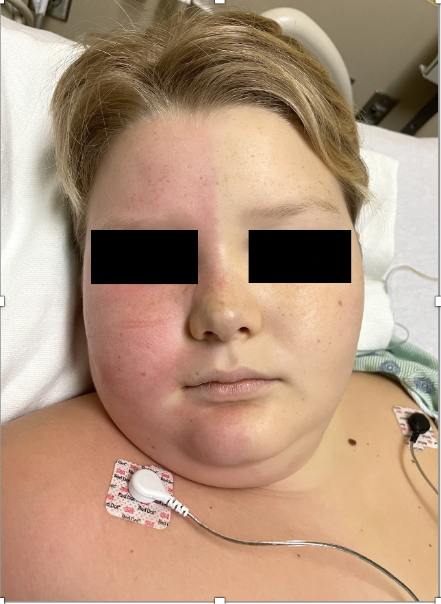
Harlequin Syndrome. This clinical image shows unilateral facial flushing sharply demarcated in the midline from the contralateral pallor in a hospitalized patient with monitoring electrodes.
Contributed by Hem Joshi, MD.
References
Lance JW, Drummond PD, Gandevia SC, Morris JG. Harlequin syndrome: the sudden onset of unilateral flushing and sweating. Journal of neurology, neurosurgery, and psychiatry. 1988 May:51(5):635-42 [PubMed PMID: 3155385]
Level 3 (low-level) evidenceHalle A, De Becdelievre A, Funalot B, Labrèze C, Morice-Picard F, Boralevi F. SCN10A variants associated with congenital harlequin syndrome. The British journal of dermatology. 2022 Jun:186(6):1039-1041. doi: 10.1111/bjd.21011. Epub 2022 Mar 22 [PubMed PMID: 35007332]
Cheshire WP Jr, Low PA. Harlequin syndrome: still only half understood. Journal of neuro-ophthalmology : the official journal of the North American Neuro-Ophthalmology Society. 2008 Sep:28(3):169-70. doi: 10.1097/WNO.0b013e318183c1a0. Epub [PubMed PMID: 18769277]
Carroll CB, Zajicek JP. The 'harlequin' sign in association with multiple sclerosis. Journal of neurology. 2004 Sep:251(9):1145-6 [PubMed PMID: 15372263]
Level 3 (low-level) evidenceValerio E, Barlotta A, Lorenzon E, Antonazzo L, Cutrone M. Harlequin Color Change: Neonatal Case Series and Brief Literature Review. AJP reports. 2015 Apr:5(1):e73-6. doi: 10.1055/s-0035-1545671. Epub 2015 Mar 2 [PubMed PMID: 26199804]
Level 2 (mid-level) evidenceNELIGAN GA, STRANG LB. A "harlequin" colour change in the newborn. Lancet (London, England). 1952 Nov 22:2(6743):1005-7 [PubMed PMID: 12991682]
Sharma B, Sharma P, Ahuja I, Pemawat A. Harlequin Syndrome and Autonomic Seizures - A Rare Association. Annals of Indian Academy of Neurology. 2023 Sep-Oct:26(5):839-841. doi: 10.4103/aian.aian_252_23. Epub 2023 Oct 18 [PubMed PMID: 38022446]
Velayuthan S, Sankararaman S. Visual diagnosis: newborn who has unilateral color change. Diagnosis: Harlequin color change. Pediatrics in review. 2013 Jul:34(7):e25-6. doi: 10.1542/pir.34-7-e25. Epub [PubMed PMID: 23818088]
Persson RM, Tellnes K, Hoven H, Haaverstad R, Svendsen ØS. Harlequin syndrome associated with thoracic epidural anaesthesia. Anaesthesia reports. 2022 Jan-Jun:10(1):. doi: 10.1002/anr3.12144. Epub 2022 Jan 25 [PubMed PMID: 35118419]
Miedl M, Baumgartner P, Disse LR, Weber KP, Pohl H, Wegener S. Harlequin syndrome in a patient with probable hemicrania continua and exertional headache - is there a link? a case report. BMC neurology. 2024 Jul 17:24(1):247. doi: 10.1186/s12883-024-03731-y. Epub 2024 Jul 17 [PubMed PMID: 39020271]
Level 3 (low-level) evidencePadilla H, Cheshire WP Jr, Benarroch EE, Berini SE, Cutsforth-Gregory JK, Mauermann ML, Sandroni P, Shouman K, Low PA, Singer W, Coon EA. Harlequin syndrome: using clinical features and autonomic testing to unmask the disorder. Clinical autonomic research : official journal of the Clinical Autonomic Research Society. 2025 Dec:35(6):839-845. doi: 10.1007/s10286-025-01151-7. Epub 2025 Jul 28 [PubMed PMID: 40721556]
Mavroudis I, Balmus IM, Ciobica A, Luca AC, Chowdhury R, Iordache AC, Gorgan DL, Radu I. Mini-Review on the Harlequin Syndrome-A Rare Dysautonomic Manifestation Requiring Attention. Medicina (Kaunas, Lithuania). 2022 Jul 15:58(7):. doi: 10.3390/medicina58070938. Epub 2022 Jul 15 [PubMed PMID: 35888657]
Manhães RK, Spitz M, Vasconcellos LF. Botulinum toxin for treatment of Harlequin syndrome. Parkinsonism & related disorders. 2016 Feb:23():112-3. doi: 10.1016/j.parkreldis.2015.11.030. Epub 2015 Dec 18 [PubMed PMID: 26750113]
Sribnick EA, Boulis NM. Treatment of Harlequin syndrome by costotransversectomy and sympathectomy: case report. Neurosurgery. 2011 Jul:69(1):E257-9. doi: 10.1227/NEU.0b013e3182186829. Epub [PubMed PMID: 21796071]
Level 3 (low-level) evidence