 Tools
Tools
 Contact Lenses: Principles, Uses and Related Complications
Introduction
Contact lenses are artificial prosthetic devices placed over the anterior ocular surface to replace or modify the cornea's refractive function and improve visual performance. Since their introduction, contact lenses have evolved from simple refractive aids into sophisticated optical, therapeutic, cosmetic, diagnostic, and rehabilitative devices that play an integral role in contemporary ophthalmic practice. Modern contact lenses are used not only to correct refractive errors such as myopia, hyperopia, astigmatism, anisometropia, aphakia, and presbyopia, but also to treat irregular corneal disorders, ocular surface diseases, facilitate postoperative rehabilitation, and provide cosmetic enhancement (see Image. Leucomatous Corneal Opacity, Contact Lens).[1] With ongoing advances in biomaterials, lens design, manufacturing technology, and ocular imaging, contact lens use has increased substantially worldwide, making contact lenses among the most commonly prescribed ophthalmic devices in modern clinical practice.[2]
Globally, an estimated 140 million individuals wear contact lenses, and this number continues to rise due to growing awareness, rising cosmetic demand, improved lens comfort, and the desire for spectacle independence. Contact lenses provide several advantages over spectacles, including a wider field of view, improved peripheral vision, reduced image distortion, improved cosmesis, enhanced sports performance, and greater convenience in occupational settings such as aviation, law enforcement, and athletics. Additionally, contact lenses have become increasingly important in pediatric ophthalmology, refractive rehabilitation, keratoconus management, and postoperative visual correction after refractive or corneal transplantation.[3]
Indications for contact lens use continue to expand. Optically, contact lenses are used to correct refractive errors and irregular astigmatism, particularly in patients with keratoconus, pellucid marginal degeneration, postrefractive surgery ectasia, and posttraumatic corneal irregularities.[4] Therapeutically, bandage contact lenses are used for recurrent corneal erosion, persistent epithelial defects, bullous keratopathy, neurotrophic keratitis, dry eye disease, corneal perforation, and postoperative corneal healing.[5] Contact lenses also serve important diagnostic and surgical roles in gonioscopy, electroretinography, laser procedures, vitreoretinal surgery, and anterior segment visualization. Cosmetic lenses may improve the appearance of corneal scars, iris defects, leukocoria, or phthisical eyes, whereas occupational lenses are increasingly used by athletes, military personnel, pilots, and individuals requiring enhanced visual performance.[6]
Contact lenses are manufactured using a wide variety of materials and designs. Based on material composition, contact lenses may be broadly divided into focons and filcons. Focons include rigid hydrophobic materials such as polymethyl methacrylate and rigid gas-permeable lenses, whereas filcons are hydrophilic hydrogels and silicone hydrogels.[7] Contact lenses can also be classified according to water content, oxygen permeability, anatomical position, wear schedule, and refractive purpose. The major types include soft contact lenses, rigid gas permeable lenses, scleral lenses, semiscleral lenses, hybrid lenses, orthokeratology lenses, toric lenses, cosmetic lenses, and multifocal or bifocal presbyopia-correcting lenses.[8]
An ideal contact lens should possess excellent optical clarity, high oxygen permeability, good wettability, dimensional stability, resistance to deposits, adequate flexibility, biocompatibility, durability, and patient comfort. Modern silicone hydrogel lenses provide significantly greater oxygen transmission than earlier hydrogel materials, thereby reducing hypoxia-related complications such as corneal edema, limbal hyperemia, endothelial changes, and corneal neovascularization.[7] The interaction among the contact lens, tear film, eyelids, conjunctiva, and cornea is highly complex and influences lens comfort, movement, tear exchange, and ocular surface health. Parameters such as overall diameter, optic zone diameter, base curve, edge design, center thickness, peripheral curves, and refractive power should therefore be individualized during lens fitting.[1]
One of the most significant advances in contact lens technology has been the development of presbyopia-correcting contact lenses. Presbyopia is an age-related physiologic reduction in accommodation resulting from progressive loss of crystalline lens elasticity and reduced accommodative amplitude, leading to gradual deterioration of near vision. Fienbloom first described bifocal contact lenses for presbyopia correction in 1938. Subsequently, Freeman, Williamson, Wesley, Jessen, and De Carle introduced simultaneous-vision, concentric bifocal, translating, and multifocal lens concepts. Modern presbyopia-correcting contact lenses include bifocal, multifocal, concentric, aspheric, diffractive, translating, monovision, and modified monovision designs. These lenses aim to provide functional near, intermediate, and distance vision while preserving binocular visual function, contrast sensitivity, and stereopsis.[2]
Despite their many benefits, contact lenses remain foreign bodies on the ocular surface and may be associated with a broad spectrum of complications. Contact lens–related complications may be infectious or noninfectious and may involve the conjunctiva, cornea, tear film, eyelids, limbus, or adnexal structures (see Image. Corneal Ulcer, Infected Contact Lens). Common conjunctival complications include allergic conjunctivitis, giant papillary conjunctivitis, contact lens–induced papillary conjunctivitis, and superior limbic keratoconjunctivitis. Corneal complications include epithelial edema, microcysts, abrasions, superficial punctate keratitis, sterile infiltrates, peripheral ulcerative keratitis, microbial keratitis, corneal neovascularization, corneal warpage, endothelial polymegathism, and limbal stem cell deficiency.[3]
Among these complications, microbial keratitis remains one of the most severe and vision-threatening consequences of contact lens wear. Bacterial keratitis, particularly Pseudomonas aeruginosa, fungal keratitis, and Acanthamoeba keratitis, have been strongly associated with improper lens hygiene, overnight wear, contaminated lens solutions, and poor patient compliance. Mechanical complications such as tight lens syndrome, lens deposits, lens decentration, giant papillary conjunctivitis, and contact lens intolerance may further compromise comfort and long-term wearability. Hypoxia-induced complications, though reduced with silicone hydrogel lenses, continue to occur in extended wear and poorly fitted lenses.[4]
Advances in contact lens biomaterials, manufacturing methods, and hygiene protocols have substantially reduced the incidence of many complications. Manufacturing techniques such as lathe cutting, spin casting, and molding have enabled the production of lenses with superior optical quality, oxygen permeability, wettability, and reproducibility. Furthermore, improvements in lens care systems, patient education, ocular surface evaluation, and digital imaging technologies have enhanced the safety and effectiveness of contact lens wear. Nevertheless, successful contact lens practice requires a detailed understanding of corneal physiology, tear film dynamics, lens material science, fitting strategies, presbyopia correction, complication recognition, and interprofessional patient management.[5]
Because contact lenses span the fields of cornea, refractive surgery, ocular surface disease, optometry, and preventive ophthalmology, healthcare professionals involved in contact lens practice must stay up to date on evolving technologies, fitting principles, ocular surface interactions, and evidence-based management strategies. This activity comprehensively reviews the anatomy and physiology of contact lens wear; indications and contraindications; materials science; classification; fitting techniques; presbyopia-correcting designs; complications; clinical significance; and interprofessional management associated with modern contact lenses.[6]
Anatomy and Physiology
Register For Free And Read The Full Article
 Search engine and full access to all medical articles
- 10 free questions in your specialty
- Free CME/CE Activities
- Free daily question in your email
- Save favorite articles to your dashboard
- Emails offering discounts
Learn more about a Subscription to StatPearls Point-of-Care
Anatomy and Physiology
Tear Film and Contact Lens Relationship
The functional integrity, transparency, and metabolic activity of the cornea are maintained primarily by the precorneal tear film, which plays a critical role in lubrication, nourishment, protection, optical quality, and epithelial homeostasis.[6] The tear film is composed of lipid, aqueous, and mucin layers that together provide a smooth refractive surface and maintain ocular surface stability. Because the cornea is avascular, it relies heavily on atmospheric oxygen and tear film-mediated nutrient exchange to meet its metabolic requirements. Contact lenses directly interact with the tear film and alter the normal physiologic relationship between the tear film, eyelids, conjunctiva, and cornea. When this delicate equilibrium is disturbed, it can adversely affect corneal metabolism, oxygenation, tear exchange, ocular comfort, and contact lens tolerance.[8]
The tear film performs several essential functions during contact lens wear: acts as a lubricating interface between the lens and the ocular surface, minimizes frictional trauma from blinking, facilitates oxygen transport, removes metabolic waste products, and stabilizes the optical surface. The tear film surrounding the lens is divided into the prelens and postlens tear films. The prelens tear film coats the anterior lens surface and influences wettability, comfort, and visual quality, whereas the postlens tear film lies between the lens and cornea and is essential for tear exchange, corneal nourishment, and debris removal.[7]
Disruption of tear film homeostasis during contact lens wear may result in tear hyperosmolarity, increased evaporation, ocular surface inflammation, dry eye symptoms, and reduced lens tolerance. Modern silicone hydrogel lenses and surface-modified materials have therefore been developed to improve wettability and maintain tear film stability. Tear film compatibility remains one of the most important determinants of successful long-term contact lens wear.[9]
The tear film associated with contact lens wear is divided into prelens and postlens tear films. The prelens tear film coats the anterior surface of the lens and influences wettability, optical quality, evaporation resistance, and patient comfort, whereas the postlens tear film lies between the posterior lens surface and cornea and plays a critical role in oxygen delivery, corneal nourishment, metabolic waste removal, and debris clearance. Disturbance of these tear film interfaces may result in dry eye symptoms, tear hyperosmolarity, ocular surface inflammation, lens intolerance, and reduced contact lens acceptability.[10]
Contact Lens Fitting Over the Cornea
Contact lenses are not physically clamped or attached to the corneal surface. Instead, they remain positioned over the cornea due to the complex interplay among tear film surface tension, viscosity, lid pressure, capillary attraction, and blinking dynamics.[11] The tear film acts as a thin adhesive interface between the posterior lens surface and the anterior corneal surface. Surface tension generated by the tear film creates an attractive force that stabilizes the lens over the cornea while still allowing controlled movement with blinking.[11]
When the lens is fitted over the cornea, the conjunctival mucus layer spreads across the lens surface, forming a continuous fluid sheet that surrounds the lens. Cohesive forces between tear molecules and adhesive interactions between the tear film and lens surface help maintain lens centration and stability. A negative hydrostatic pressure develops between the posterior lens surface and the cornea, contributing further to lens adherence. Simultaneously, blinking and eyelid motion create a tear-pump mechanism that enables tear exchange beneath the lens.[12]
The fit between the contact lens and the cornea is critical for successful wear. Excessively tight lenses may reduce tear exchange, impair oxygen transmission, and increase the risk of hypoxic complications, whereas excessively loose lenses may result in poor centration, discomfort, unstable vision, and mechanical irritation. Lens movement, centration, edge lift, sagittal depth, base curve, and tear exchange must therefore be carefully evaluated during fitting. Corneal topography and anterior segment imaging have significantly improved the precision of modern contact lens fitting, especially in the management of irregular corneal disorders and in scleral lens practice.[13]
Experimental studies have demonstrated that approximately 11 grams of force may be required to displace a properly fitted contact lens from the ocular surface. When the tear film covering the lens breaks, internal adhesion mechanisms become increasingly important. Negative pressure develops at the interface between the posterior lens surface, the cornea, and the tear film, creating a collar-like reservoir effect around the lens edge that stabilizes the contact lens on the corneal surface.[14]
Tear Pump Mechanism and Blink Dynamics
Blinking plays a central role in maintaining contact lens physiology and ocular surface health. The blink mechanism continuously renews the tear film, redistributes oxygenated tears beneath the lens, removes debris, and facilitates the elimination of metabolic waste. During blinking, eyelid pressure compresses the lens against the ocular surface, causing tears to move beneath the lens and creating a “tear pump” effect. This process is particularly important in rigid gas permeable lenses, where tear exchange contributes significantly to corneal oxygenation.[7]
The efficiency of the tear pump mechanism depends on several factors, including:
- Tear volume
- Blink frequency
- Lens movement
- Lens material
- Percentage tear exchange with each blink
- Lid tension
- Lens fitting characteristics [15]
In soft contact lenses, tear exchange is generally lower because the lens drapes more closely over the corneal surface, whereas rigid gas-permeable (RGP) lenses exhibit greater tear exchange due to increased movement during blinking. Reduced blinking frequency, especially during prolonged digital screen use, may lead to tear stagnation, dryness, discomfort, and deposit accumulation during contact lens wear. The blinking mechanism acts as a physiologic pump that continuously renews the postlens tear interface. Lid pressure compresses the contact lens against the corneal surface, forcing old tears outward and permitting fresh oxygenated tears to enter beneath the lens. Reduced blink frequency, prolonged digital screen use, and tight lens fitting may impair this tear pump mechanism, resulting in tear stagnation, hypoxia, discomfort, and deposit accumulation.[14]
Visual and Optical Considerations
The interaction between the cornea, tear film, and contact lens creates a refractive system known as the tear lens. The tear lens forms because tears occupy the space between the posterior lens surface and the anterior corneal curvature. The refractive index of tears is approximately 1.336, whereas that of the cornea is approximately 1.376. This difference allows the tear lens to neutralize certain corneal irregularities and improve optical quality.[16]
RGP lenses are particularly effective in correcting irregular astigmatism because the tear lens fills corneal surface irregularities, creating a smooth refractive interface. Contact lenses can correct approximately 5 diopters of corneal astigmatism with minimal residual refractive error. In keratoconus and post-refractive surgery ectasia, RGP and scleral lenses provide superior optical rehabilitation compared with spectacles because they mask irregular corneal surfaces by forming a tear lens. Contact lenses also reduce prismatic effects, peripheral distortion, magnification differences, and image minification associated with spectacles. These advantages are especially beneficial in anisometropia, aphakia, sports vision, and occupational visual demands.[17]
The tear lens-postlens tear film interface plays a major role in improving the optical performance of contact lenses. The posterior tear film fills corneal surface irregularities and creates a smooth refractive interface, while the anterior surface of the tear lens is shaped by the posterior curvature of the contact lens. If the refractive indices of the cornea and tears were identical, theoretically, all corneal irregularities would be perfectly neutralized. However, higher degrees of astigmatism and corneal irregularity produce proportionately greater residual astigmatism.[18]
Effects of Contact Lenses on Corneal Physiology
Contact lenses alter normal corneal physiology in multiple ways. While they provide refractive and therapeutic benefits, they also influence oxygen transport, epithelial metabolism, tear evaporation, and corneal hydration. Contact lenses may:
- Reduce oxygen delivery to the cornea
- Trap metabolic waste products
- Interfere with epithelial cell turnover
- Alter tear osmolarity
- Increase mechanical friction
- Reduce tear evaporation in selected therapeutic settings
Hard contact lenses cover approximately 50% to 80% of the corneal surface and rely heavily on tear exchange for oxygenation. Soft lenses cover a larger surface area and may reduce oxygen transmission depending on lens thickness and material. Hypoxia caused by inadequate oxygen permeability may result in epithelial edema, microcysts, endothelial polymegathism, stromal swelling, limbal hyperemia, and corneal neovascularization. Modern silicone hydrogel lenses have significantly improved oxygen permeability (Dk/t values) and reduced hypoxic complications compared with earlier hydrogel lenses. Nevertheless, prolonged wear, overnight use, tight lens fitting, and poor tear exchange may still result in metabolic stress and ocular surface compromise.[19]
Under static conditions, oxygen at the postlens tear interface may be depleted in approximately 90 seconds with conventional hydrogel contact lenses. This leads to anaerobic glycolysis, lactic acid accumulation, impaired corneal metabolism, stromal edema, and corneal haze. The anaerobic metabolic pathway is estimated to be only approximately one-eighteenth as efficient as aerobic metabolism, thereby highlighting the importance of adequate oxygen transmission during contact lens wear.[20]
Corneal Metabolism and Oxygen Dynamics
The cornea derives oxygen primarily from the atmosphere through diffusion across the tear film. During contact lens wear, oxygen transmission depends on lens material, water content, thickness, and tear exchange. Open-eye oxygenation is generally adequate with modern lenses; however, closed-eye conditions during sleep significantly reduce oxygen availability and increase the risk of hypoxia. Corneal hypoxia results in anaerobic glycolysis and accumulation of lactate within the stroma, leading to osmotic imbalance and corneal edema. Chronic hypoxia may induce endothelial stress, polymegathism, epithelial compromise, and neovascularization. Extended-wear contact lenses, therefore, require high oxygen transmissibility to maintain normal corneal metabolism.[21]
Accommodation and Presbyopia Physiology
Presbyopia is a physiological, age-related insufficiency of accommodation, characterized by a progressive decline in near vision due to reduced accommodative amplitude. Although presbyopia is universally experienced with aging, the precise mechanism underlying it remains controversial. Proposed theories are broadly divided into lenticular and extralenticular mechanisms. The lenticular theory proposes that presbyopia results from progressive sclerosis and reduced elasticity of the crystalline lens, lens capsule, and zonular apparatus. Extralenticular theories suggest contributions from ciliary muscle dysfunction, reduced zonular elasticity, altered vitreous dynamics, and biomechanical changes within the accommodative system.[22]
Helmholtz Theory
Helmholtz proposed the classical theory of accommodation, which states that the crystalline lens is relatively flat during distance viewing. During accommodation, ciliary muscle contraction reduces zonular tension, allowing the elastic lens to assume a more convex shape and increase refractive power. Age-related hardening of the lens reduces its ability to change shape, resulting in presbyopia.[23]
Coleman Theory
Coleman proposed the catenary theory, suggesting that accommodation results from pressure gradients between the anterior and posterior chambers. During ciliary muscle contraction, pressure differences alter lens curvature, increasing central lens steepening and accommodative power.[24]
Schachar Theory
Schachar proposed that ciliary muscle contraction increases equatorial zonular tension, thereby altering lens shape and accommodative power. According to this theory, progressive lens growth throughout life reduces zonular elasticity and accommodative efficiency, contributing to the development of presbyopia.[25]
Dysfunctional Lens Syndrome
Dysfunctional lens syndrome refers to the progressive aging of the crystalline lens.
- Stage 1 corresponds to presbyopia and early accommodative loss.
- Stage 2 involves increasing higher-order aberrations and reduced contrast sensitivity associated with dysfunctional lens syndrome and early cataract formation.
- Stage 3 represents a visually significant cataract with progressive optical deterioration affecting daily visual function.[26]
See Tables 1–3 below to learn more about the components and functions of the tear film, the physiologic effects of contact lens wear on the cornea, and the theories of accommodation and presbyopia.
Table 1. Components and Functions of the Tear Film
|
Tear Film Layer |
Composition |
Primary Function |
|
Lipid layer |
Meibomian gland secretions |
Prevents evaporation and stabilizes the tear film |
|
Aqueous layer |
Lacrimal gland secretions |
Provides oxygen, nutrients, and antimicrobial factors |
|
Mucin layer |
Goblet cell mucins |
Enhances wettability and tear adherence |
Table 2. Physiologic Effects of Contact Lens Wear on the Cornea
|
Physiologic Effect |
Clinical Consequence |
|
Reduced oxygen transmission |
Corneal hypoxia and edema |
|
Altered tear exchange |
Debris accumulation and dryness |
|
Increased mechanical friction |
Epithelial erosions and SPKs |
|
Tear film instability |
Dry eye symptoms |
|
Reduced epithelial metabolism |
Microcysts and epithelial compromise |
|
Chronic hypoxia |
Neovascularization and endothelial stress |
Table 3. Theories of Accommodation and Presbyopia
|
Theory |
Proposed Mechanism |
|
Helmholtz theory |
Reduced zonular tension increases lens curvature |
|
Coleman theory |
Pressure gradient alters lens shape |
|
Schachar theory |
Increased equatorial zonular tension changes lens curvature |
|
Dysfunctional lens syndrome |
Progressive lens aging reduces accommodation |
Indications
The indications for contact lens use are broad and continually expanding due to advances in lens materials, optics, fitting techniques, and ocular surface rehabilitation strategies (see Table 4). Contact lenses are prescribed not only for refractive correction but also for therapeutic, preventive, cosmetic, diagnostic, operative, occupational, and rehabilitative purposes. The choice of contact lens depends on several patient-specific factors, including visual requirements, occupational needs, binocularity, stereopsis, presbyopic add requirement, ocular surface status, motivation for spectacle independence, lifestyle demands, affordability, and patient compliance.
Successful contact lens wear requires proper patient motivation and realistic expectations. Individuals seeking a spectacle-free lifestyle, improved cosmesis, better peripheral vision, or enhanced sports performance are often ideal candidates. In presbyopic individuals, factors such as binocular visual function, stereopsis, near visual demands, occupational requirements, and tolerance for multifocal optics or monovision correction significantly influence contact lens selection.[27]
Optical Indications
Optical indications constitute the most common reason for contact lens prescription. Contact lenses provide superior optical correction compared with spectacles in several refractive and corneal conditions because they minimize image distortion, reduce magnification differences, improve peripheral vision, and neutralize corneal irregularities.[28]
High myopia
In high myopia, contact lenses reduce retinal image minification and eliminate the peripheral aberrations (eg, ring scotoma and distortion) associated with thick minus spectacle lenses, thereby improving visual quality, peripheral vision, and cosmesis.[29]
Unilateral aphakia
Contact lenses are especially useful in unilateral aphakia because they minimize aniseikonia and improve binocular vision compared with aphakic spectacles.[30]
Anisometropia
Contact lenses help reduce retinal image size differences between the 2 eyes and improve stereopsis and binocular function.[31]
Keratoconus and irregular astigmatism
RGP and scleral lenses are considered the gold standard for visual rehabilitation in keratoconus and irregular corneal astigmatism. The tear lens formed beneath the contact lens neutralizes corneal surface irregularities, improving visual acuity.[32]
Corneal scars
Contact lenses may improve optical quality in selected corneal scars by masking anterior surface irregularities.[33]
Refractive errors
Myopia, hyperopia, astigmatism, and presbyopia are commonly corrected with contact lenses because of their optical advantages and cosmetic acceptability.[34]
Therapeutic Indications
Therapeutic or bandage contact lenses are extensively used in corneal and ocular surface disorders because they protect the epithelium, reduce pain, promote healing, and maintain ocular surface stability.[35]
Corneal pathologies
Bandage contact lenses help protect the corneal epithelium from mechanical trauma and facilitate epithelial healing.[36]
Pseudophakic bullous keratopathy
Contact lenses reduce pain caused by ruptured epithelial bullae and improve patient comfort.[37]
Persistent epithelial defects and nonresolving corneal ulcers
Therapeutic lenses create a stable healing environment and reduce friction from blinking.[38]
Recurrent corneal erosion syndrome
Bandage contact lenses reduce epithelial trauma and promote adhesion of the regenerating epithelium.[39]
Dry eye disease
Scleral lenses create a fluid reservoir over the cornea and are increasingly used in severe ocular surface disease and dry eye syndromes.[40]
Post-keratoplasty and post-superficial keratectomy
Contact lenses improve epithelial healing and visual rehabilitation after corneal surgery.[41]
Corneal perforation and microleaks
Bandage lenses may temporarily tamponade small perforations and assist in tectonic stabilization.[42]
Postcorneal tear repair
Contact lenses may protect the wound and reduce postoperative discomfort.[43]
Iris Pathology Indications
Cosmetic and prosthetic contact lenses are valuable in managing iris abnormalities.[44]
Aniridia
Tinted or prosthetic lenses reduce glare and photophobia and improve cosmetic appearance.[45]
Iris coloboma
Custom iris lenses improve cosmesis and reduce visual disturbances.[45]
Albinism
Tinted contact lenses reduce photophobia and improve visual comfort by decreasing excess light transmission.[46]
Glaucoma-Related Indications
Contact lenses are increasingly being explored as drug delivery systems in glaucoma management. Drug-eluting contact lenses may provide sustained release of antiglaucoma medications and improve patient compliance compared with topical eye drops.[47]
Pediatric Indications
Contact lenses play an important role in pediatric ophthalmology.[1]
Amblyopia
Opaque contact lenses may be used for occlusion therapy in amblyopia management.[48]
Pediatric aphakia
Contact lenses are frequently prescribed after congenital cataract surgery to provide optical rehabilitation and prevent amblyopia.[49]
High refractive errors
Children with anisometropia or high ametropia may achieve better binocular vision and visual development with contact lenses.[48]
Retinal Indications
Contact lenses are occasionally used after vitreoretinal surgery, particularly in patients with postoperative epithelial defects or ocular surface compromise. Special contact lens systems are also used during retinal laser procedures and posterior segment visualization.[50]
Diagnostic Indications
Several specialized contact lenses are used in ophthalmic examination and diagnostic procedures.
Gonioscopy
Gonioscopy lenses permit visualization of the anterior chamber angle.[51]
Electroretinography
Contact lens electrodes are used during electroretinographic recordings.
Fundus examination
Diagnostic contact lenses improve posterior segment visualization in patients with astigmatism or irregular corneas.[52]
Fundus photography
Specialized contact lenses improve retinal imaging quality.
Goldman three-mirror examination
Three-mirror lenses permit detailed evaluation of the peripheral retina, pars plana, and angle structures.[53]
Preventive Indications
Contact lenses may be used prophylactically to protect the ocular surface and prevent further damage.
Trichiasis
Bandage contact lenses reduce mechanical trauma caused by misdirected eyelashes.[52]
Exposure keratitis
Therapeutic lenses help maintain ocular surface hydration and epithelial protection.[54]
Prevention of symblepharon
Contact lenses may prevent the formation of adhesions between the conjunctival surfaces following burns or severe ocular surface disease.
Occupational Indications
Contact lenses are preferred in many occupations that require wide visual fields, freedom of movement, and reduced dependence on spectacles.[55]
Sports personnel
Athletes benefit from improved peripheral vision, stability, and reduced risk of spectacle-related injury.
Police and military personnel
Contact lenses improve mobility and tactical performance.[56]
Pilots
Pilots prefer contact lenses because they minimize peripheral distortion and improve compatibility with headgear and helmets.
Cosmetic Indications
Cosmetic and prosthetic contact lenses improve appearance and patient confidence.[57]
Corneal scar
Tinted or cosmetic lenses improve cosmesis in visible corneal opacities.
Phthisis bulbi
Custom prosthetic lenses improve cosmetic appearance in disfigured eyes.[58]
Operative Indications
Specialized contact lenses are used intraoperatively during several ophthalmic procedures.
Pars plana vitrectomy
Wide-angle viewing systems use contact lenses to improve retinal visualization.[59]
Retinal photocoagulation
Contact lenses stabilize the eye and enhance laser delivery.[60]
Goniotomy
Goniotomy lenses permit visualization of the anterior chamber angle during pediatric glaucoma surgery.[61]
Table 4. Major Indications of Contact Lens Use
|
Category |
Common Indications |
|
Optical |
Myopia, hyperopia, keratoconus, anisometropia, aphakia |
|
Therapeutic |
Bullous keratopathy, epithelial defects, dry eye, and perforation |
|
Cosmetic |
Corneal scar, aniridia, phthisis bulbi |
|
Diagnostic |
Gonioscopy, electroretinography, fundus examination |
|
Preventive |
Exposure keratitis, trichiasis |
|
Occupational |
Sports, police, pilots |
|
Pediatric |
Amblyopia, pediatric aphakia |
|
Operative |
Vitrectomy, photocoagulation, and goniotomy |
Contraindications
Although contact lenses are widely used for refractive correction, therapeutic ocular surface management, keratoconus rehabilitation, presbyopia correction, and cosmetic purposes, they are not suitable for every patient. Successful contact lens wear requires adequate tear film stability, corneal sensation, eyelid function, ocular surface integrity, patient motivation, manual dexterity, hygiene compliance, and regular follow-up. Contraindications vary by the type of contact lens prescribed, including soft lenses, RGP lenses, scleral lenses, orthokeratology lenses, therapeutic bandage lenses, multifocal lenses, cosmetic lenses, and extended-wear lenses. Failure to identify contraindications may result in contact lens intolerance, microbial keratitis, chronic inflammation, corneal scarring, or permanent visual loss.[62] Contraindications are broadly divided into absolute and relative contraindications (see Table 5).
Absolute Contraindications
Absolute contraindications are conditions in which contact lens wear is generally unsafe because of significantly increased risk of infection, inflammation, epithelial compromise, poor compliance, or severe ocular morbidity.[1]
Cerebral palsy and individuals with intellectual disability
Patients with severe cerebral palsy, intellectual disability, poor cognitive function, or inability to cooperate may not be suitable candidates for contact lens wear because proper insertion, removal, hygiene, and compliance cannot be ensured. These patients may unintentionally damage the lens, rub their eyes excessively, or fail to recognize symptoms of infection or corneal injury. However, in selected pediatric aphakia or therapeutic cases, contact lenses may still be used under strict caregiver supervision and close follow-up.[59]
Particularly problematic for:
- Soft lenses
- Extended-wear lenses
- Orthokeratology lenses
- Cosmetic lenses [63]
Chronic dacryocystitis
Chronic dacryocystitis represents a major contraindication because the lacrimal sac acts as a persistent reservoir of microorganisms. Continuous bacterial contamination significantly increases the risk of:
- Conjunctivitis
- Microbial keratitis
- Corneal ulceration
- Therapeutic lens contamination [64]
Contact lenses should generally be avoided until the lacrimal infection has been treated surgically or medically.
Particularly risky for:
- Therapeutic bandage lenses
- Soft lenses
- Extended-wear lenses
- Scleral lenses [65]
Dry eye disease and tear film insufficiency
Adequate tear film quantity and quality are essential for successful contact lens wear. Severe dry eye disease causes:
- Reduced lubrication
- Increased friction
- Tear hyperosmolarity
- Lens intolerance
- Epithelial breakdown
- Increased infection risk [66]
Patients with the following may not tolerate conventional soft or rigid lenses.[63]
- Sjögren syndrome
- Ocular cicatricial pemphigoid
- Severe meibomian gland dysfunction
- Graft-versus-host disease
- Stevens–Johnson syndrome
Lens-specific considerations:
- Soft hydrogel lenses may worsen dryness because of dehydration.
- RGP lenses may increase foreign body sensation.
- Extended-wear lenses increase hypoxic stress.
- Multifocal soft lenses may worsen visual fluctuation in dry eye.
However, scleral lenses may be therapeutic in severe dry eye disease by creating a fluid reservoir over the cornea.[7]
Hordeolum internum and hordeolum externum
Active lid infections, such as internal and external hordeolum, are contraindications because they increase bacterial contamination and inflammation around the ocular surface. Contact lens wear during active lid infection increases the risk of:
- Corneal infiltration
- Microbial keratitis
- Lens contamination
- Giant papillary conjunctivitis
Lens wear should be discontinued until the infection resolves completely.[67]
Blepharitis
Blepharitis and meibomian gland dysfunction significantly impair tear film quality and promote:
- Lens deposits
- Surface contamination
- Contact lens discomfort
- Dry eye symptoms
- Giant papillary conjunctivitis [7]
Chronic blepharitis also increases the risk of marginal keratitis and microbial keratitis.
Particularly problematic for:
- Soft lenses
- Extended-wear lenses
- Multifocal lenses
- Scleral lenses with poor hygiene
Blepharitis should be treated before lens fitting.[14]
Trichiasis
Misdirected eyelashes mechanically traumatize the lens and ocular surface, causing:
- Corneal staining
- Epithelial defects
- Lens damage
- Reduced tolerance
Soft lenses may occasionally be used therapeutically after trichiasis is treated, but untreated trichiasis remains a contraindication.[16]
Conjunctivitis
Active bacterial, viral, allergic, or toxic conjunctivitis is an absolute contraindication to contact lens wear because the inflammatory process increases:
- Microbial adherence
- Lens contamination
- Corneal involvement
- Ocular surface instability [14]
Lens wear should only resume after complete resolution.
Particularly contraindicated for:
- Soft lenses
- Cosmetic lenses
- Extended-wear lenses
Corneal dystrophies and degenerations
Corneal dystrophies and degenerations may impair epithelial integrity and corneal metabolism. Contact lens wear in such patients may worsen:
- Recurrent erosions
- Epithelial edema
- Hypoxia
- Corneal haze
However, scleral and RGP lenses may sometimes be therapeutically useful in selected corneal dystrophies under specialist supervision.[7]
Uveitis
Active uveitis is a contraindication because contact lens wear may:
- Increase inflammation
- Reduce comfort
- Aggravate photophobia
- Increase risk of secondary infection
Lens wear should be avoided until inflammation is completely controlled.
Episcleritis and scleritis
These inflammatory disorders are associated with ocular pain, vascular congestion, and ocular surface inflammation. Contact lens wear may worsen inflammation and increase discomfort. Therapeutic lenses should be used only in selected cases of ocular surface protection under specialist supervision.[14]
Fifth nerve palsy and corneal anesthesia
Patients with trigeminal nerve dysfunction or neurotrophic keratopathy have impaired corneal sensation and are unable to detect:
- Corneal abrasions
- Hypoxia
- Ulceration
- Infection
This significantly increases the risk of painless corneal melt and perforation.
Especially dangerous with:
- Extended-wear lenses
- Orthokeratology lenses
- Therapeutic lenses without monitoring [6]
Seventh nerve palsy
Facial nerve palsy impairs blinking and tear distribution, causing:
- Exposure keratopathy
- Corneal drying
- Poor tear exchange
- Lens instability
Soft lenses may occasionally be used therapeutically in exposure keratopathy, but routine lens wear is generally contraindicated without close supervision.
Allergic conjunctivitis
Allergic conjunctivitis predisposes to:
- Itching
- Eye rubbing
- Lens deposits
- Giant papillary conjunctivitis
- Reduced lens tolerance
Soft lenses, especially extended-wear and older hydrogel lenses, are particularly poorly tolerated in allergic patients.
Daily disposable lenses may occasionally improve tolerance in mild allergy.[6]
Superficial punctate keratitis and subepithelial keratitis
These conditions indicate compromised epithelial integrity and ocular surface inflammation. Contact lens wear may worsen:
- Corneal staining
- Epithelial trauma
- Infection risk
- Dry eye symptoms
Lens wear should be withheld until complete epithelial recovery.
Corneal ulcer
Active corneal ulceration is an absolute contraindication to routine contact lens wear because of the risk of progression and perforation. However, therapeutic bandage lenses may occasionally be used for the following conditions under strict supervision by a corneal specialist.[6]
- Small perforations
- Corneal melts
- Persistent epithelial defects
Relative Contraindications
Relative contraindications are conditions in which contact lenses may still be prescribed with caution, careful selection, patient counseling, and close follow-up.
Pregnancy
Hormonal changes during pregnancy may alter:
- Tear film stability
- Corneal curvature
- Corneal thickness
- Refraction
- Lens tolerance
Patients may develop transient contact lens intolerance, dryness, or fluctuating vision.[16]
Particularly affected:
- Soft lenses
- Multifocal lenses
- Extended-wear lenses
Giant papillary conjunctivitis
Giant papillary conjunctivitis is a hypersensitivity reaction caused by:
- Lens deposits
- Mechanical irritation
- Protein accumulation
- Preservative sensitivity [6]
Symptoms include:
- Itching
- Mucus discharge
- Lens awareness
- Reduced wearing time
Most common with:
- Soft lenses
- Extended-wear lenses
- Older hydrogel lenses
Daily disposable or silicone hydrogel lenses may improve tolerance.
Strabismus
Patients with strabismus may have:
- Poor binocularity
- Reduced stereopsis
- Suppression
- Poor adaptation to multifocal optics
Particularly important in:
- Monovision lenses
- Multifocal lenses
- Translating bifocal lenses
These patients require careful binocular vision assessment before fitting.[68]
Additional Modern Relative Contraindications
Poor hygiene and noncompliance
Poor hygiene significantly increases the risk of:
- Microbial keratitis
- Lens contamination
- Corneal ulceration
This is particularly dangerous with:
- Extended-wear lenses
- Cosmetic lenses
- Orthokeratology lenses [14]
Large pupils
Large pupils exacerbate the following symptoms, particularly with multifocal and simultaneous-vision lenses.[69]
- Halos
- Glare
- Dysphotopsia
- Reduced night vision
Occupational Risks
Certain occupations involving the following may reduce lens tolerance and increase the risk of complications.
- Dust
- Smoke
- Chemical exposure
- Dry environments
Contact sports and water exposure
Swimming and water exposure increase the risk of:
- Acanthamoeba keratitis
- Lens contamination
- Corneal infection
Patients should be counseled appropriately.[7]
Table 5. Contraindications According to Lens Type
|
Condition |
Soft Lens |
RGP Lens |
Scleral Lens |
Orthokeratology |
Therapeutic Lens |
|
Severe dry eye |
Poor tolerance |
Moderate intolerance |
May be beneficial |
Contraindicated |
Sometimes useful |
|
Blepharitis |
High deposit risk |
Moderate |
Moderate |
Contraindicated |
Caution |
|
Corneal anesthesia |
Unsafe |
Unsafe |
High risk |
Contraindicated |
Only supervised |
|
Allergic conjunctivitis |
Poor tolerance |
Better than soft |
Moderate |
Poor tolerance |
Caution |
|
Active infection |
Contraindicated |
Contraindicated |
Contraindicated |
Contraindicated |
Only therapeutic use |
|
Poor compliance |
High risk |
Moderate risk |
High maintenance |
Unsafe |
Unsafe |
|
Large pupils |
Halos with multifocal |
Better optics |
Better optics |
Usually acceptable |
Depends |
|
Strabismus |
Variable |
Variable |
Variable |
Usually acceptable |
Not relevant |
|
Pregnancy |
Temporary intolerance |
Better than soft |
Variable |
Caution |
Variable |
Equipment
Contact lens equipment and classification have evolved tremendously, driven by advances in biomaterials, corneal imaging, ocular surface rehabilitation, refractive technology, and drug-delivery systems. Modern contact lenses are no longer limited to refractive correction alone and are now extensively used in keratoconus management, ocular surface disease, postoperative rehabilitation, glaucoma drug delivery, presbyopia correction, myopia control, retinal visualization, and therapeutic ocular surface protection. Contact lenses may therefore be classified according to anatomical position, material, oxygen permeability, water content, wear schedule, optical design, therapeutic application, and specialty indications.[14]
Classification Based on Anatomical Position
Corneal contact lenses
Corneal contact lenses cover only the corneal surface and are the most commonly used contact lenses in routine ophthalmic practice. These lenses may be soft, rigid gas permeable, toric, multifocal, or therapeutic. They are widely used for refractive correction, presbyopia, myopia control, and cosmetic applications.[6]
Scleral contact lenses
Scleral lenses vault over the cornea and limbus and rest on the sclera and conjunctiva. These lenses create a fluid reservoir between the lens and cornea and are particularly useful in:
- Keratoconus
- Pellucid marginal degeneration
- Post-laser-assisted in situ keratomileusis ectasia
- Severe dry eye disease
- Stevens–Johnson syndrome
- Ocular cicatricial pemphigoid
- Neurotrophic keratopathy
- Post-keratoplasty irregular astigmatism
Modern mini-scleral and large-diameter scleral lenses have revolutionized ocular surface rehabilitation and visual rehabilitation in advanced corneal ectatic disorders.[13]
Semi-scleral contact lenses
Semi-scleral lenses bridge the limbus and partially rest on the conjunctiva and sclera while covering the cornea. They provide improved comfort and centration compared with traditional corneal RGP lenses and are useful in irregular corneas and moderate ocular surface disease.[16]
Classification Based on Water Content
Water content significantly influences oxygen transmission, flexibility, wettability, and mechanical characteristics of hydrogel lenses.[69] See Table 6.
Table 6. Water Content and Percentage of Hydrogel Lenses
|
Water Content |
Percentage |
|
Low water content |
0%–40% |
|
Medium water content |
40%–55% |
|
High water content |
>55% |
Higher water content generally improves oxygen transmission in conventional hydrogel lenses but may also increase dehydration and the formation of deposits.[70]
Classification Based on Wear Schedule
Daily wear lenses
These lenses are worn during the day and removed before sleep. Daily wear lenses remain the safest modality because overnight hypoxia and microbial risk are minimized.
Extended wear lenses
Extended-wear lenses are approved for overnight use, with durations varying by material and oxygen permeability. Silicone hydrogel lenses are commonly used because of their high oxygen permeability. However, overnight wear remains one of the strongest risk factors for microbial keratitis.[71]
Disposable contact lenses
Disposable lenses are designed for scheduled replacement:
- Daily disposable
- Weekly disposable
- Biweekly disposable
- Monthly disposable
- Quarterly disposable
Daily disposable lenses reduce deposit accumulation, solution-related toxicity, and the risk of infection.[15]
Classification Based on Material
Soft hydrogel contact lenses
These lenses are composed primarily of hydroxyethyl methacrylate and related hydrophilic polymers. Soft lenses are flexible, comfortable, and widely accepted by patients. Modern silicone hydrogel lenses additionally provide significantly improved oxygen permeability.
Advantages
- Better initial comfort
- Rapid adaptation
- Stable centration
- Good for sports and occasional wear
Limitations
- Reduced tear exchange
- Increased deposit formation
- Higher microbial adherence [72]
RGP contact lenses
RGP lenses are made from silicone-containing polymers and cellulose acetate butyrate derivatives that permit oxygen transmission while maintaining rigidity.
Advantages
- Excellent optics
- Better correction of irregular astigmatism
- Reduced deposits
- Better oxygen transmission
- Longer durability
Indications
- Keratoconus
- Irregular corneas
- High astigmatism
- Orthokeratology
- Presbyopia [16]
Rigid non-gas permeable contact lenses
These traditional PMMA lenses provide excellent optical quality but have poor oxygen transmission and are rarely used today due to hypoxic complications.[4]
Specialty Contact Lenses
Therapeutic/bandage contact lenses
Therapeutic lenses protect the ocular surface and promote epithelial healing.
- Persistent epithelial defects
- Bullous keratopathy
- Corneal perforation
- Recurrent corneal erosion
- Neurotrophic keratitis
- Post-keratoplasty healing
- Post-superficial keratectomy
- Corneal trauma
Silicone hydrogel bandage lenses are commonly preferred because of superior oxygen permeability.[26]
Keratoconus contact lenses
Contact lens rehabilitation remains the mainstay of visual correction in keratoconus.
Types Used
- RGP lenses
- Hybrid lenses
- Piggyback lenses
- Scleral lenses
- Semi-scleral lenses
- Custom wavefront-guided lenses
Scleral lenses have dramatically improved visual rehabilitation in advanced ectatic disease by vaulting the irregular corneal surface and creating a uniform refractive interface.[73]
Hybrid contact lenses
Hybrid lenses combine:
- A rigid gas-permeable central optic zone
- A soft peripheral skirt
These lenses provide:
- Better comfort than RGP lenses
- Superior optics compared with soft toric lenses
They are useful in keratoconus, irregular astigmatism, and post-refractive surgery ectasia.[21]
Orthokeratology lenses
Orthokeratology lenses are specially designed reverse-geometry rigid gas permeable lenses worn overnight to temporarily reshape the cornea and reduce myopia.
Uses include:
- Myopia correction
- Myopia progression control in children [74]
Drug-eluting contact lenses
Drug-eluting contact lenses are an emerging technology that enables sustained ocular drug delivery.
Advantages
- Prolonged drug contact time
- Improved bioavailability
- Better patient compliance
- Reduced dosing frequency
Applications
- Glaucoma
- Dry eye disease
- Postoperative inflammation
- Antibiotic delivery
- Anti-allergic therapy [75]
Drug-eluting glaucoma lenses are being investigated for sustained release of:
- Timolol
- Latanoprost
- Brimonidine
These lenses may overcome poor compliance associated with topical medications.
Smart and biosensor contact lenses
Modern biosensor lenses are capable of:
- Monitoring intraocular pressure
- Measuring glucose levels
- Detecting ocular biomarkers [76]
Smart lenses are currently being investigated for:
- Glaucoma monitoring
- Diabetes screening
- Ocular telemetry
- Augmented reality systems
Designs of Contact Lenses
Single-cut lenses
These lenses have:
- A single continuous anterior curve
- A posterior base curve with a peripheral curve
These are relatively simple designs used in conventional lens systems.[19]
Lenticular cut lenses
These lenses possess:
- A central optical zone
- A thinner peripheral carrier zone
The flatter peripheral portion reduces lens thickness and weight while improving centration and comfort.[22]
Presbyopia-Correcting Contact Lenses
Bifocal contact lenses
Bifocal contact lenses contain separate optical zones for distance and near vision and may be available as:
- Rigid gas permeable lenses
- Hydrogel lenses
- Silicone hydrogel lenses
These may function using:
- Simultaneous vision
- Alternating/translating optics [20]
Simultaneous vision contact lenses
These lenses simultaneously present images at both distance and near to the retina, after which the brain selectively processes the desired image.
Types
- Concentric
- Aspheric
- Diffractive
Advantages
- Better comfort
- Easier fitting
- Less dependence on lens movement
- Similar fitting characteristics to single-vision lenses
Disadvantages
- Reduced contrast sensitivity
- Ghosting
- Halos
- Difficulty in low illumination
- Reduced intermediate vision
- Adaptation difficulty [77]
Concentric simultaneous lenses
These lenses contain concentric rings of near- and distance-correction. Their effectiveness depends heavily on pupil size and centration.[78]
Aspheric multifocal lenses
These progressive lenses gradually change power across the lens surface and improve:
- Depth of focus
- Intermediate vision
- Functional near vision
Diffractive contact lenses
Diffractive lenses contain concentric optical rings, similar to Fresnel prisms, and provide high-resolution, simultaneous vision with minimal dependence on pupil size.[10]
Alternating or translating bifocal lenses
These lenses contain separate distance and near zones that shift position during downgaze.
Requirements
- Good lens translation
- Stable orientation
- Inferior centration in downgaze
- Adequate pupil coverage [12]
Contraindications
- Ptosis
- Lax lids
- Large pupils
- Low blink rate
- Poor motivation
- RGP intolerance [8]
Monovision contact lenses
Monovision corrects:
- One eye for distance
- One eye for near
This approach is widely used because of its simplicity and affordability.
Advantages
- Easy fitting
- High success rate
- Good for occasional users
- Lower cost [17]
Disadvantages
- Reduced stereoacuity
- Reduced contrast sensitivity
- Night vision difficulty
- Reduced intermediate vision
Contraindications
- Amblyopia
- Poor binocularity
- High stereopsis demand
- Occupations requiring precise depth perception [79]
Modified monovision
Modified monovision combines:
- Single-vision correction in one eye
- Multifocal/bifocal correction in the fellow eye
This improves binocularity and stereopsis compared with conventional monovision.
Terminology Related to Contact Lens Material Wettability
Wettability refers to the ability of a liquid to spread over a solid surface despite its cohesive forces; it is inversely proportional to the wetting angle. (see Table 7).[80]
Table 7. Wetting Type and Angles
|
Wetting Type |
Angle |
|
No wetting |
150° |
|
Partial wetting |
70° |
|
Complete wetting |
0° |
Water content
Water content is the percentage of water in the lens matrix. Increased water content generally:
- Improves oxygen transmission
- Increases flexibility
- Increases lens thickness
- Influences the mechanical strength [80]
Oxygen permeability
Oxygen permeability describes the ability of a lens material to transmit oxygen and is represented by Dk:
- D = diffusion coefficient
- K = solubility coefficient [80]
Oxygen transmission
Oxygen transmissibility refers to the passage of oxygen through a lens of a specific thickness and is represented by Dk/L.[80] (See Table 8.)
Table 8. Comprehensive Classification of Contact Lenses
|
Classification |
Types |
|
By anatomical position |
Corneal, scleral, semi-scleral |
|
By material |
Hydrogel, silicone hydrogel, rigid gas-permeable, polymethyl methacrylate |
|
By water content |
Low, medium, high water content |
|
By wear schedule |
Daily wear, extended wear, disposable |
|
By design |
Single-cut, lenticular, toric, multifocal |
|
By function |
Optical, therapeutic, cosmetic, diagnostic |
|
By specialty use |
Keratoconus, orthokeratology, drug-eluting, biosensor |
|
By presbyopia correction |
Bifocal, multifocal, monovision, modified monovision |
|
By replacement schedule |
Daily, biweekly, monthly, quarterly |
Personnel
Successful contact lens practice requires a multidisciplinary and interprofessional approach involving ophthalmologists, cornea and contact lens specialists, optometrists, contact lens technicians, mid-level ophthalmic personnel, nursing staff, and trained paramedical professionals. Each team member plays an important role in patient evaluation, lens fitting, education, hygiene counseling, recognition of complications, follow-up, and long-term visual rehabilitation.[28]
The ophthalmologist or cornea and contact lens specialist is expected to possess a detailed understanding of:
- Ocular anatomy and physiology
- Tear film dynamics
- Corneal biomechanics
- Contact lens optics
- Lens materials and oxygen permeability
- Principles of contact lens fitting
- Contact lens-related pharmacology
- Ocular surface disease
- Presbyopia correction strategies
- Keratoconus rehabilitation
- Therapeutic lens applications
- Management of contact lens-related complications [80]
The clinician must also be familiar with:
- Contact lens nomenclature
- Corneal topography
- Keratometry
- Tear film assessment
- Lens movement analysis
- Fluorescein evaluation
- Contact lens hygiene systems
- Ocular surface imaging
- Specialty lens selection
- Presbyopic lens fitting
- Scleral lens fitting
- Orthokeratology
- Drug-eluting and biosensor lens technologies [80]
The optometrist or contact lens practitioner is primarily responsible for:
- Refraction
- Lens selection
- Trial lens fitting
- Over-refraction
- Lens centration assessment
- Evaluation of movement and comfort
- Patient counseling
- Education regarding insertion/removal
- Hygiene training
- Follow-up monitoring [80]
Mid-level ophthalmic personnel and contact lens technicians assist in:
- Trial lens handling
- Patient training
- Cleaning and maintenance education
- Contact lens inventory
- Lens ordering
- Appointment coordination
- Preliminary ocular surface assessment
- Documentation of complications [80]
Specialized contact lens clinics increasingly require expertise in:
- Corneal tomography
- Scleral lens fitting
- Hybrid lens systems
- Multifocal and monovision fitting
- Keratoconus rehabilitation
- Pediatric contact lens fitting
- Orthokeratology
- Ocular surface rehabilitation
- Myopia control strategies
- Digital imaging analysis [80]
Healthcare professionals prescribing contact lenses must additionally understand:
- Indications and contraindications
- Occupational visual requirements
- Cost considerations
- Compliance issues
- Infection prevention
- Sterilization protocols
- Lens replacement schedules
- Emergency management of microbial keratitis [80]
Patient education remains one of the most important responsibilities of the contact lens team. Patients must be counseled regarding:
- Hand hygiene
- Lens cleaning
- Lens storage
- Solution replacement
- Avoidance of overnight wear when inappropriate
- Avoidance of water exposure
- Swimming precautions
- Replacement schedules
- Early warning symptoms of complications [80]
Early recognition and referral of complications such as microbial keratitis, giant papillary conjunctivitis, corneal hypoxia, and severe dry eye disease are essential to prevent permanent visual morbidity.
Nomenclature
Contact lens nomenclature refers to the standardized terminology used to describe the dimensions, curves, optical zones, thickness, and structural characteristics of contact lenses. Understanding nomenclature is essential for accurate lens prescription, fitting, troubleshooting, communication between practitioners, and customization of specialty lenses.[4] (See Tables 9 and 10.)
The major parameters used in contact lens nomenclature include:
- Overall diameter
- Optic zone diameter
- Base curve
- Peripheral curve
- Front surface curve
- Edge design
- Lens power
- Central thickness
- Tint or color
- Sagittal depth
- Peripheral width
- Oxygen permeability
- Oxygen transmissibility [80]
Basic Components of Contact Lens Nomenclature
Diameter of the lens: Overall diameter
The overall diameter is the total width of the contact lens, measured from one edge to the opposite edge. Lens diameter significantly influences:
- Lens centration
- Comfort
- Tear exchange
- Lens movement
- Corneal coverage [80]
Soft lenses generally have larger diameters than rigid gas-permeable lenses, while scleral lenses possess the largest diameters.
Optic zone diameter
The optic zone diameter is the central optical portion of the lens responsible for refractive correction. An inadequate optic zone may produce:
- Halos
- Glare
- Ghosting
- Poor night vision
- Reduced optical quality [80]
This parameter is especially important in:
- Multifocal lenses
- Orthokeratology
- Large pupil patients
- Keratoconus lenses
Curves of the lens base curve
The base curve represents the primary posterior curvature of the lens and is one of the most critical fitting parameters; this determines:
- Lens alignment
- Corneal relationship
- Movement
- Tear exchange
- Comfort [80]
Steeper base curves may result in tight fitting and hypoxia, whereas flatter curves may produce excessive movement and decentration.
Peripheral curve
Peripheral curves flatten progressively toward the lens edge, and help optimize:
- Tear exchange
- Edge lift
- Lens comfort
- Peripheral alignment [80]
Central anterior curve/front curve
The front surface curvature contributes to:
- Optical power
- Lens thickness profile
- Multifocal optics
- Tear film interaction
Intermediate anterior curve
Intermediate curves help create smooth transition zones between the central and peripheral lens areas, improving lens optics and comfort.
Blend zones
Blend zones are transition regions between different lens curves.[80]
Types
- Light blend
- Medium blend
- Heavy blend
Blending influences:
- Lens comfort
- Tear film flow
- Edge profile
- Mechanical interaction with eyelids [80]
Poor blending may result in discomfort, edge awareness, and increased deposit formation.[80]
Edge design
The edge of the contact lens is a critical determinant of comfort and tear exchange.
Edge characteristics influence:
- Lid interaction
- Blink comfort
- Tear exchange
- Lens awareness
- Mechanical staining
- Peripheral corneal health [80]
Improper edge design may contribute to:
- 3 and 9 o’clock staining
- Superior epithelial arcuate lesions
- Mechanical conjunctival irritation
- Lens intolerance [80]
Lens power
Contact lens power refers to the refractive correction incorporated into the lens.
Lens power may be:
- Spherical
- Cylindrical
- Multifocal
- Aspheric
- Toric
- Prism-ballasted [80]
Vertex distance considerations become increasingly important in:
- High myopia
- High hyperopia
- Aphakia
Central thickness
Central thickness affects:
- Oxygen transmission
- Lens rigidity
- Mechanical stability
- Flexural properties
- Handling characteristics [80]
Thicker lenses generally reduce oxygen transmission but may improve handling and structural integrity.
Tint or color
Tinting may be:
- Visibility tint
- Handling tint
- Cosmetic tint
- Prosthetic tint
- Therapeutic filter tint [80]
Colored and prosthetic lenses are used in:
- Cosmetic enhancement
- Aniridia
- Iris coloboma
- Albinism
- Corneal scars
- Phthisis bulbi [80]
Additional modern design parameters
Sagittal depth
Sagittal depth describes the vaulting relationship between the posterior lens surface and the anterior ocular surface.
It is especially important in:
- Scleral lenses
- Keratoconus lenses
- Orthokeratology
- Irregular corneas
Modulus of elasticity
The modulus describes lens stiffness and flexibility.
Higher modulus lenses:
- Maintain shape better
- May improve handling
- May increase mechanical complications [80]
Lower modulus lenses:
- Improve comfort
- Drape better over the cornea [80]
Surface wettability
Surface wettability influences:
- Comfort
- Tear film stability
- Deposit resistance
- Visual quality [80]
Poor wettability contributes to:
- Dryness
- Deposits
- Reduced wearing time
- Contact lens discomfort [80]
Table 9. Nomenclature Types of Contact Lenses
|
S. No |
Characteristic |
British Nomenclature |
American Nomenclature |
|
1 |
Optic zone |
Back optic zone |
Optic zone |
|
2 |
Radius |
Back optic zone radius (BOZR) |
Optic zone radius (OZR) |
|
Back central optic radius (BCOR) |
Base curve radius (BCR) |
||
|
3 |
Diameter |
Back optic zone diameter (BOZD) |
Optic zone diameter (OZD) |
|
4 |
Central optic portion |
Back central optic portion (BCOP) |
Base curve (BC) or central posterior curve (CPC) |
|
5 |
Peripheral optic portion |
Back peripheral optic portion (BPOP) |
Peripheral curve |
|
6 |
Peripheral radius |
Back peripheral radius (BPR) |
Peripheral curve radius (PCR) |
|
7 |
Frontal optic portion |
Frontal central optic portion (FCOP) |
Optic cap |
|
8 |
Central thickness |
Geometrical central thickness (GCT) |
Thickness |
|
9 |
Width |
Peripheral curve width (PCW) |
Peripheral curve diameter (PCD) |
|
10 |
Overall diameter |
Total diameter (TD) |
Overall diameter (OD) |
|
11 |
Level |
Front level |
— |
Table 10. Additional Modern Contact Lens Design Terminologies
|
Parameter |
Clinical Importance |
|
Sagittal depth |
Determines corneal vaulting |
|
Dk value |
Oxygen permeability |
|
Dk/t |
Oxygen transmissibility |
|
Modulus |
Lens stiffness |
|
Edge lift |
Tear exchange and comfort |
|
Prism ballast |
Lens rotational stability |
|
Eccentricity |
Peripheral corneal alignment |
|
Toricity |
Astigmatism correction |
|
Multifocal add |
Presbyopic correction |
|
Wavefront-guided optics |
Aberration correction |
Preparation
Preparation for contact lens prescription requires a structured evaluation of the patient, ocular surface, refractive status, visual demands, lens material, lens design, hygiene ability, and long-term follow-up needs. The aim is not only to provide a clear vision but also to ensure comfort, safety, physiological compatibility, and prevention of contact lens-related complications. Preparation is especially important in patients requiring presbyopia correction, keratoconus lenses, therapeutic lenses, pediatric lenses, scleral lenses, and extended-wear lenses.[63]
Pre-Fitting Patient Evaluation
A complete history should be obtained before selecting a contact lens. This should include the patient’s age, occupation, visual requirements, duration of near work, digital screen exposure, outdoor activity, sports participation, cosmetic expectations, previous contact lens use, history of intolerance, systemic disease, allergy, medication use, ocular surgery, and motivation for contact lens wear. Patients should also be assessed for their ability to handle lenses, understand hygiene instructions, follow replacement schedules, and attend follow-up visits.[5]
The ocular history should include symptoms of dryness, redness, watering, allergy, photophobia, recurrent infection, blepharitis, meibomian gland dysfunction, previous microbial keratitis, corneal trauma, refractive surgery, keratoplasty, or ocular surface disease. Patients with poor compliance, inadequate hygiene, severe dry eye, active inflammation, or recurrent infection require careful counseling before lens prescription.[13]
Visual and Refractive Assessment
The baseline visual assessment should include uncorrected and corrected distance, intermediate, and near visual acuity. In presbyopic patients, the near add requirement should be documented carefully. High- and low-contrast visual acuity, measured in logarithm of the minimum angle of resolution units, is preferred for assessing clinical visual performance, particularly with multifocal or presbyopia-correcting contact lenses.
Contrast sensitivity, stereopsis, binocular single vision, vergence status, ocular dominance, and dysphotopsia should also be evaluated when prescribing multifocal, monovision, or modified monovision lenses. Fernandes et al reported that high-contrast visual acuity with multifocal lenses was better than in earlier studies using Proclear multifocal lenses, while high-contrast near visual acuity was comparable to that reported by Ferrer-Blasco and Madrid-Costa. In practical fitting, simultaneous-vision bifocal lenses may be preferred for patients with smaller pupils, whereas translating lenses may be more suitable for patients with larger pupils.[67]
Ocular Examination
A detailed anterior and posterior segment examination must be performed before a contact lens prescription. Slit-lamp examination should assess the eyelids, lid margin, eyelashes, conjunctiva, tear film, corneal epithelium, corneal curvature, limbus, anterior chamber, iris, and lens. The posterior segment should be evaluated to rule out retinal or optic nerve disease that may limit visual potential.[81]
Particular attention should be given to:
- Tear film stability
- Meibomian gland function
- Blepharitis
- Allergic conjunctivitis
- Corneal scars
- Corneal neovascularization
- Epithelial defects
- Corneal sensation
- Lid position
- Blink pattern
- Pupil size
- Ocular surface staining
Baseline fluorescein staining, tear breakup time, Schirmer testing, meibography, and ocular surface disease index assessment may be useful in patients with suspected dry eye or contact lens discomfort.[6]
Corneal Measurements and Imaging
Keratometry and corneal topography are essential before fitting rigid gas permeable, toric, scleral, orthokeratology, and keratoconus lenses. Corneal tomography helps evaluate irregular astigmatism, ectasia, keratoconus severity, post-LASIK ectasia, and post-keratoplasty corneal irregularity. Anterior segment optical coherence tomography may assist in scleral lens fitting by measuring vault, limbal clearance, landing zone alignment, and tear reservoir depth.[50]
Important measurements include:
- Keratometry readings
- Corneal diameter
- Horizontal visible iris diameter
- Pupil size in photopic and mesopic conditions
- Corneal topography/tomography
- Tear meniscus height
- Sagittal depth
- Corneal eccentricity
- Lid aperture
- Lower lid position [3]
Material Preparation and Lens Selection
Lens material selection is an important component of preparation. Earlier contact lenses were made of glass but were limited by weight, brittleness, and manufacturing challenges. Obrig and Muller introduced transparent methyl methacrylate lenses in 1938; Tuohy developed plastic corneal lenses in 1948; and Wichterle introduced soft, hydrophilic contact lenses in 1961, changing the landscape of contact lens prescribing.
Contact lens materials may be broadly divided into filcons and focons. Filcons are hydrophilic, non-rigid soft lens materials, including silicone rubber elastomers and hydrogels, while focons are hydrophobic, rigid materials, including PMMA and RGP lenses. An ideal contact lens material should be biocompatible, stable, moldable, sterilizable, comfortable, wettable, optically transparent, and sufficiently gas permeable to maintain corneal oxygenation. Material selection should be individualized based on refractive error, ocular surface condition, tear film quality, corneal shape, oxygen requirements, occupational demands, and patient-handling ability.[9]
Common Contact Lens Materials
Silicone hydrogel lenses
Silicone hydrogel lenses have high oxygen permeability and relatively low water content. Their oxygen transmission depends largely on the silicone component of the lens. These lenses are useful for daily wear, extended wear in selected patients, therapeutic bandage use, and patients at risk of hypoxia.[82]
Hydrogel lenses
Hydrogel lenses, including hydroxyethyl methacrylate lenses, are widely used soft contact lenses. They are resistant to chemical and enzymatic degradation and can tolerate chemical and thermal sterilization. HEMA-VP (2-hydroxyethyl methacrylate-vinyl pyrrolidone) lenses were designed to achieve higher hydration than conventional HEMA lenses and may reach approximately 45% hydration; however, they may change color with age.[44]
High water content lenses
High-water-content lenses increase corneal exposure to atmospheric oxygen and may improve oxygen delivery when combined with reduced lens thickness. However, very thin lenses may be fragile and susceptible to mechanical damage from blinking and handling.[83]
Rigid gas permeable lenses
Rigid gas-permeable lenses, also called semisoft lenses, are more oxygen-permeable than PMMA lenses and provide superior optical quality for irregular astigmatism. Earlier RGP materials included cellulose acetate butyrate, silicone, and styrene. Modern RGP lenses include silicone acrylate and fluoropolymer materials.
Rigid non-gas permeable lenses
PMMA lenses were the first commercially available plastic hard lenses. Although stable, light, optically clear, and moldable, they have poor oxygen transmission, reduced tolerance, risk of corneal abrasion, and poor wettability. They are rarely used in modern practice.[6]
Trial Lens Selection
Trial lens selection depends on the patient’s refractive error, keratometry, ocular surface status, lens purpose, and visual demands. Soft spherical lenses may be selected for uncomplicated refractive errors; toric lenses for astigmatism; multifocal lenses for presbyopia; RGP or scleral lenses for keratoconus and irregular corneas; and bandage lenses for therapeutic indications.
During trial fitting, the practitioner should evaluate:
- Lens centration
- Lens movement
- Comfort
- Over-refraction
- Tear exchange
- Edge alignment
- Rotation in toric lenses
- Vault in scleral lenses
- Visual acuity at distance, intermediate, and near
- Fluorescein pattern in RGP lenses
- Corneal and conjunctival response after settling [71]
Presbyopia-Specific Preparation
Presbyopic contact lens fitting requires additional assessment because the final outcome depends on visual needs, pupil size, ocular dominance, binocularity, stereopsis, contrast sensitivity, and adaptation ability (see Table 11). Patients should be counseled that multifocal and monovision lenses may require adaptation and may not provide perfect vision at every distance.[84]
Table 11. Fitting Strategy Based on Degree of Presbyopia
|
Presbyopia Category |
Near Add Required |
Contact Lens Category |
Type of Correction Needed |
|
Early presbyopia |
Up to +1.00 D |
Simultaneous vision contact lenses |
Full correction in both eyes |
|
Early presbyopia |
Up to +1.00 D |
Monovision contact lenses |
Full distance and near correction |
|
Mid presbyopia |
+1.25 to +2.00 D |
Simultaneous vision contact lenses |
Full correction |
|
Mid presbyopia |
+1.25 to +2.00 D |
Monovision contact lenses |
Full correction |
|
Mid presbyopia |
+1.25 to +2.00 D |
Translating contact lenses |
Full distance and near correction |
|
Late presbyopia |
+2.25 to +3.00 D |
Translating contact lenses |
Full correction |
|
Late presbyopia |
+2.25 to +3.00 D |
Monovision contact lenses |
Partial distance and near correction |
|
Late presbyopia |
+2.25 to +3.00 D |
Simultaneous vision contact lenses |
Modified monovision correction |
Patient Counseling and Informed Consent
Before dispensing contact lenses, the patient should receive detailed counseling regarding benefits, limitations, cost, replacement schedule, expected adaptation, visual compromises, and potential complications. Presbyopic individuals should be warned about possible halos, glare, ghosting, reduced contrast sensitivity, and difficulty with night driving. Patients with monovision should be counseled regarding reduced stereopsis and adaptation time.
The patient should be taught:
- Hand washing before lens handling
- Correct insertion and removal
- Cleaning and disinfection
- Storage case hygiene
- Avoidance of tap water
- Avoidance of swimming with lenses
- Avoidance of sleeping in lenses unless prescribed
- Replacement schedule
- Warning signs requiring urgent consultation [6]
Follow-Up Preparation
Follow-up visits should be scheduled after initial dispensing to assess comfort, wearing time, visual performance, ocular surface response, and compliance (see Table 12). A typical schedule may include review after 1 week, 1 month, 3 months, and then every 6 to 12 months, depending on lens type and risk profile. Therapeutic, scleral, orthokeratology, pediatric, and extended-wear lenses require closer monitoring.
At follow-up, evaluate:
- Visual acuity
- Over-refraction
- Lens movement and centration
- Corneal staining
- Conjunctival response
- Lens deposits
- Lens damage
- Tear film stability
- Compliance with hygiene
- Wearing schedule
- Symptoms of discomfort or dryness [6]
Table 12. Key Components of Contact Lens Preparation
|
Component |
Purpose |
|
History |
Identify visual needs, occupation, compliance, and contraindications |
|
Refraction |
Determine baseline optical correction |
|
Distance/intermediate/near vision |
Essential for presbyopic and multifocal fitting |
|
Contrast sensitivity |
Detect quality-of-vision issues |
|
Stereopsis/binocularity |
Important for monovision and multifocal selection |
|
Slit-lamp examination |
Rule out ocular surface disease |
|
Tear film assessment |
Predict tolerance and dryness risk |
|
Keratometry/topography |
Guide RGP, toric, scleral, keratoconus fitting |
|
Pupil size |
Guides multifocal and translating lens selection |
|
Trial lens fitting |
Assess movement, comfort, vision, and centration |
|
Patient counseling |
Improve safety and compliance |
|
Follow-up plan |
Detect complications early |
Technique or Treatment
Contact Lens Manufacturing Techniques
The evolution of contact lens technology has been closely linked to advances in manufacturing techniques, polymer chemistry, precision optics, and surface engineering. Modern manufacturing methods aim to produce contact lenses with high optical quality, excellent dimensional accuracy, smooth surface finish, superior oxygen permeability, optimal wettability, reproducibility, and patient comfort. The major manufacturing techniques include lathe cutting, molding, and spin casting. Each technique has specific advantages and is selected based on the type of contact lens being manufactured, including soft, RGP, scleral, multifocal, toric, and specialty therapeutic lenses.[5]
Lathe cutting
Lathe cutting is one of the oldest and most precise methods of contact lens manufacturing. In this technique, specialized lathes are used to sculpt the anterior and posterior surfaces of the contact lens from a solid lens blank. Separate lathes may be used to machine the lens's front and back surfaces to achieve the desired optical and fitting parameters.[85]
Rigid and hard contact lenses are polished using:
- Convex wax laps for the posterior surface
- Concave wax laps for the anterior surface [86]
Soft contact lenses are polished using:
- Aluminum oxide polishing compounds
- Distilled paraffin lubrication systems [13]
After the primary curves are generated, additional modifications are performed on:
- Peripheral curves
- Intermediate curves
- Edge profiles
- Lens diameter
- Surface smoothness
The lens is then verified and modified according to required parameters such as:
- Base curve
- Diameter
- Optic zone
- Thickness
- Edge lift
- Optical power [2]
Lathe cutting offers several important advantages:
- Excellent dimensional accuracy
- Superior optical quality
- High-quality surface finishing comparable to PMMA lenses
- Ability to engrave or customize soft lenses
- Ease of modifying specialty lenses
- Precise control over peripheral design
- Customization for keratoconus and scleral lenses
- Accurate dry-state evaluation of lenses
Modern computer-controlled precision lathes have further improved the precision of custom rigid gas-permeable, scleral, wavefront-guided, and keratoconus contact lenses.[85]
Molding technique
Molding is a manufacturing technique in which the contact lens material is heated to a malleable or molten state and then shaped using a pre-designed mold. This technique is widely used for the mass production of soft disposable contact lenses because of its speed, reproducibility, and cost-effectiveness.
In molding:
- The polymer or monomer mixture is introduced into a mold cavity.
- The material is heated or polymerized.
- The mold shapes the lens according to predetermined optical and geometric parameters.
- The lens is hydrated, sterilized, and packaged.[8]
Advantages of molding include:
- Large-scale manufacturing capability
- Excellent reproducibility
- Reduced manufacturing time
- Lower production cost
- Smooth surface quality
- Suitable for disposable lenses
- Uniform lens thickness
- Better consistency between lenses
Molding is commonly used in:
- Daily disposable lenses
- Silicone hydrogel lenses
- Cosmetic lenses
- Multifocal soft lenses
- Colored contact lenses [87]
Spin casting
Spin casting is a specialized manufacturing technique primarily used for hydrogel soft contact lenses. In this method, centrifugal forces generated by rapid spinning determine the lens's shape and thickness profile.
The process involves:
- Placement of monomer components, cross-linking agents, and polymerization initiators into a rotating concave mold.
- Constant rotation of the mold during polymerization.
- Formation of the lens shape due to centrifugal force.
- Creation of:
- A spherical outer surface
- An aspherical inner surface [88]
The curvature and thickness of the lens depend on:
- Shape of the mold
- Speed of rotation
- Polymer composition
- Duration of spinning
After polymerization:
- The lens is hydrated
- Unpolymerized material is removed
- The lens is soaked in water for approximately 24 hours [89]
Advantages of spin casting include:
- Production of thin, lightweight lenses
- Excellent centration characteristics
- Smooth surface formation
- Consistent soft lens geometry
- Suitable for mass production
Spin casting was historically important in the development of soft hydrogel contact lenses and contributed significantly to the widespread adoption of disposable lenses.[90]
Stages in Contact Lens Manufacturing
The manufacturing of contact lenses involves multiple stages to ensure dimensional precision, optical quality, and physiologic compatibility.
Stage 1: Contact lens blank
This stage includes preparation of a clear PMMA, ethyl acrylate, or methyl-ethyl acrylate copolymer rod or sheet.
Typical dimensions:
- Diameter: 12.7 to 15.9 mm
- Thickness: 3 to 6.5 mm
This serves as the raw material for lens fabrication.[91]
Stage 2: Semi-finished blank (posterior curve stage)
At this stage:
- The posterior curve is generated
- Initial corneal fitting characteristics are established
- Base curve and posterior geometry are defined
This stage is critical in determining:
- Lens fit
- Tear exchange
- Corneal alignment [92]
Stage 3: Semi-finished optical blank
At this stage:
- Both anterior and posterior curves are created
- The optical surface is polished
- Optical quality is achieved
- Lens power is refined
This stage determines:
- Refractive correction
- Surface smoothness
- Optical performance [93]
Stage 4: Finished corneal contact lens
This is the final stage in which:
- Peripheral and intermediate posterior curves are modified
- Edge finishing is performed
- Diameter is finalized
- Surface quality is verified
- Final polishing and sterilization are completed
The finished lens is then inspected for:
- Power accuracy
- Surface defects
- Diameter
- Thickness
- Wettability
- Optical clarity [94]
Contact Lens Fitting Strategies in Presbyopia
Presbyopia-correcting contact lens fitting represents one of the most challenging areas in contact lens practice because it requires balancing:
- Distance vision
- Intermediate vision
Near vision
- Contrast sensitivity
- Binocular function
- Stereopsis
- Patient adaptation [95]
Presbyopic individuals should be counseled thoroughly before fitting because multifocal and monovision lenses may not provide perfect vision at all distances simultaneously.
The fitting process should include:
- Extended trial period
- Realistic patient expectations
- Careful evaluation of visual needs
- Assessment of occupational requirements
- Ocular dominance testing
- Pupil size evaluation
- Binocular vision assessment
- Contrast sensitivity testing
Disposable lenses are generally preferred in presbyopic patients because they:
- Reduce deposit accumulation
- Improve comfort
- Reduce allergic reactions
- Improve compliance
- Minimize infection risk [96]
The trial lens power should closely approximate the final required correction, and manufacturer-specific fitting guidelines should be followed carefully during the trial period.
Tinted lenses may assist elderly patients in:
- Handling
- Insertion
- Removal
- Orientation
During fitting, the practitioner must reconcile:
- Near vision performance
- Intermediate visual function
- Distance clarity
- Patient lifestyle demands
- Night driving requirements
- Computer usage needs
- Reading habits [97]
Presbyopic Contact Lens Modalities
Simultaneous vision lenses
These lenses present both distance and near images simultaneously to the retina.
Advantages
- Better centration
- Easier adaptation
- Less dependence on lens movement
- Suitable for small pupils
Limitations
- Reduced contrast sensitivity
- Halos and glare
- Reduced night vision quality
- Ghost images [95]
Translating lenses
These lenses function similarly to bifocal spectacles.
Mechanism
- Distance optics are used in primary gaze
- Near segment moves into the visual axis during downgaze
Advantages
- Better near vision
- Better contrast sensitivity
- Useful for larger pupils
Limitations
- Requires stable lens positioning
- More difficult fitting
- Dependent on lid interaction [98]
Monovision
Monovision corrects:
- Dominant eye for distance
- Non-dominant eye for near (see Table 13)
Advantages
- Simple fitting
- Lower cost
- Good adaptation in selected patients
Limitations
- Reduced stereopsis
- Reduced depth perception
- Night-driving difficulties
- Reduced contrast sensitivity [99]
Modified monovision
This combines:
- Multifocal correction in one eye
- Single vision correction in the fellow eye
This approach may improve:
- Binocular function
- Intermediate vision
- Contrast sensitivity
- Stereoacuity [98]
Table 13. Quattro Suggested Power Selection for Dominant and Non-Dominant Eye
|
S. No |
Power Add |
Age (Years) |
Dominant Eye Treatment |
Non-Dominant Eye Treatment |
|
1 |
+1.25 |
≤46 |
+1.00 |
+1.25 |
|
2 |
+1.50 |
47–48 |
+1.00 |
+1.25 |
|
3 |
+1.75 |
49–50 |
+1.25 |
+1.50 |
|
4 |
+2.00 |
51–52 |
+1.50 |
+1.75 |
|
5 |
+2.25 |
53–54 |
+1.75 |
+2.00 |
|
6 |
+2.50 |
≥55 |
+1.75 |
+2.25 |
Clinical Pearls in Presbyopic Contact Lens Fitting
- Small pupils generally respond better to simultaneous vision multifocal lenses.
- Large pupils may benefit more from translating bifocal lenses.
- Computer users often require enhanced intermediate correction.
- Night drivers may complain of halos and glare with multifocal lenses.
- Monovision may reduce stereopsis and should be avoided in occupations requiring precise depth perception.
- Patients with dry eye disease may tolerate scleral multifocal lenses better than conventional soft multifocals.
- Extended adaptation periods are often necessary in advanced presbyopia.
- Realistic counseling significantly improves patient satisfaction and compliance.
Complications
Contact lens-related complications may be broadly classified as corneal, conjunctival, mechanical, optical, hypoxic, toxic, infective, inflammatory, and lens-related complications (see Table 14). Although advances in contact lens materials, oxygen permeability, manufacturing techniques, and hygiene systems have reduced the frequency of many adverse events, complications remain clinically important because they may cause discomfort, reduced wearing time, corneal scarring, microbial keratitis, and permanent visual loss. Contact lenses act as a foreign body on the ocular surface and may disrupt the normal relationship among the tear film, eyelids, conjunctiva, and cornea.
Complications are more common with poor hygiene, overnight or extended wear, improper lens fitting, contaminated storage cases, poor compliance, dry eye disease, blepharitis, and use of inappropriate cleaning solutions. The uploaded complications document lists corneal complications, including epithelial edema, microcysts, abrasions, superficial punctate keratitis, peripheral corneal staining, sterile infiltrates, corneal neovascularization, microbial keratitis, fungal keratitis, Acanthamoeba keratitis, warpage, and endothelial changes, as well as conjunctival and lens-related complications.[7]
Corneal Complications:
Epithelial edema
Epithelial edema is one of the earliest and most common corneal complications of contact lens wear; it usually occurs secondary to corneal hypoxia, particularly with poorly fitted, thick, low oxygen-transmission, or extended-wear lenses. The patient may complain of blurred vision, halos, mild discomfort, or reduced tolerance to contact lenses. Clinically, epithelial edema may appear as microcystic epithelial changes or mild corneal haze. This condition is usually reversible after discontinuing lens wear and correcting the underlying cause. Management includes a temporary cessation of contact lens wear, refitting with a higher-oxygen-permeable lens, avoiding overnight wear, and treating associated ocular surface disease.[3]
Epithelial microcysts
Epithelial microcysts result from chronic reduction in corneal epithelial metabolism. They are seen more commonly in soft contact lens users and extended-wear lens users. These appear as small, dot-like epithelial lesions of variable density and may resemble Cogan microcystic dystrophy. They usually clear after discontinuing lens wear, switching from extended wear to daily wear, or switching from low-Dk lenses to gas-permeable or silicone hydrogel lenses. Persistent microcysts indicate chronic hypoxic stress and should not be ignored.[100]
Corneal abrasions
Corneal abrasions occur due to mechanical trauma during lens insertion, removal, poor lens fit, damaged lens edges, foreign bodies trapped beneath the lens, or fingernail injury. Patients typically present with pain, redness, watering, photophobia, foreign body sensation, and reduced contact lens tolerance. Fluorescein staining helps delineate the epithelial defect.
Management includes immediate discontinuation of contact lens wear, topical broad-spectrum antibiotics, lubricants, and close follow-up. Patching is generally avoided in contact lens-related abrasions because of the risk of infection, particularly Pseudomonas keratitis. Contact lens wear should be restarted only after complete epithelial healing and reassessment of lens fit and hygiene practices.[101]
Superficial punctate keratitis
Superficial punctate keratitis may result from mechanical friction, dry eye, poor lens fit, chemical toxicity, solution hypersensitivity, inadequate rinsing after enzymatic cleaners, or incomplete neutralization of hydrogen peroxide disinfectants. This condition may also be associated with blepharitis, meibomian gland dysfunction, or tear film instability. Patients may complain of irritation, burning, foreign body sensation, watering, and reduced wearing time. Management includes temporary discontinuation of lenses, preservative-free lubricants, treatment of blepharitis or dry eye, avoidance of toxic solutions, and refitting if required.[102]
3 and 9 o’clock staining
Three and nine o’clock staining occurs due to localized corneal desiccation at the nasal and temporal limbus. This is classically associated with rigid gas permeable lenses and is caused by poor lens edge lift, limited lens movement, low-riding lenses, reduced blink rate, pinguecula, poor tear distribution, or tear film instability. Fluorescein staining reveals peripheral epithelial staining at the horizontal meridians. Management includes improving lens edge design, increasing tear exchange, treating dry eye, optimizing blink habits, using lubricants, and refitting the lens if necessary.[103]
Sterile corneal infiltrates
Sterile infiltrates are inflammatory lesions caused by an immune response to antigens, lens deposits, bacterial toxins, or preservatives in lens care solutions. They appear as small epithelial, subepithelial, or anterior stromal opacities, often peripheral and minimally symptomatic. They may be detected during routine examination.
However, sterile infiltrates must always be differentiated from early microbial keratitis. Pain, epithelial defect, discharge, anterior chamber reaction, and progressive infiltrate suggest infection rather than sterile inflammation. Management includes discontinuation of lens wear, preservative-free lubricants, careful monitoring, and topical anti-inflammatory therapy only after infection has been excluded.[104]
Contact lens-related peripheral ulceration/marginal keratitis
Peripheral corneal ulceration presents as a peripheral, crescentic, or oval epithelial defect with adjacent stromal infiltrate near the limbus. This is commonly related to staphylococcal toxin-mediated hypersensitivity rather than direct microbial invasion. Unlike microbial keratitis, it usually has milder symptoms, limited stromal melt, and relative sparing of the visual axis. Management includes discontinuation of lens wear, lid hygiene, treatment of blepharitis, topical antibiotics, and cautious anti-inflammatory therapy when infection is excluded.[105]
Corneal neovascularization
Corneal neovascularization occurs due to chronic hypoxia, prolonged lens wear, tight or thick lenses, extended-wear schedules, or low-oxygen-permeable materials. It may be superficial, deep, sectoral, or circumferential. A small amount of peripheral superficial vascularization may be seen with extended wear or therapeutic lenses, but progression beyond 2 mm, deep stromal involvement, or central extension is abnormal. Management includes discontinuing extended wear, switching to high-Dk silicone hydrogel or RGP lenses, improving fit, reducing wearing time, treating inflammation, and regular follow-up.[106]
Infective Complications
Contact lens-induced microbial keratitis
Contact lens-induced microbial keratitis is one of the most serious and sight-threatening complications of contact lens wear. This may occur with any lens type but is more common with soft lenses, extended wear, overnight wear, poor hygiene, contaminated storage cases, exposure to tap water, and inadequate lens disinfection. The infection usually begins with epithelial compromise due to trauma, hypoxia, or dryness, followed by microbial adherence and stromal invasion. Patients present with pain, redness, photophobia, watering, discharge, decreased vision, corneal infiltrate, epithelial defect, and sometimes hypopyon. Management requires immediate cessation of lens wear; urgent corneal scraping for smear and culture when indicated; intensive topical broad-spectrum fortified antibiotics or fluoroquinolones; cycloplegia; and close monitoring.[107]
Pseudomonas aeruginosa keratitis
Pseudomonas aeruginosa is one of the most common and aggressive causes of microbial keratitis in contact lens wearers; this may progress rapidly and can cause stromal melt and perforation. Patients often present with severe pain, redness, discharge, rapidly enlarging infiltrate, ring infiltrate, epithelial defect, and hypopyon. Risk factors include overnight wear, contaminated lens cases, poor hygiene, swimming with lenses, and using tap water. Management requires urgent intensive antipseudomonal therapy, often with fortified antibiotics or fourth-generation fluoroquinolones, and close follow-up.[108]
Acanthamoeba keratitis
Acanthamoeba keratitis is strongly associated with exposure to contaminated water, swimming with contact lenses, homemade saline use, tap-water rinsing, poor hygiene, and overnight wear. A classical feature is pain disproportionate to clinical signs. Early findings may include epithelial irregularity, pseudodendrites, patchy stromal infiltrates, and perineural infiltrates. Late findings include radial keratoneuritis, ring infiltrate, stromal abscess, and severe keratitis. Diagnosis may require corneal scraping, culture, confocal microscopy, polymerase chain reaction (PCR), or biopsy. Management is prolonged and includes biguanides such as polyhexamethylene biguanide or chlorhexidine, diamidines where available, pain control, and avoidance of steroids in active disease unless carefully supervised.[109]
Fungal keratitis
Fungal keratitis may occur in contact lens wearers, particularly with poor hygiene, trauma with vegetative matter, extended wear, contaminated solutions, or immunosuppression. Clinical features include a gray-white stromal infiltrate, feathery margins, satellite lesions, an endothelial plaque, a hypopyon, and slow progression. Common organisms include Fusarium, Aspergillus, and Candida. Diagnosis is made by corneal scraping, smear, culture, and sometimes confocal microscopy or PCR. Treatment includes topical antifungals such as natamycin, amphotericin B, voriconazole, or itraconazole, depending on the organism and severity. Non-resolving cases may require therapeutic keratoplasty.[101]
Hypoxic and Metabolic Complications
Acute hypoxia
Acute hypoxia occurs when oxygen delivery to the cornea is suddenly reduced, usually due to tight lenses, thick lenses, extended wear, sleeping in lenses, or low-Dk materials. Patients may experience halos, blurred vision, discomfort, and reduced wearing time. Clinical signs include corneal edema, epithelial microcysts, limbal hyperemia, and reduced corneal clarity. Management involves discontinuing lens wear, refitting with high-oxygen-permeable lenses, and avoiding overnight wear.[110]
Chronic hypoxia
Chronic hypoxia may cause epithelial microcysts, stromal edema, corneal neovascularization, endothelial changes, and chronic inflammation. This condition is more common with older hydrogel lenses, PMMA lenses, extended-wear lenses, and poor fitting. Long-term management requires reducing wear time, avoiding overnight wear, improving oxygen transmissibility, and conducting regular corneal assessments.[111]
Corneal warpage
Corneal warpage is a reversible or occasionally persistent alteration in corneal shape induced by long-term contact lens wear, especially rigid or poorly fitted lenses. It can induce irregular astigmatism, fluctuating refraction, reduced best-corrected visual acuity, and abnormal corneal topography. Distinguishing warpage from keratoconus or ectasia. Management involves discontinuing lens wear for an adequate washout period, repeating refraction/topography, and refitting if needed.[112]
Corneal endothelial changes
Contact lens wearers may develop endothelial changes due to chronic hypoxia, lactic acid accumulation, elevated carbon dioxide, and reduced pH. Short-term changes include endothelial blebs, which may appear within minutes of lens wear and usually resolve after lens removal. Long-term changes include endothelial polymegathism and pleomorphism. These changes may increase the risk of corneal decompensation after intraocular surgery. Daily-wear lenses, higher oxygen-transmission materials, and avoidance of extended wear may reduce risk.[113]
Conjunctival Complications
Allergic conjunctivitis
Allergic conjunctivitis may occur due to hypersensitivity to preservatives such as thiomersal or to other components of contact lens solutions. Symptoms include itching, redness, burning, watering, and discomfort. Examination may show conjunctival hyperemia and papillary reaction. Management includes discontinuation of the offending solution, switching to preservative-free systems, lubricants, antihistamine/mast cell stabilizers, and, in selected cases, short-tapering topical steroids.[67]
Giant papillary conjunctivitis
Giant papillary conjunctivitis is an immunological response to lens deposits, denatured proteins, mechanical irritation, lens material, or care solutions. This condition is more common with soft lenses and in patients with atopy, asthma, hay fever, or animal allergy. Symptoms include itching, mucus discharge, photophobia, foreign body sensation, reduced lens tolerance, and lens movement. The upper tarsal conjunctiva shows large cobblestone papillae. Management includes discontinuing lens wear for 1 to 2 months in significant cases, replacing old lenses, improving cleaning, using daily disposables, avoiding heat disinfection, treating allergies with mast cell stabilizers or topical steroids, and refitting after resolution.[114]
Superior limbic keratoconjunctivitis
Superior limbic keratoconjunctivitis may be related to hypersensitivity to preservatives or mechanical interaction between the lens and superior limbus. This condition is usually bilateral but asymmetric. Symptoms include pain, redness, foreign body sensation, photophobia, and contact lens intolerance. Signs include inflammation and hypertrophy of the superior bulbar conjunctiva, positive Rose Bengal staining, superior punctate epithelial erosions, superior corneal filaments or microfilaments, and papillary hypertrophy of the superior tarsal conjunctiva. Management includes discontinuing lens wear, using frequent lubricants, switching to preservative-free solutions, treating inflammation, and refitting with a new lens after inflammation resolves.[115]
Mechanical and Lens-Related Complications
Tight lens syndrome
Tight lens syndrome occurs when the lens is too steep, too large, or adherent to the ocular surface, resulting in reduced tear exchange and hypoxia. Patients may report discomfort, redness, blurred vision, and lens awareness after lens removal. Clinical signs include conjunctival indentation, reduced lens movement, corneal edema, and limbal hyperemia. Management includes lens removal, refitting with flatter or more appropriate parameters, and improving tear exchange.[116]
Lens deposits
Lens deposits may be proteinaceous, lipid-laden, mucinous, calcium-based, environmental, cosmetic-related, or drug-induced. They are more common in extended-wear lenses, soft lenses, RGP lenses, older lenses, poor hygiene, blepharitis, meibomian gland dysfunction, dry eye, and altered tear composition. Deposits reduce wettability, oxygen transmission, optical quality, and comfort. Symptoms include blurred vision, halos, photophobia, lens awareness, and reduced wearing time. Management includes improved cleaning, enzymatic cleaners where appropriate, switching to daily disposables, treating blepharitis/dry eye, and replacing lenses when deposits are significant.[115]
Physical damage to the lens
Physical damage includes lens breakage, chipping, cracking, edge defects, warping, surface scratches, and biochemical degradation. Damaged lenses may cause epithelial abrasions, discomfort, visual distortion, and recurrent inflammation. Patients should be instructed never to wear chipped or cracked lenses.
Lens discoloration
Lens discoloration may result from systemic or topical drugs, environmental exposure, cosmetics, smoking, deposits, or chemical interactions with lens solutions. Drugs such as rifampicin, fluorescein, and phenylephrine have been implicated. Management includes identifying and removing the cause, changing lens care systems, and replacing the lens.[116]
Lost lens
Lens loss is more common among children, rigid-lens users, and extended-wear users. A displaced lens may lodge in the superior fornix and cause a foreign-body sensation, irritation, or a granulomatous reaction. Careful lid eversion and slit-lamp examination are needed if a lens is suspected to be retained.[115]
Foreign body track formation, dellen, vacuoles, mucin balls, and dimple veiling
Foreign body tracks may form when debris becomes trapped beneath the lens and is dragged across the epithelium. Corneal dellen may occur due to localized dehydration, especially adjacent to elevated conjunctival or limbal areas. Vacuoles, mucin balls, and dimple veiling are more frequently associated with soft silicone hydrogel lenses and may cause transient visual symptoms or epithelial impressions. Management includes lubrication, improving fit, changing lens material, and reducing wearing time.[101]
Contact lens-related discomfort
Contact lens-related discomfort is defined as intermittent or persistent adverse ocular sensations associated with lens wear, with or without visual disturbance, resulting from a reduced harmony between the lens and the ocular surface environment. This may lead to reduced wearing time or discontinuation. Lens-related factors include material, edge design, modulus, surface wettability, deposits, fit, and wearing schedule. Patient-related factors include dry eye, meibomian gland dysfunction, allergy, medications, occupation, low humidity, prolonged digital work, and poor compliance. Management requires identifying modifiable factors, optimizing ocular surface health, improving lens materials or designs, using lubricants, treating blepharitis, reducing wear time, and reinforcing hygiene.[101]
Presbyopic and Multifocal Contact Lens-Related Complications
Presbyopic contact lenses may cause optical symptoms even when the ocular surface is healthy. Multifocal, bifocal, simultaneous-vision, translating, and monovision designs may produce glare, halos, ghosting, reduced contrast sensitivity, difficulty with night driving, reduced intermediate vision, image jump, dysphotopsia, and adaptation problems. Monovision may reduce stereoacuity, contrast sensitivity, distance acuity, and fine depth perception. These issues are more relevant in patients with large pupils, high visual demands, poor binocularity, amblyopia, strabismus, or occupations requiring precise stereopsis. Management includes careful patient selection, realistic counseling, trial fitting, over-refraction, modified monovision, changing lens design, or abandoning the approach if unacceptable.[104]
Serious Warning Signs Requiring Urgent Review
Patients should be educated to stop contact lens wear and seek urgent care if they develop pain, redness, photophobia, discharge, sudden blurred vision, white corneal opacity, persistent foreign body sensation, marked watering, or reduced lens tolerance. Any painful red eye in a contact lens wearer should be treated as microbial keratitis until proven otherwise.[104]
Table 14. Comprehensive Classification of Contact Lens-Related Complications
|
Category |
Complications |
|
Epithelial |
Edema, microcysts, abrasions, SPK, epithelial defects |
|
Peripheral corneal |
3 and 9 o’clock staining, marginal keratitis, peripheral ulceration |
|
Infective |
Bacterial, Pseudomonas, Acanthamoeba, fungal keratitis |
|
Hypoxic |
Acute hypoxia, chronic hypoxia, neovascularization, and edema |
|
Stromal/structural |
Warpage, dellen, scarring, infiltrates |
|
Endothelial |
Endothelial blebs, polymegathism, pleomorphism |
|
Conjunctival |
Allergic conjunctivitis, GPC, SLK |
|
Mechanical/lens-related |
Tight lens syndrome, lens deposits, discoloration, damage, and loss of the lens |
|
Optical/presbyopic |
Ghosting, glare, halos, reduced contrast, reduced stereopsis |
|
Comfort-related |
Contact lens discomfort, dry eye, and reduced wearing time |
GPC, giant papillary conjunctivitis; SLK, superior limbic keratoconjunctivitis; SPK, superficial punctate keratitis
Mechanical and Lens-Related Complications
Physical damage to the lens
Contact lenses may deteriorate physically with prolonged use and repeated handling. Lens damage may occur during insertion, removal, cleaning, or storage, including breakage, chipping, cracking, edge defects, warping, or surface scratches. Biochemical degradation of lens material over time may further compromise lens integrity and optical quality. Damaged lenses may cause epithelial abrasions, foreign-body sensation, irritation, unstable vision, recurrent inflammation, and reduced patient comfort. Patients should be instructed to discontinue use of damaged lenses immediately and replace them as appropriate.[116]
Lens discoloration
Lens discoloration may occur secondary to systemic medications, topical drugs, environmental pollutants, cosmetic products, or biochemical changes within the lens material. Drugs such as rifampicin, fluorescein, and phenylephrine have been implicated in lens staining and discoloration. Lens discoloration may reduce optical clarity and cosmetic acceptability and occasionally interfere with lens performance. Management includes replacing the affected lens and, whenever possible, eliminating the precipitating factor.[116]
Loss of lens
Loss of contact lenses is more common among pediatric individuals, RGP lens users, and extended-wear contact lens users. Poor lens centration, excessive lens mobility, improper handling, eye rubbing, and blinking abnormalities may predispose to accidental lens displacement or loss. Occasionally, a displaced lens may become trapped within the superior fornix and present with chronic irritation, foreign body sensation, or conjunctival inflammation. Careful eyelid eversion and slit-lamp examination are essential when a retained lens is suspected.[116]
Deposits over the lens
Lens deposits are among the most common long-term complications associated with contact lens wear. Deposits are more frequently encountered with extended-wear lenses, daily-wear soft lenses, rigid gas-permeable lenses, and older lens materials.[20] Deposits may originate from the tear film, cosmetics, topical medications, environmental pollutants, dust, fumes, and improper lens handling.[116]
Predisposing factors include:
- Poor personal hygiene
- Lens surface defects
- Material degradation
- Aging of lens material
- Blepharitis
- Meibomian gland dysfunction
- Lagophthalmos
- Dry eye disease
- Tear film instability [25]
Lens deposits may be:
- Proteinaceous
- Lipid-laden
- Mucinous
- Calcium-containing
- Environmental or cosmetic-related
Clinically significant deposits may result in:
- Blurred or hazy vision
- Halos and glare
- Polyopia
- Photophobia
- Optical aberrations
- Reduced oxygen transmission
- Physiological impairment of gas diffusion
- Lens intolerance
- Increased microbial adherence
- Surface roughness and mechanical trauma [48]
Management includes meticulous lens hygiene, enzymatic cleaners when indicated, treatment of associated ocular surface disease, regular replacement schedules, switching to daily-disposable lenses when necessary, and replacing lenses once deposits become clinically significant. Daily-wear lenses are generally recommended to be replaced every 6 months or earlier, depending on the material and deposit accumulation.
Clinical Significance
Contact lenses have transformed the field of ophthalmology, optometry, refractive rehabilitation, and ocular surface disease management. Their clinical significance extends far beyond simple refractive correction and now includes therapeutic ocular surface protection, visual rehabilitation in irregular corneas, presbyopia management, pediatric aphakia correction, myopia control, cosmetic rehabilitation, drug delivery, biosensing technology, and postoperative visual restoration. Over the decades, contact lenses have evolved from early glass and rigid PMMA lenses to modern silicone hydrogel, scleral, hybrid, multifocal, orthokeratology, and smart biosensor lenses, dramatically improving patient comfort, safety, oxygen transmission, and long-term clinical outcomes.
Historically, the earliest contact lenses were made of blown glass and were limited by weight, brittleness, poor oxygen permeability, manufacturing difficulties, and extremely poor tolerance. The introduction of PMMA hard lenses marked a major milestone, offering improved optical clarity, dimensional stability, durability, and reproducibility. However, PMMA lenses had major physiological limitations because they were rigid, non-gas-permeable, and allowed minimal oxygen transmission to the cornea. Chronic hypoxia associated with PMMA lenses frequently resulted in:
- Corneal edema
- Epithelial microcysts
- Corneal neovascularization
- Reduced wearing tolerance
- Corneal abrasions
- Contact lens discomfort
- Endothelial changes [16]
Despite these limitations, PMMA lenses significantly improved optical rehabilitation for high refractive errors and irregular astigmatism and laid the foundation for modern rigid gas-permeable lens technology. Their excellent optical quality allowed superior correction of keratoconus and irregular corneal surfaces compared with spectacles. The development of hydrogel soft contact lenses by Wichterle in 1961 revolutionized contact lens practice (see Image. Normal Contact Lens, Digital Image). Hydrophilic soft lenses offered:
- Better comfort
- Rapid patient adaptation
- Improved centration
- Reduced foreign body sensation
- Easier fitting
- Greater cosmetic acceptability [71]
This dramatically increased global contact lens use and expanded indications to include sports vision, pediatric use, therapeutic bandage applications, and cosmetic correction. However, early hydrogel lenses still had limitations related to oxygen permeability, deposit formation, dehydration, and infection risk, particularly during extended wear. The subsequent introduction of silicone hydrogel lenses represented another major advancement, as these lenses significantly improved oxygen permeability while maintaining flexibility and comfort. Silicone hydrogel technology reduced hypoxia-related complications such as:
- Corneal edema
- Limbal hyperemia
- Microcysts
- Neovascularization
- Endothelial stress [73]
Modern daily disposable silicone hydrogel lenses additionally reduced:
- Lens deposits
- Solution-related toxicity
- Giant papillary conjunctivitis
- Allergic reactions
- Microbial contamination [7]
Today, contact lenses play a major role in refractive correction for:
- Myopia
- Hyperopia
- Astigmatism
- Presbyopia
- Anisometropia
- Aphakia
Contact lenses offer several optical advantages over spectacles, including:
- Wider visual fields
- Reduced peripheral distortion
- Better image quality
- Reduced prismatic effects
- Improved binocular vision
- Better stereopsis
- Improved cosmesis [16]
These advantages become particularly important in:
- High myopia
- High hyperopia
- Unilateral aphakia
- Irregular astigmatism
- Keratoconus
Rigid gas permeable and scleral lenses have become indispensable in the management of keratoconus and other corneal ectatic disorders. The tear lens formed beneath the rigid lens neutralizes irregular astigmatism and significantly improves visual acuity. Modern scleral lenses have transformed the management of:
- Advanced keratoconus
- Pellucid marginal degeneration
- Post-LASIK ectasia
- Post-keratoplasty irregularity
- Severe dry eye disease
- Stevens–Johnson syndrome
- Ocular cicatricial pemphigoid
- Neurotrophic keratitis [7]
Scleral lenses create a fluid reservoir over the cornea, improving both optical rehabilitation and ocular surface protection. Their ability to simultaneously improve vision and ocular surface stability has made them one of the most significant advances in modern contact lens practice. Therapeutic or bandage contact lenses have significant clinical value in corneal and ocular surface diseases. These lenses protect the corneal epithelium, reduce pain, promote epithelial healing, and stabilize the tear film. Therapeutic lenses are now routinely used in:
- Recurrent corneal erosion syndrome
- Persistent epithelial defects
- Bullous keratopathy
- Neurotrophic keratitis
- Corneal perforations
- Postoperative epithelial healing
- Corneal trauma
- Chemical injuries [13]
The use of silicone hydrogel bandage lenses has significantly improved oxygenation during prolonged therapeutic wear and reduced the complications previously associated with older hydrogel therapeutic lenses. Presbyopia-correcting contact lenses represent another major area of clinical significance. Multifocal, bifocal, translating, simultaneous-vision, and monovision contact lenses provide spectacle independence for near and intermediate tasks and improve quality of life in aging populations. Modern multifocal lens technology has improved:
- Intermediate vision
- Computer vision
- Binocular visual function
- Contrast sensitivity
- Patient satisfaction
Although presbyopic contact lens fitting remains challenging because of dysphotopsia, reduced contrast sensitivity, and adaptation issues, advances in aspheric optics, diffractive designs, and customized multifocal systems continue to improve outcomes. Orthokeratology lenses have gained substantial clinical significance for myopia control in pediatric and adolescent populations. Overnight corneal reshaping lenses temporarily flatten the cornea and reduce daytime myopia without spectacles or surgery. Increasing evidence suggests that orthokeratology may slow axial elongation and progression of myopia in children.[7]
Contact lenses are also clinically important in occupational and sports vision. Athletes, military personnel, police officers, pilots, and individuals requiring wide peripheral vision benefit from:
- Better field of vision
- Reduced fogging
- Improved mobility
- Better compatibility with protective equipment
- Reduced spectacle-related injury risk [72]
Cosmetic and prosthetic contact lenses have major psychosocial significance in patients with:
- Corneal scars
- Aniridia
- Iris coloboma
- Albinism
- Leukocoria
- Phthisis bulbi [117]
These lenses improve self-esteem, cosmesis, and social confidence while reducing glare and photophobia under certain conditions.
Modern contact lens technology is now extending into advanced biomedical applications. Drug-eluting contact lenses are being investigated for sustained delivery of:
- Antiglaucoma medications
- Antibiotics
- Anti-inflammatory agents
- Anti-allergic drugs
These systems may improve compliance and bioavailability compared with conventional topical therapy.
Smart and biosensor contact lenses represent a future frontier in ophthalmology. Experimental biosensor lenses are capable of:
- Monitoring intraocular pressure
- Measuring glucose concentration
- Detecting tear biomarkers
- Providing augmented reality interfaces [3]
These innovations may significantly impact glaucoma monitoring, diabetes management, teleophthalmology, and personalized medicine in the coming years.
Despite these advances, contact lens-related complications remain clinically significant. Microbial keratitis, particularly Pseudomonas and Acanthamoeba keratitis, remains one of the most serious vision-threatening complications associated with contact lens wear. Poor hygiene, overnight wear, exposure to contaminated water, and poor compliance remain major risk factors. Therefore, patient education, hygiene reinforcement, appropriate lens selection, and regular follow-up are critical components of safe contact lens practice.[117]
The evolution from early glass and PMMA lenses to modern silicone hydrogel, scleral, multifocal, orthokeratology, therapeutic, and biosensor lenses illustrates the enormous clinical importance of contact lenses in modern ophthalmology. Today, contact lenses are not merely refractive devices but multifunctional therapeutic and rehabilitative tools that continue to expand the boundaries of ocular surface management, refractive correction, drug delivery, and visual rehabilitation.[98] In presbyopia contact lenses, both foci are formed simultaneously, so the lens does not have to move. The lens designs are also axially symmetrical, so axial rotation is not a problem. The fitting of the contact lenses can be remembered by the acronym RISONS.
- R- Refraction
- I- Initial trial of lenses, depending on the refraction and fitting number
- S- Setting time of a minimum of 15 to 20 minutes
- O- Overrefraction, if needed
- N- Near assessment
- S- Send away after a trial [98]
The patient should undergo an assessment for binocular vision. The fitting recommendation should be followed. The patient should undergo a trial of extended-wear lenses. The balance between near and distant should be maintained. Adequate adaptation time should be allowed to ensure the patient achieves excellent visual performance.
Patients should be carefully screened for single-lens wearers and for bifocal lenses. Monovision should remain a second option.[118] The patient should have realistic expectations before starting a trial fitting. The patient's success depends on understanding the patient's needs, using a wide range of contact lens fitting options, listening to and considering patient feedback, and the enthusiasm of the treating ophthalmologist or optometrist.[119]
Enhancing Healthcare Team Outcomes
Contact lens practice requires a highly coordinated interprofessional approach involving ophthalmologists, cornea and contact lens specialists, optometrists, advanced ophthalmic practitioners, nursing staff, contact lens technicians, pharmacists, counselors, and mid-level ophthalmic personnel. Effective collaboration among healthcare professionals is essential to ensure accurate diagnosis, appropriate lens selection, patient education, prevention of complications, long-term compliance, and optimal visual rehabilitation. Because contact lenses are increasingly used not only for refractive correction but also for therapeutic, cosmetic, pediatric, presbyopic, keratoconus, and ocular-surface indications, coordinated, patient-centered care has become fundamental to successful outcomes.[67]
Patients presenting for routine ocular examination and diagnosed with refractive errors are among the most common candidates for contact lens prescription. However, specialized indications such as keratoconus, irregular astigmatism, recurrent corneal erosion syndrome, pediatric aphakia, severe dry eye disease, ocular surface disorders, and presbyopia require advanced clinical evaluation and long-term multidisciplinary management. Early recognition of patient expectations, occupational requirements, visual demands, tear film abnormalities, ocular surface disease, and compliance limitations significantly improves patient satisfaction and reduces contact lens-related complications.
The ophthalmologist or cornea and contact lens specialist plays the primary role in:
- Comprehensive ocular examination
- Diagnosis of refractive and ocular surface disorders
- Corneal evaluation
- Tear film assessment
- Lens selection
- Management of complications
- Monitoring long-term corneal health
- Therapeutic decision-making
- Surgical planning in advanced disease [2]
Specialized cornea and contact lens experts are additionally responsible for fitting:
- Rigid gas permeable lenses
- Scleral lenses
- Keratoconus lenses [72]
- Orthokeratology lenses
- Multifocal and presbyopic lenses
- Therapeutic bandage lenses
- Drug-eluting and specialty contact lenses [4]
The optometrist or contact lens practitioner plays a crucial role in:
- Refraction
- Keratometry
- Trial lens fitting
- Over-refraction
- Evaluation of lens movement and centration
- Assessment of visual quality
- Presbyopic fitting strategies
- Patient counseling
- Follow-up monitoring [71]
Optometrists are often the first healthcare professionals to recognize:
- Early lens intolerance
- Dry eye disease
- Giant papillary conjunctivitis
- Corneal staining
- Hypoxic changes
- Early microbial keratitis
- Lens hygiene problems [29]
Their role in early referral and interprofessional communication is essential for preventing vision-threatening complications.
Contact lens-trained nursing staff and ophthalmic assistants contribute significantly by:
- Educating patients regarding lens insertion and removal
- Demonstrating cleaning and storage techniques
- Reinforcing hygiene protocols
- Monitoring compliance
- Counseling regarding replacement schedules
- Identifying warning symptoms
- Coordinating follow-up visits
- Assisting during specialty lens fitting procedures [71]
Proper patient education by nursing personnel substantially reduces the risk of:
- Microbial keratitis
- Solution toxicity
- Lens contamination
- Noncompliance
- Improper overnight wear
- Contact lens-related dry eye disease [110]
Counselors and patient educators play an important role in improving long-term adherence and patient satisfaction. Patients should be informed regarding:
- Different lens options
- Advantages and disadvantages of each modality
- Cost implications
- Need for replacement schedules
- Potential complications
- Adaptation period
- Expected visual compromises in multifocal or monovision lenses [72]
Presbyopic individuals in particular should understand that:
- Presbyopia is progressive
- Lens power requirements will change with age
- Multifocal adaptation may require time
- Reading performance may vary under different lighting conditions
- Future changes in correction may become necessary [100]
Pharmacists contribute by ensuring the availability and proper dispensing of:
- Lubricants
- Cleaning solutions
- Disinfecting systems
- Preservative-free medications
- Therapeutic ocular medications
- Emergency antimicrobial agents in contact lens-related infections [120]
Pharmacists also assist in counseling patients regarding:
- Correct solution usage
- Avoidance of expired products
- Drug-lens interactions
- Preservative hypersensitivity
- Storage precautions
Interprofessional communication becomes particularly important in patients presenting with contact lens-related complications. Any painful red eye in a contact lens wearer should be treated as microbial keratitis until proven otherwise. Rapid communication between the optometrist, cornea specialist, microbiology laboratory, nursing staff, and pharmacy team is essential to initiate timely therapy and prevent permanent visual loss.[121]
Patients with severe complications such as:
- Pseudomonas keratitis
- Acanthamoeba keratitis
- Corneal ulceration
- Corneal perforation
- Severe giant papillary conjunctivitis
- Contact lens-induced dry eye disease
- Limbal stem cell deficiency [109]
Often require multidisciplinary management involving:
- Cornea specialists
- Ocular surface experts
- Microbiologists
- Pharmacists
- Nursing teams
- Counselors
- Sometimes rheumatologists or immunologists in systemic disease
Ethical considerations in contact lens practice are equally important. Healthcare professionals must ensure:
- Appropriate patient selection
- Informed consent
- Realistic expectation setting
- Avoidance of unnecessary lens prescription
- Proper counseling regarding risks
- Transparency regarding costs
- Safe prescribing practices
- Prompt referral of complications [122]
Patients should never be encouraged to pursue inappropriate extended-wear schedules or cosmetic lens use without adequate education and follow-up. Cosmetic contact lenses, particularly over-the-counter decorative lenses, have been associated with serious complications when prescribed without professional supervision.
Team-based management significantly improves:
- Patient compliance
- Lens tolerance
- Visual outcomes
- Early complication detection
- Infection prevention
- Long-term ocular surface health
- Quality of life
- Patient satisfaction
Modern contact lens practice increasingly requires integration of advanced technologies such as:
- Corneal topography
- Anterior segment optical coherence tomography
- Tear film imaging
- Meibography
- Digital fitting software
- Teleophthalmology follow-up
- Artificial intelligence-assisted fitting systems
Healthcare teams must therefore continuously update their knowledge regarding modern biomaterials, specialty lenses, ocular surface disease management, infection control, and evolving contact lens technologies. Ultimately, successful contact lens outcomes depend not only on lens material or fitting technique but also on coordinated interprofessional communication, patient-centered counseling, ethical prescribing, meticulous follow-up, and collaborative long-term care.[123]
Nursing, Allied Health, and Interprofessional Team Interventions
Nursing staff, allied health professionals, optometrists, contact lens technicians, pharmacists, counselors, and ophthalmologists collectively play a crucial role in ensuring safe, effective, and patient-centered contact lens care. Successful contact lens practice extends far beyond lens prescription alone and depends heavily on patient education, hygiene compliance, early identification of complications, regular follow-up, and coordinated interprofessional communication. Because contact lenses are increasingly used not only for refractive correction but also for therapeutic ocular surface management, keratoconus rehabilitation, pediatric aphakia, presbyopia correction, and severe dry eye disease, multidisciplinary collaboration has become essential to achieve optimal clinical outcomes and minimize preventable vision-threatening complications.[3]
Ophthalmic nurses and allied healthcare personnel are often responsible for the initial patient interaction and play a major role in counseling and practical training. Patients must be educated regarding the different types of contact lenses available, their indications, advantages, limitations, expected adaptation period, and long-term maintenance requirements. Nursing staff and trained contact lens personnel assist patients in understanding proper lens insertion and removal techniques, cleaning protocols, disinfection methods, storage procedures, and replacement schedules.
Demonstration-based training significantly improves patient confidence and reduces handling errors, particularly among first-time contact lens users, elderly presbyopic individuals, pediatrics, and patients with keratoconus who require scleral or rigid gas-permeable lenses. Reinforcement of strict hand hygiene, avoidance of water exposure, prevention of overnight wear when contraindicated, and maintenance of storage case hygiene are essential components of patient education because poor compliance remains one of the strongest risk factors for microbial keratitis and other contact lens-related complications.[124]
Optometrists and contact lens practitioners contribute substantially to refractive assessment, keratometry, corneal topography, trial lens fitting, over-refraction, and evaluation of lens movement, centration, and visual quality. They are frequently the first healthcare professionals to recognize early signs of contact lens intolerance, dry eye disease, giant papillary conjunctivitis, corneal staining, hypoxic stress, or early microbial keratitis. Early recognition and timely referral to a cornea specialist are essential in preventing progression to severe corneal damage. Optometrists additionally play a key role in fitting multifocal and presbyopic contact lenses, where careful assessment of stereopsis, ocular dominance, contrast sensitivity, pupil size, and occupational visual requirements is necessary to achieve satisfactory outcomes.[9]
Cornea and contact lens specialists are primarily responsible for advanced diagnostic and therapeutic decision-making in patients with keratoconus, irregular astigmatism, ocular surface disease, therapeutic lens requirements, orthokeratology, scleral lens fitting, and contact lens-related microbial keratitis. Complex cases often require close coordination between cornea specialists, ocular surface disease experts, microbiologists, pharmacists, rheumatologists, pediatric ophthalmologists, and allied health personnel. In patients with severe complications such as Pseudomonas keratitis, Acanthamoeba keratitis, limbal stem cell deficiency, or severe ocular surface disease, rapid interprofessional communication becomes critical to preserve vision and ocular integrity.[124]
Pharmacists also play an important supportive role by ensuring the availability and appropriate dispensing of lubricants, disinfecting solutions, preservative-free medications, topical antibiotics, anti-inflammatory agents, and antiallergic medications. They also help counsel patients on proper solution use, avoiding expired products, preservative-related toxicity, medication interactions with contact lenses, and safe storage practices. Contact lens technicians and paramedical contact lens experts contribute by assisting with lens-handling training, trial-lens preparation, scleral-lens filling techniques, inventory management, and equipment sterilization.[8]
Counselors and patient educators further enhance long-term outcomes by discussing realistic expectations, occupational visual demands, lifestyle considerations, adaptation difficulties, cost implications, and the progressive nature of presbyopia. Presbyopic individuals, in particular, should understand that near-add requirements increase with age and that multifocal or monovision lenses may compromise contrast sensitivity, night vision, or intermediate visual performance. Careful counseling improves adaptation, patient satisfaction, and long-term compliance.[82]
Interprofessional communication becomes especially important during emergency situations. Any painful red eye in a contact lens wearer should be treated as microbial keratitis until proven otherwise. Immediate coordination between nursing staff, optometrists, microbiology laboratories, pharmacists, and cornea specialists is necessary for prompt diagnosis, corneal scraping, initiation of antimicrobial therapy, and prevention of corneal perforation or permanent visual loss. Healthcare teams must additionally address ethical aspects of contact lens care, including appropriate patient selection, informed consent, realistic counseling, avoidance of unsafe lens practices, and prevention of unsupervised cosmetic lens use.[125]
As contact lens technology continues to evolve with silicone hydrogel materials, scleral lenses, multifocal systems, orthokeratology, drug-eluting lenses, biosensor lenses, and artificial intelligence-assisted fitting systems, continuous education and training of healthcare professionals remain essential. Ultimately, successful contact lens practice depends on coordinated, multidisciplinary care focused on patient safety, visual rehabilitation, hygiene compliance, early detection of complications, ocular surface preservation, and long-term patient satisfaction.[40]
Nursing, Allied Health, and Interprofessional Team Monitoring
Continuous monitoring by an interprofessional healthcare team is essential for ensuring safe, effective, and long-term successful contact lens wear. Contact lenses are associated with a broad spectrum of complications ranging from mild dryness and discomfort to severe microbial keratitis, corneal perforation, and permanent visual loss. Effective monitoring, therefore, requires coordinated participation from ophthalmologists, cornea and contact lens specialists, optometrists, ophthalmic nurses, contact lens technicians, pharmacists, counselors, and allied healthcare personnel. Regular follow-up, patient education, reinforcement of hygiene practices, and early recognition of warning signs significantly improve patient outcomes and reduce preventable complications.[62]
The ophthalmologist or cornea and contact lens specialist plays the primary role in long-term ocular health surveillance. During follow-up visits, detailed assessment of visual acuity, refractive stability, lens centration, movement, edge alignment, tear film stability, corneal integrity, conjunctival response, limbal vascularization, and epithelial health must be performed. Slit-lamp biomicroscopy is essential to identify early signs of hypoxia, corneal staining, infiltrates, neovascularization, giant papillary conjunctivitis, limbal stem cell stress, or microbial keratitis. Patients using specialty lenses such as scleral lenses, orthokeratology lenses, therapeutic bandage lenses, rigid gas-permeable lenses, and multifocal lenses require more frequent and meticulous monitoring because these modalities present unique fitting and physiological challenges.[70]
Optometrists and contact lens practitioners are frequently the first healthcare professionals to detect subtle signs of contact lens intolerance or early complications. They monitor lens fit, over-refraction, tear exchange, lens deposits, lens damage, visual quality, contrast sensitivity, and patient comfort during routine follow-up. Early identification of punctate epithelial erosions, dry eye disease, allergic reactions, reduced tolerance to wear, or early inflammatory changes enables timely referral to a corneal specialist before significant corneal damage occurs. In presbyopic patients, optometrists also monitor stereopsis, dysphotopsia, glare, halos, reduced contrast sensitivity, and changing near-add requirements as presbyopia progresses with age.[59]
Ophthalmic nursing staff and allied healthcare personnel play a vital role in reinforcing patient education and monitoring compliance with hygiene protocols. Patients must be counseled repeatedly on proper handwashing, contact lens insertion and removal, cleaning and disinfection methods, storage case hygiene, replacement schedules, and avoidance of water exposure and inappropriate overnight wear. Nursing personnel should actively inquire about symptoms such as redness, pain, photophobia, discharge, watering, blurred vision, reduced wearing time, or foreign body sensation, all of which may indicate early contact lens-related pathology. Reinforcement of these preventive measures is particularly important in pediatric patients, elderly individuals, therapeutic lens users, and patients with poor compliance.[1]
Contact lens technicians and paramedical personnel assist in monitoring the physical condition of lenses, including detecting scratches, cracks, warpage, discoloration, deposits, and edge defects that may compromise comfort and ocular surface integrity. In scleral lens wearers, assessment of vault, limbal clearance, conjunctival compression, tear reservoir debris, and midday fogging is essential to prevent chronic hypoxia, conjunctival prolapse, or ocular surface inflammation. Patients wearing orthokeratology lenses require close monitoring of corneal topography, epithelial integrity, and refractive stability because overnight lens wear increases the risk of microbial keratitis and corneal complications.[63]
Pharmacists contribute to monitoring the safe and appropriate use of lubricants, disinfecting systems, lens-cleaning solutions, preservative-free medications, and topical ocular therapies. They help identify preservative toxicity, expired products, inappropriate solution use, and potential medication interactions affecting contact lens tolerance. Pharmacists also reinforce patient counseling regarding proper storage techniques, solution replacement, and safe handling practices.[4]
Interprofessional communication becomes critically important when contact lens-related complications develop. Any painful red eye in a contact lens wearer should immediately raise suspicion for microbial keratitis until proven otherwise. Prompt communication among optometrists, ophthalmologists, microbiologists, pharmacists, nursing staff, and contact lens personnel is essential for urgent diagnosis, corneal scraping, microbiological evaluation, initiation of antimicrobial therapy, and close follow-up. Delayed referral or inadequate communication may lead to corneal ulceration, perforation, therapeutic keratoplasty, or irreversible vision loss.[2]
Patients with systemic diseases such as diabetes mellitus, autoimmune disorders, Sjögren syndrome, Stevens–Johnson syndrome, graft-versus-host disease, or severe ocular surface disease require specialized multidisciplinary monitoring because these conditions significantly increase the risk of lens intolerance, epithelial breakdown, infection, and chronic inflammation. Such patients may require coordinated management involving cornea specialists, ocular surface disease experts, rheumatologists, and allied healthcare personnel.[4]
Modern contact lens monitoring increasingly incorporates advanced diagnostic technologies, including corneal topography, anterior segment optical coherence tomography, tear-film analysis, meibography, ocular surface imaging, and digital fitting software. Continuing education and training of healthcare professionals on newer silicone hydrogel materials, scleral lens systems, multifocal optics, orthokeratology, drug-eluting lenses, biosensor lenses, and artificial intelligence-assisted fitting technologies remain essential to maintaining high standards of patient care. Ultimately, effective interprofessional monitoring improves patient safety, enhances visual rehabilitation, facilitates early detection of complications, preserves ocular surface health, strengthens patient compliance, and significantly improves long-term patient satisfaction with contact lens wear.[5]
Media
(Click Image to Enlarge)
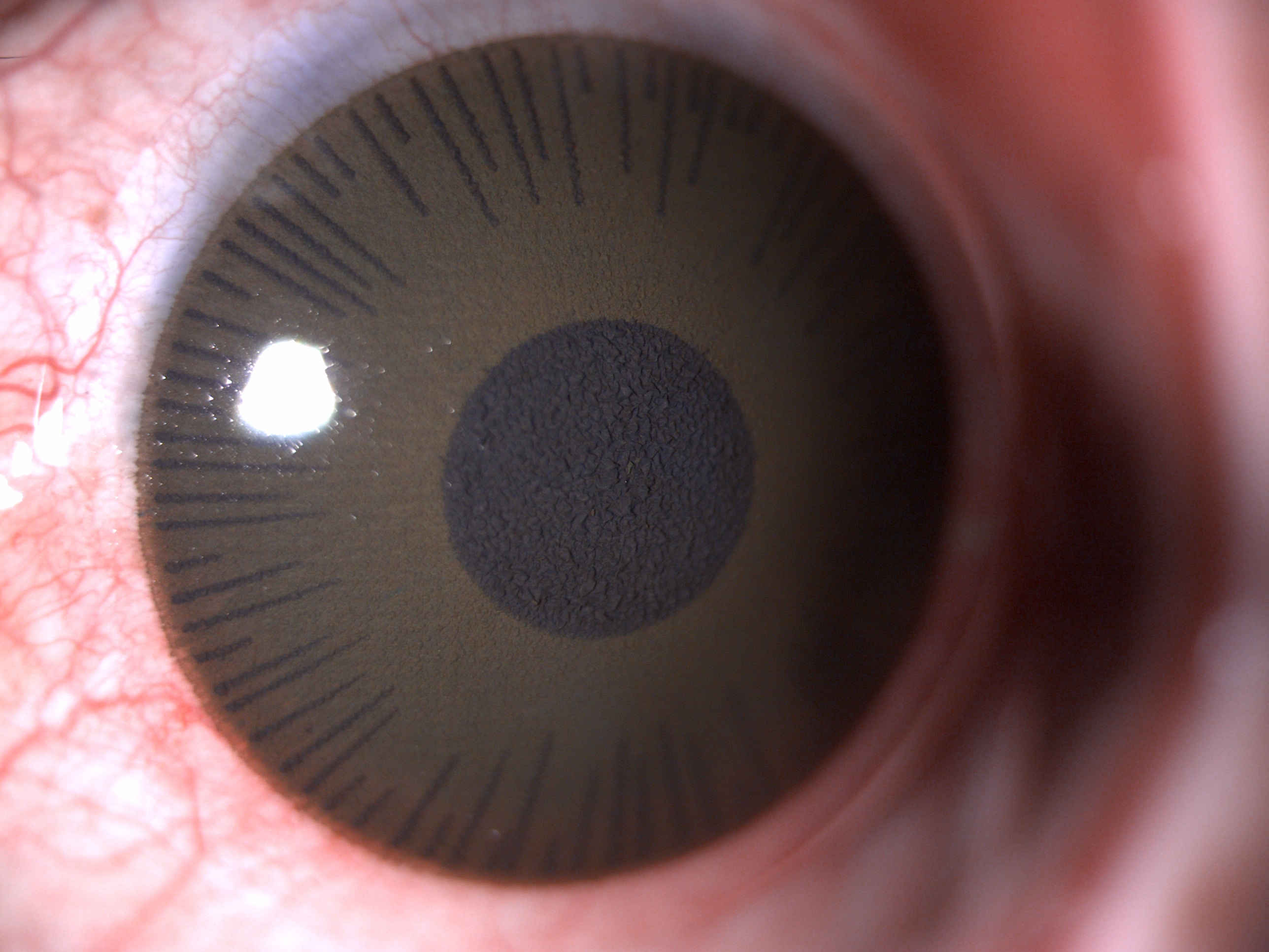
Leucomatous Corneal Opacity, Contact Lens. This digital image depicts a cosmetic contact lens in a patient with leucomatous corneal opacity.
Contributed by B Gurnani, MBBS, DNB, FCRS, FICO, MRCS Ed, MNAMS
(Click Image to Enlarge)
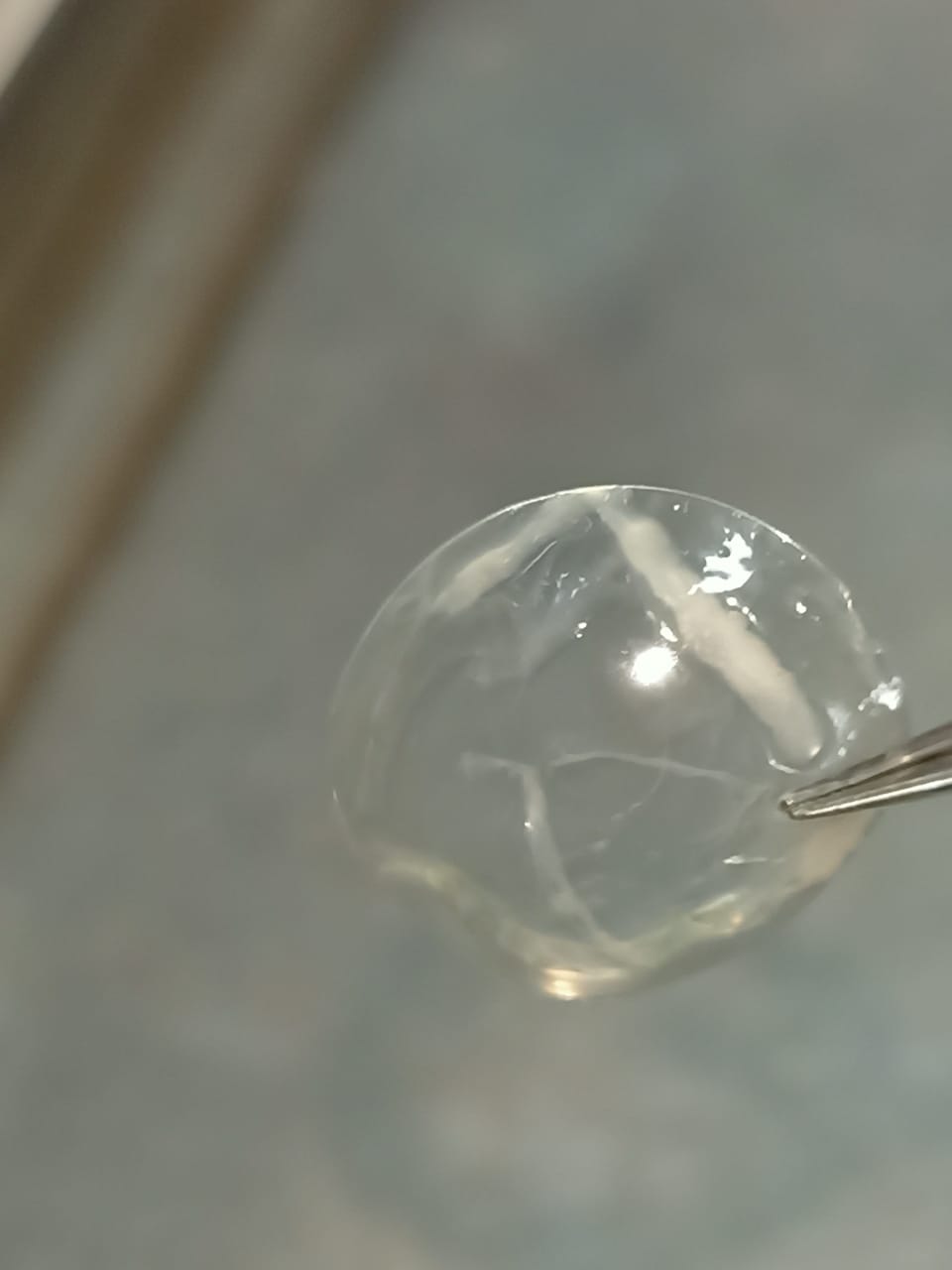
Corneal Ulcer, Infected Contact Lens. This digital slit lamp image is of an infected soft contact lens in a patient with a corneal ulcer.
Contributed by B Gurnani, MBBS, DNB, FCRS, FICO, MRCS Ed, MNAMS
(Click Image to Enlarge)
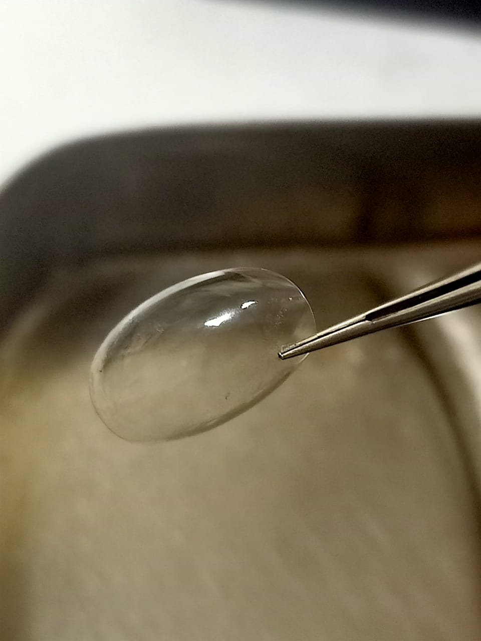
Normal Contact Lens, Digital Image.
Contributed by B Gurnani, MBBS, DNB, FCRS, FICO, MRCS Ed, MNAMS
References
Erdinest N, Bura N. Comfort and Ocular Surface Impact of Verofilcon A Daily Disposable Contact Lenses in Satisfied Senofilcon A Daily Disposable Lens Wearers: A Prospective Non-Interventional Study [Letter]. Clinical optometry. 2026:18():619170. doi: 10.2147/OPTO.S619170. Epub 2026 May 9 [PubMed PMID: 42137635]
Level 3 (low-level) evidenceFacchin A, Vampo M, Boccardo L. Psychometric evaluation of the SPEED questionnaire: Factor structure and measurement invariance across contact lens wearers and non-wearers, sex and age. The ocular surface. 2026 Jul:41():80-86. doi: 10.1016/j.jtos.2026.05.008. Epub 2026 May 13 [PubMed PMID: 42134505]
Gurnani B, Kaur K. Refractive Surprises After Cataract Surgery: Identification and Management in Optometric Practice- A Comprehensive Review. Clinical optometry. 2026:18():572733. doi: 10.2147/OPTO.S572733. Epub 2026 Apr 29 [PubMed PMID: 42083643]
Harthan JS, Nau A, Shorter E, Nau CB, Schornack M, Fogt JS. Presence of Eyelid Disease in Habitual Scleral Lens Wearers. Journal of clinical medicine. 2026 Apr 22:15(9):. doi: 10.3390/jcm15093181. Epub 2026 Apr 22 [PubMed PMID: 42122914]
Goto S, Singh S, Zhu Q, Vyas SA, Wildsoet C. Development of an Optical Defocus-Induced Myopia Model for Guinea Pigs Using Rigid Gas Permeable Contact Lenses. Eye & contact lens. 2026 Jul 1:52(7):306-314. doi: 10.1097/ICL.0000000000001279. Epub 2026 May 13 [PubMed PMID: 42121359]
Dashti Rahmatabadi Z, Naroo SA, Hatami F, Abdi S, Mohammad-Rabei H, Mehravin M, Kheiri B, Farjami M, Sheibani K, Momeni-Moghaddam H, Sabbaghi H. The impact of contact lenses on binocular vision in patients with Keratoconus. Journal of optometry. 2026 Jul-Sep:19(3):100610. doi: 10.1016/j.optom.2026.100610. Epub 2026 May 12 [PubMed PMID: 42119242]
Valsamidis TN, Mohr KM, Hjortdal JØ. The potential role of artificial intelligence in the diagnosis of Acanthamoeba keratitis-a scoping review. Contact lens & anterior eye : the journal of the British Contact Lens Association. 2026 Jun:49(3):102661. doi: 10.1016/j.clae.2026.102661. Epub 2026 May 11 [PubMed PMID: 42114398]
Level 2 (mid-level) evidenceGurnani B, Kaur K. Differentiating Pythium Keratitis From Fungal and Acanthamoeba Keratitis: A Practical Guide. StatPearls. 2026 Jan:(): [PubMed PMID: 42044243]
Gurnani B, Patel D. Advancing vision care in children - bridging science, policy, and practice. BMC pediatrics. 2026 Apr 16:26(1):. doi: 10.1186/s12887-026-06745-2. Epub 2026 Apr 16 [PubMed PMID: 41987103]
Gurnani B, Kaur K. Advancing diagnostics in Pythium insidiosum keratitis: the emerging role of point-of-care imaging and biosensor-based detection devices. Expert review of medical devices. 2025 Dec:22(12):1287-1292. doi: 10.1080/17434440.2025.2582616. Epub 2025 Nov 2 [PubMed PMID: 41147335]
Gurnani B, Kaur K. Post-market surveillance of ophthalmic implants: role of real-world evidence. Expert review of medical devices. 2025 Dec:22(12):1303-1308. doi: 10.1080/17434440.2025.2579578. Epub 2025 Oct 24 [PubMed PMID: 41123186]
Gurnani B, Kaur K. Teleophthalmology-enabled devices: bridging the gap in rural eye care. Expert review of medical devices. 2025 Nov:22(11):1167-1172. doi: 10.1080/17434440.2025.2566741. Epub 2025 Oct 15 [PubMed PMID: 40990172]
Wang J, Guo YM, Wei J, Liu G, Han Y, Ye L. Retrospective Comparative Analysis of Myopia Control Efficacy: Orthokeratology, Defocus-Incorporated Soft Contact Lenses and Their Combination with 0.01% Atropine in Chinese Children and Adolescents. Ophthalmic & physiological optics : the journal of the British College of Ophthalmic Opticians (Optometrists). 2026 Aug:46(4):881-888. doi: 10.1007/s44402-026-00104-5. Epub 2026 May 7 [PubMed PMID: 42098575]
Level 2 (mid-level) evidenceHeitz F, Scheider A, Harter P, Groeben M. Ocular Side Effects of Antibody-Drug Conjugates for Cancer Treatment. Deutsches Arzteblatt international. 2026 Jul 10:123(14):397-402. doi: 10.3238/arztebl.m2026.0060. Epub 2026 Jul 10 [PubMed PMID: 42095422]
Grzybowski A, Lanca C. Management of myopia: a mini-review. Medical hypothesis, discovery & innovation ophthalmology journal. 2026 Spring:15(1):19-39. doi: 10.51329/mehdiophthal1538. Epub 2026 Apr 22 [PubMed PMID: 42083561]
Sager S, Prieto PM, Artal P. Effect of spherical aberration on the vergence-accommodation conflict. Biomedical optics express. 2026 Apr 1:17(4):1782-1793. doi: 10.1364/BOE.593240. Epub 2026 Mar 10 [PubMed PMID: 42111011]
Gurnani B, Kaur K. Contact Lens–Related Complications. StatPearls. 2026 Jan:(): [PubMed PMID: 36512659]
Gurnani B, Kaur K. Corneal cross-linking for keratoconus: a global economic and clinical perspective. Journal of medical economics. 2025 Dec:28(1):2119-2124. doi: 10.1080/13696998.2025.2587416. Epub 2025 Nov 30 [PubMed PMID: 41320236]
Level 3 (low-level) evidenceMaghsoudlou P, Sood G, Gurnani B, Akhondi H. Cornea Transplantation. StatPearls. 2026 Jan:(): [PubMed PMID: 30969512]
Gurnani B, Kaur K. Quality of life in patients with keratoconus. Indian journal of ophthalmology. 2022 Aug:70(8):3155. doi: 10.4103/ijo.IJO_927_22. Epub [PubMed PMID: 35919000]
Level 2 (mid-level) evidenceGurnani B, Kaur K, Lalgudi VG, Kundu G, Mimouni M, Liu H, Jhanji V, Prakash G, Roy AS, Shetty R, Gurav JS. Role of artificial intelligence, machine learning and deep learning models in corneal disorders - A narrative review. Journal francais d'ophtalmologie. 2024 Sep:47(7):104242. doi: 10.1016/j.jfo.2024.104242. Epub 2024 Jul 15 [PubMed PMID: 39013268]
Level 3 (low-level) evidenceGurnani B, Kaur K, Narayana S. Corneal ectasia, cerulean (blue dot) cataract with acute hydrops in a child with Down's syndrome and hypothyroidism - a rare presentation. GMS ophthalmology cases. 2023:13():Doc07. doi: 10.3205/oc000215. Epub 2023 Mar 1 [PubMed PMID: 37034411]
Level 3 (low-level) evidencePinhas A. The evolution of healthcare through the eye: From ancient superstition to the ophthalmoscope. Frontiers in medicine. 2026:13():1775739. doi: 10.3389/fmed.2026.1775739. Epub 2026 Mar 3 [PubMed PMID: 41852533]
Coleman DJ, Fish SK. Presbyopia, accommodation, and the mature catenary. Ophthalmology. 2001 Sep:108(9):1544-51 [PubMed PMID: 11535447]
Schachar RA, Schachar IH, Fabio J, Balicki D, Sufi N, Pierscionek BK, Hunter B. Isolated human lens interferometric surface radius of curvatures: Implications for the mechanism of accommodation. PloS one. 2025:20(6):e0327028. doi: 10.1371/journal.pone.0327028. Epub 2025 Jun 24 [PubMed PMID: 40554627]
Chen Y, Kaper HJ, van Kooten T, Sharma PK. Importance of Type and Direction of Eyelid Sliding to Mimic Blinking In Vitro for Dry Eye Disease and Contact Lens Research. Investigative ophthalmology & visual science. 2026 Apr 1:67(4):56. doi: 10.1167/iovs.67.4.56. Epub [PubMed PMID: 42028975]
Falahati-Marvast F, Alipour F, Farokhzadian J, Ahmadian L. Determining the information needs of contact lens wearers for better education and more support: a qualitative study. BMC ophthalmology. 2021 Sep 7:21(1):325. doi: 10.1186/s12886-021-02085-0. Epub 2021 Sep 7 [PubMed PMID: 34493240]
Level 2 (mid-level) evidencePucker AD, Tichenor AA. A Review of Contact Lens Dropout. Clinical optometry. 2020:12():85-94. doi: 10.2147/OPTO.S198637. Epub 2020 Jun 25 [PubMed PMID: 32612404]
Alipour F, Khaheshi S, Soleimanzadeh M, Heidarzadeh S, Heydarzadeh S. Contact Lens-related Complications: A Review. Journal of ophthalmic & vision research. 2017 Apr-Jun:12(2):193-204. doi: 10.4103/jovr.jovr_159_16. Epub [PubMed PMID: 28540012]
Khan MH, Mubeen SM, Chaudhry TA, Khan SA. Contact lens use and its compliance for care among healthcare workers in Pakistan. Indian journal of ophthalmology. 2013 Jul:61(7):334-7. doi: 10.4103/0301-4738.97552. Epub [PubMed PMID: 23552347]
Level 2 (mid-level) evidenceZimmerman AB, Nixon AD, Rueff EM. Contact lens associated microbial keratitis: practical considerations for the optometrist. Clinical optometry. 2016:8():1-12. doi: 10.2147/OPTO.S66424. Epub 2016 Jan 29 [PubMed PMID: 30214344]
Dupuis P, Prokopich CL, Hynes A, Kim H. A contemporary look at allergic conjunctivitis. Allergy, asthma, and clinical immunology : official journal of the Canadian Society of Allergy and Clinical Immunology. 2020:16():5. doi: 10.1186/s13223-020-0403-9. Epub 2020 Jan 21 [PubMed PMID: 31993069]
Zhang X, M VJ, Qu Y, He X, Ou S, Bu J, Jia C, Wang J, Wu H, Liu Z, Li W. Dry Eye Management: Targeting the Ocular Surface Microenvironment. International journal of molecular sciences. 2017 Jun 29:18(7):. doi: 10.3390/ijms18071398. Epub 2017 Jun 29 [PubMed PMID: 28661456]
Mann A, Tighe B. Contact lens interactions with the tear film. Experimental eye research. 2013 Dec:117():88-98. doi: 10.1016/j.exer.2013.07.013. Epub 2013 Jul 23 [PubMed PMID: 23886658]
Panaser A, Tighe BJ. Function of lipids − their fate in contact lens wear: an interpretive review. Contact lens & anterior eye : the journal of the British Contact Lens Association. 2012 Jun:35(3):100-11. doi: 10.1016/j.clae.2012.01.003. Epub 2012 Feb 10 [PubMed PMID: 22317829]
Kumari A, Sharma PK, Garg VK, Garg G. Ocular inserts - Advancement in therapy of eye diseases. Journal of advanced pharmaceutical technology & research. 2010 Jul:1(3):291-6. doi: 10.4103/0110-5558.72419. Epub [PubMed PMID: 22247860]
Gurnani B, Kaur K. Pseudophakic Bullous Keratopathy. StatPearls. 2026 Jan:(): [PubMed PMID: 34662019]
Gurnani B, Kaur K. Immune Camouflage in Pythium insidiosum Keratitis: A Hypothesis on Molecular Mimicry and Host Pattern Recognition Receptor Evasion. Clinical ophthalmology (Auckland, N.Z.). 2025:19():4855-4873. doi: 10.2147/OPTH.S560311. Epub 2025 Dec 19 [PubMed PMID: 41446892]
Gurnani B, Feroze KB, Patel BC. Neurotrophic Keratitis. StatPearls. 2026 Jan:(): [PubMed PMID: 28613758]
Gurnani B, Kaur K. Redefining Dry Eye Disease Management: Emerging Pharmacologic and Device-Based Therapies in the Era of Precision Care: A Narrative Review. Ophthalmology and therapy. 2026 May:15(5):1561-1584. doi: 10.1007/s40123-026-01354-9. Epub 2026 Mar 23 [PubMed PMID: 41866434]
Level 3 (low-level) evidenceTripathi M, Markan A, Gurnani B. Salzmann Nodular Corneal Degeneration. StatPearls. 2026 Jan:(): [PubMed PMID: 32809519]
Gurnani B, Kaur K, Venugopal A, Srinivasan B, Bagga B, Iyer G, Christy J, Prajna L, Vanathi M, Garg P, Narayana S, Agarwal S, Sahu S. Pythium insidiosum keratitis - A review. Indian journal of ophthalmology. 2022 Apr:70(4):1107-1120. doi: 10.4103/ijo.IJO_1534_21. Epub [PubMed PMID: 35325996]
Gurnani B, Kaur K. Traumatic Iris Reconstruction. StatPearls. 2026 Jan:(): [PubMed PMID: 35201728]
Mohseni M, Gurnani B, Blair K. Blunt Eye Trauma. StatPearls. 2026 Jan:(): [PubMed PMID: 29261988]
Kaur K, Zeppieri M, Gurnani B. Primary Congenital Glaucoma. StatPearls. 2026 Jan:(): [PubMed PMID: 34662067]
Marshood AA. Retinal Detachment in the eye of Albino - A review. Romanian journal of ophthalmology. 2026 Jan-Mar:70(1):35-41. doi: 10.22336/rjo.2026.05. Epub [PubMed PMID: 42146956]
So YH, Mishra D, Gite S, Sonawane R, Waite D, Shaikh R, Vora LK, Thakur RRS. Emerging trends in long-acting sustained drug delivery for glaucoma management. Drug delivery and translational research. 2025 Jun:15(6):1907-1934. doi: 10.1007/s13346-024-01779-4. Epub 2025 Jan 9 [PubMed PMID: 39786666]
Nagendran I, Roy L, Ravikumar P, Gireesh P, Kaur K, Gurnani B. The Evolution of Vision Therapy Software and Its Impact on Vision Care - A Comprehensive Major Review. Romanian journal of ophthalmology. 2025 Oct-Dec:69(4):495-511. doi: 10.22336/rjo.2025.78. Epub [PubMed PMID: 41971215]
Sedhom CR, Bahgat N, Awadein A, Abdelhamid M, Abdelaziz MS, Shokier G. Yamane Technique for The Management of Cataract in Children with Insufficient Capsular Support: A Prospective Study. Current eye research. 2026 Jul:51(7):863-872. doi: 10.1080/02713683.2026.2649979. Epub 2026 Apr 6 [PubMed PMID: 41937488]
Gurnani B, Kaur K. Artificial intelligence in ophthalmology: from innovation to clinical integration. Frontiers in ophthalmology. 2026:6():1839194. doi: 10.3389/fopht.2026.1839194. Epub 2026 Apr 30 [PubMed PMID: 42147450]
Riva I, Micheletti E, Oddone F, Bruttini C, Montescani S, De Angelis G, Rovati L, Weinreb RN, Quaranta L. Anterior Chamber Angle Assessment Techniques: A Review. Journal of clinical medicine. 2020 Nov 25:9(12):. doi: 10.3390/jcm9123814. Epub 2020 Nov 25 [PubMed PMID: 33255754]
Kanai K, Igawa Y, Tachibana M, Yoshikawa Y, Katsumoto T, Shibuya M, Kawagoe T, Makita J, Shinoda K. Electroretinograms Recorded with Skin Electrodes from Post-Vitrectomy Silicone Oil Filled Eyes with Proliferative Diabetic Retinopathy. Clinical ophthalmology (Auckland, N.Z.). 2024:18():2167-2174. doi: 10.2147/OPTH.S468759. Epub 2024 Jul 30 [PubMed PMID: 39100695]
Panwar N, Huang P, Lee J, Keane PA, Chuan TS, Richhariya A, Teoh S, Lim TH, Agrawal R. Fundus Photography in the 21st Century--A Review of Recent Technological Advances and Their Implications for Worldwide Healthcare. Telemedicine journal and e-health : the official journal of the American Telemedicine Association. 2016 Mar:22(3):198-208. doi: 10.1089/tmj.2015.0068. Epub 2015 Aug 26 [PubMed PMID: 26308281]
Level 3 (low-level) evidenceTripathi M, Markan A, Gurnani B. Cytomegalovirus Corneal Endotheliitis. StatPearls. 2026 Jan:(): [PubMed PMID: 35201702]
Kaufman HE, Thomas EL. Prevention and treatment of symblepharon. American journal of ophthalmology. 1979 Sep:88(3 Pt 1):419-23 [PubMed PMID: 484669]
Mishra A, Verma AK. Sports related ocular injuries. Medical journal, Armed Forces India. 2012 Jul:68(3):260-6. doi: 10.1016/j.mjafi.2011.12.004. Epub 2012 May 31 [PubMed PMID: 24532883]
Tredici TJ, Flynn WJ. The use of contact lenses by USAF aviators. Aviation, space, and environmental medicine. 1987 May:58(5):438-43 [PubMed PMID: 3297018]
Shetty R, Lasrado K, Hussain S, Shetty M. Aesthetic and Functional Rehabilitation of Phthisis Bulbi Through Custom Scleral Shell Prosthesis: A Technical Case Report. Cureus. 2025 Sep:17(9):e92676. doi: 10.7759/cureus.92676. Epub 2025 Sep 18 [PubMed PMID: 41122573]
Level 3 (low-level) evidenceLee H, Byun S, Noh H. Nanocomposite hydrogels with temperature-accelerated antioxidant activity for ocular applications. Nanoscale. 2026 Jun 18:18(23):12326-12333. doi: 10.1039/d6nr00711b. Epub 2026 Jun 18 [PubMed PMID: 42149130]
Wang Y, Wang L, Zheng Z, Xu J, Zhang Z. Comparison of the effectiveness of ranibizumab monotherapy vs. combined panretinal photocoagulation on the regression of neovascularization in proliferative diabetic retinopathy. Frontiers in medicine. 2026:13():1808957. doi: 10.3389/fmed.2026.1808957. Epub 2026 Apr 28 [PubMed PMID: 42131595]
Seresirikachorn K, Vu DM, Perez AM, Thiamthat W, Peter Chang TC. Childhood Angle Closure: Etiology and Treatment Outcomes from a Large Series Spanning over 20 Years. Ophthalmology. Glaucoma. 2026 May 15:():. pii: S2589-4196(26)00071-2. doi: 10.1016/j.ogla.2026.05.005. Epub 2026 May 15 [PubMed PMID: 42142661]
Wu C, Wu J, Li K, Li C, Li D, Wang L, Du Y, Liu G, Yu L. Hyaluronic acid-based corneal epithelium-mimicking coating for therapeutic contact lenses to manage dry eye disease and prevent bacterial keratitis. International journal of biological macromolecules. 2026 Jun:367():152594. doi: 10.1016/j.ijbiomac.2026.152594. Epub 2026 May 17 [PubMed PMID: 42150662]
Song W, Shi H, Feng J, Qian K, Dong Q, Sun Z, Ding J, Hu W. Prelithiation for Lithium-Ion Batteries: Material and Process Innovations. Advanced materials (Deerfield Beach, Fla.). 2026 May 14:():e73290. doi: 10.1002/adma.73290. Epub 2026 May 14 [PubMed PMID: 42136257]
Level 2 (mid-level) evidenceByrd LB, Gurnani B, Martin N. Corneal Ulcer. StatPearls. 2026 Jan:(): [PubMed PMID: 30969511]
Gurnani B, Kaur K. Biosensor-embedded wearables for dry eye monitoring and management. Expert review of medical devices. 2025 Oct:22(10):1073-1079. doi: 10.1080/17434440.2025.2555485. Epub 2025 Sep 1 [PubMed PMID: 40884106]
Kaur RP, Gurnani B, Kaur K. Intricate insights into immune response in dry eye disease. Indian journal of ophthalmology. 2023 Apr:71(4):1248-1255. doi: 10.4103/IJO.IJO_481_23. Epub [PubMed PMID: 37026255]
Jonas JB, Jonas RA, Bikbov MM, Kazakbaeva GM, Dong L, Wang YX, Panda-Jonas S. Mechanism of myopic axial elongation related to Bruch´s membrane. Asia-Pacific journal of ophthalmology (Philadelphia, Pa.). 2026 May-Jun:15(3):100329. doi: 10.1016/j.apjo.2026.100329. Epub 2026 May 14 [PubMed PMID: 42134650]
Dahlmann-Noor A, Kessel L, Lagrèze WA, Noval S, Polling JR, Villani E, Brémond-Gignac D. Workforce challenges to implementation of myopia management in Europe: an electronic/telephone expert survey. Frontiers in ophthalmology. 2026:6():1812277. doi: 10.3389/fopht.2026.1812277. Epub 2026 Apr 22 [PubMed PMID: 42100441]
Level 3 (low-level) evidenceRen J, Zhang X, Li J, Ma K. Sleep quality as a mediator between perceived stress and dry eye severity among Chinese nurses. Frontiers in public health. 2026:14():1817895. doi: 10.3389/fpubh.2026.1817895. Epub 2026 Apr 15 [PubMed PMID: 42064899]
Level 2 (mid-level) evidenceDeng M. Letter to the editor concerning "Multifocal contact lenses and defocus incorporated multiple segments lenses slow myopic progression in Chinese children with high myopia". Journal of optometry. 2026 Jul-Sep:19(3):100619. doi: 10.1016/j.optom.2026.100619. Epub 2026 May 18 [PubMed PMID: 42150297]
Level 3 (low-level) evidenceFisher D, Iqbal A, Alonso-Caneiro D, Collins MJ, Vincent SJ. Tear Dynamics During Fenestrated Scleral Lens Wear: A Pilot Study. Ophthalmic & physiological optics : the journal of the British College of Ophthalmic Opticians (Optometrists). 2026 Aug:46(4):786-793. doi: 10.1007/s44402-026-00102-7. Epub 2026 May 6 [PubMed PMID: 42090093]
Level 3 (low-level) evidenceHe A, Chen V, Yu J. Sustained ocular drug delivery via polydopamine chitosan curcumin nanocomposite coatings. Discover nano. 2026 May 6:21(1):. doi: 10.1186/s11671-026-04556-z. Epub 2026 May 6 [PubMed PMID: 42090042]
Ndayisenga JP, Oudshoorn A, Hynie M, Mukamana D, Babenko-Mould Y, Jackson KT. Where shame meets vulnerability: unintended adolescent pregnancy within the cultural context of the rural district of Rwandan Eastern Province. BMC pregnancy and childbirth. 2026 May 5:26(1):. doi: 10.1186/s12884-026-09133-x. Epub 2026 May 5 [PubMed PMID: 42087091]
Gurnani B, Patel BC. Photorefractive Keratectomy. StatPearls. 2026 Jan:(): [PubMed PMID: 31751077]
Vohra V, Tuteja S, Gurnani B, Chawla H. Collagen Cross Linking for Keratoconus. StatPearls. 2026 Jan:(): [PubMed PMID: 32965942]
Gurnani B, Kaur K. Evolving concepts in etiopathogenesis of keratoconus: Is it quasi-inflammatory or inflammatory? Indian journal of ophthalmology. 2023 Jun:71(6):2609-2610. doi: 10.4103/ijo.IJO_783_22. Epub [PubMed PMID: 37322699]
Gurnani B, Kaur K, Tripathy K. Is there a genetic link between Keratoconus and Fuch's endothelial corneal dystrophy? Medical hypotheses. 2021 Dec:157():110699. doi: 10.1016/j.mehy.2021.110699. Epub 2021 Oct 9 [PubMed PMID: 34666260]
Gurnani B, Kaur K. Comments on: Are we treating the effect and neglecting the cause of keratoconus? Indian journal of ophthalmology. 2021 Feb:69(2):462-463. doi: 10.4103/ijo.IJO_3280_20. Epub [PubMed PMID: 33463618]
Level 3 (low-level) evidenceGurnani B, Kaur K. Contact Lenses for Presbyopia(Archived). StatPearls. 2026 Jan:(): [PubMed PMID: 36251823]
Kaur K, Gurnani B. Specular Microscopy. StatPearls. 2026 Jan:(): [PubMed PMID: 36256774]
Bakaraju RC, Constable PA, Tilia D, Patel K, Chakraborty R. Electroretinographic evaluation of single vision contact lenses with non-refractive opaque features. Clinical & experimental optometry. 2026 May 19:():1-15. doi: 10.1080/08164622.2026.2659189. Epub 2026 May 19 [PubMed PMID: 42154044]
Gurnani B, Kaur K. Artificial Intelligence and Deep Learning in the Diagnosis of Pythium Keratitis. StatPearls. 2026 Jan:(): [PubMed PMID: 42044246]
Kaur K, Kanukollu VM, Gurnani B. Strabismus. StatPearls. 2026 Jan:(): [PubMed PMID: 32809617]
Kha R, Kandel H, Watson S. Trends in the Management of Keratoconus in Australia. Ophthalmic & physiological optics : the journal of the British College of Ophthalmic Opticians (Optometrists). 2026 May:46(2):135-142. doi: 10.1007/s44402-026-00096-2. Epub 2026 May 19 [PubMed PMID: 42154407]
Saeki K, Huyan D, Sawada M, Nakamura A, Kubota S, Uno K, Ohnuma K, Shiina T. Three-dimensional measurement for spherical and nonspherical shapes of contact lenses. Applied optics. 2021 May 1:60(13):3689-3698. doi: 10.1364/AO.419721. Epub [PubMed PMID: 33983301]
Hsiao MC, Yen YC, Wang CH, Chen YN, Wang SP, Su KC. Biomechanical evaluation of different plunger size and plunger position on removing soft contact lenses and rigid gas permeable contact lenses. Technology and health care : official journal of the European Society for Engineering and Medicine. 2024:32(6):4109-4121. doi: 10.3233/THC-231983. Epub [PubMed PMID: 39058461]
Yen PY, Lin CM, Chang Chien IH. Multi-Objective Optimization of an Injection Molding Process for an Alvarez Freeform Lens Using an Integrated Optical System and Mold Flow Analyses. Polymers. 2025 Sep 10:17(18):. doi: 10.3390/polym17182453. Epub 2025 Sep 10 [PubMed PMID: 41012216]
Cseh M, Katona G, Berkó S, Budai-Szűcs M, Csóka I. A Stereolithography-Based Modified Spin-Casting Method for Faster Laboratory-Scale Production of Dexamethasone-Containing Dissolving Microneedle Arrays. Pharmaceutics. 2024 Jul 29:16(8):. doi: 10.3390/pharmaceutics16081005. Epub 2024 Jul 29 [PubMed PMID: 39204350]
Giovanzana S, Evans T, Pierscionek B. Lens internal curvature effects on age-related eye model and lens paradox. Biomedical optics express. 2017 Nov 1:8(11):4827-4837. doi: 10.1364/BOE.8.004827. Epub 2017 Oct 3 [PubMed PMID: 29188084]
Maldonado-Codina C, Efron N. Impact of manufacturing technology and material composition on the clinical performance of hydrogel lenses. Optometry and vision science : official publication of the American Academy of Optometry. 2004 Jun:81(6):442-54 [PubMed PMID: 15201718]
Musgrave CSA, Fang F. Contact Lens Materials: A Materials Science Perspective. Materials (Basel, Switzerland). 2019 Jan 14:12(2):. doi: 10.3390/ma12020261. Epub 2019 Jan 14 [PubMed PMID: 30646633]
Level 3 (low-level) evidenceAslani F, Khorrami-Nejad M, Aghazadeh Amiri M, Hashemian H, Askarizadeh F, Khosravi B. Characteristics of Posterior Corneal Astigmatism in Different Stages of Keratoconus. Journal of ophthalmic & vision research. 2018 Jan-Mar:13(1):3-9. doi: 10.4103/jovr.jovr_217_16. Epub [PubMed PMID: 29403582]
Gatinel D. An optical method for deriving the anterior and posterior curvatures of intraocular lenses using dual back-vertex power measurements. PloS one. 2025:20(12):e0337041. doi: 10.1371/journal.pone.0337041. Epub 2025 Dec 2 [PubMed PMID: 41329769]
Eliaz N, Metoki N. Calcium Phosphate Bioceramics: A Review of Their History, Structure, Properties, Coating Technologies and Biomedical Applications. Materials (Basel, Switzerland). 2017 Mar 24:10(4):. doi: 10.3390/ma10040334. Epub 2017 Mar 24 [PubMed PMID: 28772697]
Remón L, Pérez-Merino P, Macedo-de-Araújo RJ, Amorim-de-Sousa AI, González-Méijome JM. Bifocal and Multifocal Contact Lenses for Presbyopia and Myopia Control. Journal of ophthalmology. 2020:2020():8067657. doi: 10.1155/2020/8067657. Epub 2020 Mar 27 [PubMed PMID: 32318285]
Balgos MJTD, Vargas V, Alió JL. Correction of presbyopia: An integrated update for the practical surgeon. Taiwan journal of ophthalmology. 2018 Jul-Sep:8(3):121-140. doi: 10.4103/tjo.tjo_53_18. Epub [PubMed PMID: 30294526]
Ayton LN, Rizzo JF 3rd, Bailey IL, Colenbrander A, Dagnelie G, Geruschat DR, Hessburg PC, McCarthy CD, Petoe MA, Rubin GS, Troyk PR, HOVER International Taskforce. Harmonization of Outcomes and Vision Endpoints in Vision Restoration Trials: Recommendations from the International HOVER Taskforce. Translational vision science & technology. 2020 Jul:9(8):25. doi: 10.1167/tvst.9.8.25. Epub 2020 Jul 16 [PubMed PMID: 32864194]
Toshida H, Takahashi K, Sado K, Kanai A, Murakami A. Bifocal contact lenses: History, types, characteristics, and actual state and problems. Clinical ophthalmology (Auckland, N.Z.). 2008 Dec:2(4):869-77 [PubMed PMID: 19668441]
Rodriguez-Lopez V, Barcala X, Zaytouny A, Dorronsoro C, Peli E, Marcos S. Monovision Correction Preference and Eye Dominance Measurements. Translational vision science & technology. 2023 Mar 1:12(3):18. doi: 10.1167/tvst.12.3.18. Epub [PubMed PMID: 36939712]
Fathima TZ, Tamilarasi S, Sachdev G, Dandapani R. Concurrent phakic intraocular lens explantation and phacoemulsification with IOL implantation: A retrospective analysis of efficacy and safety. Indian journal of ophthalmology. 2026 May 1:74(Suppl 1):S46-S51. doi: 10.4103/IJO.IJO_1718_25. Epub 2026 Apr 30 [PubMed PMID: 42080716]
Level 2 (mid-level) evidenceAlsaif BA, Alfaraidi AT, Alshabeeb R. Fungal Keratitis Following Application of Dehydrated Amniotic Membrane (Omnigen) With (OmniLenz) Bandage Contact Lens. Case reports in ophthalmological medicine. 2026:2026():3157153. doi: 10.1155/crop/3157153. Epub 2026 Apr 28 [PubMed PMID: 42058157]
Level 3 (low-level) evidencePenzani R, Parolini B, Apuzzo A. Ocular Motility Changes After Macular Buckle for Myopic Traction Maculopathy. Journal of pediatric ophthalmology and strabismus. 2026 Apr 17:():1-5. doi: 10.3928/01913913-20260317-02. Epub 2026 Apr 17 [PubMed PMID: 41996526]
Roth J, Macedo AF, Somajo S, Tannerfeldt L, Toprak B, Lagali N. Infectious bacterial keratitis management in a Swedish region: Risk factors, treatment and time-to-resolution. Acta ophthalmologica. 2026 Apr 12:():. doi: 10.1111/aos.70141. Epub 2026 Apr 12 [PubMed PMID: 41968694]
Jin X, Wang Y, Gui MF, Guo XH, Gong Y, Zhang XJ, Zhang L. Efficacy and ocular surface safety of different correction methods for adolescents with high myopia. International journal of ophthalmology. 2026:19(4):750-756. doi: 10.18240/ijo.2026.04.14. Epub 2026 Apr 18 [PubMed PMID: 41924373]
Gurnani B, Kaur K. Renaissance of an Emerging Pathogen-Pythium insidiosum Keratitis: An Indian Perspective. Clinical ophthalmology (Auckland, N.Z.). 2025:19():4811-4830. doi: 10.2147/OPTH.S568552. Epub 2025 Dec 18 [PubMed PMID: 41446894]
Level 3 (low-level) evidenceFu L, Gurnani B, Hollick EJ. Artificial Corneal Transplantation. StatPearls. 2026 Jan:(): [PubMed PMID: 33760451]
Stapleton F, Bakkar M, Carnt N, Chalmers R, Vijay AK, Marasini S, Ng A, Tan J, Wagner H, Woods C, Wolffsohn JS. CLEAR - Contact lens complications. Contact lens & anterior eye : the journal of the British Contact Lens Association. 2021 Apr:44(2):330-367. doi: 10.1016/j.clae.2021.02.010. Epub 2021 Mar 25 [PubMed PMID: 33775382]
Kaur K, Gurnani B. Microspherophakia. StatPearls. 2026 Jan:(): [PubMed PMID: 35015420]
Gurnani B, Ronquillo Y, Moshirfar M. Acanthamoeba Keratitis. StatPearls. 2026 Jan:(): [PubMed PMID: 31751053]
Shah SS, Gurnani B. Lens-Induced Glaucoma. StatPearls. 2026 Jan:(): [PubMed PMID: 34662038]
Ko Y, Ko M, Ueffing M, Kang HJ, Kwon HJ. Organelle contact reorganization drives calcium-dependent autophagy under proteostatic stress. Autophagy. 2026 Jun 6:():1-20. doi: 10.1080/15548627.2026.2677184. Epub 2026 Jun 6 [PubMed PMID: 42152515]
Arslan N, Kakil ŞB, Aydoğdu B, Telek HH. Longitudinal evaluation of corneal stability and tear film changes in first-time soft-contact lens wearers. Arquivos brasileiros de oftalmologia. 2025:88(6):e20250118. doi: 10.5935/0004-2749.2025-0118. Epub 2025 Oct 10 [PubMed PMID: 41092216]
MacRae SM, Matsuda M, Shellans S. Corneal endothelial changes associated with contact lens wear. The CLAO journal : official publication of the Contact Lens Association of Ophthalmologists, Inc. 1989 Jan-Mar:15(1):82-7 [PubMed PMID: 2917404]
Donshik PC. Giant papillary conjunctivitis. Transactions of the American Ophthalmological Society. 1994:92():687-744 [PubMed PMID: 7886881]
Lahoti S, Weiss M, Johnson DA, Kheirkhah A. Superior limbic keratoconjunctivitis: a comprehensive review. Survey of ophthalmology. 2022 Mar-Apr:67(2):331-341. doi: 10.1016/j.survophthal.2021.05.009. Epub 2021 May 30 [PubMed PMID: 34077767]
Level 3 (low-level) evidenceAdam MP, Bick S, Mirzaa GM, Pagon RA, Wallace SE, Amemiya A, Kosho T, Yamaguchi T, Mizumoto S, Mendoza-Londono R. Musculocontractural Ehlers-Danlos Syndrome. GeneReviews(®). 1993:(): [PubMed PMID: 40373179]
Liou JC, Chen PH, Tung HC, Tung TY, Chang CW, Cheng CY. Original article preliminary exploratory finding of anti-defocus (ADF) contact lenses on binocular vision. Contact lens & anterior eye : the journal of the British Contact Lens Association. 2026 Jun:49(3):102657. doi: 10.1016/j.clae.2026.102657. Epub 2026 May 4 [PubMed PMID: 42085830]
Evans BJ. Monovision: a review. Ophthalmic & physiological optics : the journal of the British College of Ophthalmic Opticians (Optometrists). 2007 Sep:27(5):417-39 [PubMed PMID: 17718882]
El-Haddad C, Hegazi I, Hu W. Understanding Patient Expectations of Health Care: A Qualitative Study. Journal of patient experience. 2020 Dec:7(6):1724-1731. doi: 10.1177/2374373520921692. Epub 2020 Apr 28 [PubMed PMID: 33457636]
Level 2 (mid-level) evidenceLambert M, van Dijk L, Benko R, Llor C, Lykkegaard J, Mantel-Teeuwisse AK, Pont L, Taxis K. The role of the community pharmacist in antibiotic use - a commentary on current status and future perspectives. Exploratory research in clinical and social pharmacy. 2025 Dec:20():100652. doi: 10.1016/j.rcsop.2025.100652. Epub 2025 Aug 30 [PubMed PMID: 40980189]
Level 3 (low-level) evidenceTadesse YB, Sendekie AK, Mekonnen BA, Denberu FG, Kassaw AT. Pharmacists' Medication Counseling Practices and Knowledge and Satisfaction of Patients With an Outpatient Hospital Pharmacy Service. Inquiry : a journal of medical care organization, provision and financing. 2023 Jan-Dec:60():469580231219457. doi: 10.1177/00469580231219457. Epub [PubMed PMID: 38131171]
Van Bentum RE, Van den Berg JM, Wolf SE, Van der Bijl J, Tan HS, Verbraak FD, van der Horst-Bruinsma IE. Multidisciplinary management of auto-immune ocular diseases in adult patients by ophthalmologists and rheumatologists. Acta ophthalmologica. 2021 Mar:99(2):e164-e170. doi: 10.1111/aos.14548. Epub 2020 Aug 4 [PubMed PMID: 32749781]
Fogt JS, Roth M, Gardner HP. How Can We Better Inform Patients of the Importance of Contact Lens Compliance?: Current Perspectives. Clinical optometry. 2024:16():267-286. doi: 10.2147/OPTO.S405204. Epub 2024 Nov 2 [PubMed PMID: 39507399]
Level 3 (low-level) evidenceEnaholo ES, Gurnani B. Accommodative Insufficiency. StatPearls. 2026 Jan:(): [PubMed PMID: 36508537]
Singh B, Sahu V, Kumar A, Shrivastava S, Gurnani B, Kaur K. Regression Patterns of Neovascularization in Proliferative Diabetic Retinopathy Following Panretinal Photocoagulation - A Prospective, Observational Study. Clinical ophthalmology (Auckland, N.Z.). 2026:20():575268. doi: 10.2147/OPTH.S575268. Epub 2026 Mar 19 [PubMed PMID: 41891101]
Level 2 (mid-level) evidence