 Tools
Tools
 Charles Bonnet Syndrome
Introduction
Charles Bonnet Syndrome (CBS), also known as visual release hallucinations, refers to the occurrence of complex visual hallucinations in individuals with partial or complete loss of vision.[1] According to the World Health Organization International Classification of Diseases–11, “hallucinations are exclusively visual, usually temporary, and unrelated to mental and behavioural disorders.” The phenomenon was first described in the eighteenth century by Charles Bonnet, who documented vivid visual hallucinations experienced by his grandfather following significant vision loss from cataracts.[2]
CBS is a neuro-ophthalmic condition characterized by formed, often detailed visual hallucinations in psychologically intact individuals with marked visual impairment. The syndrome is of particular clinical interest because it arises from visual sensory deprivation and intersects the disciplines of ophthalmology, neurology, and psychiatry. The modern term “Charles Bonnet Syndrome” was later introduced by Georges de Morsier in the twentieth century to describe these complex hallucinations occurring in the absence of psychiatric or cognitive disease.[2]
Although once thought rare, CBS is now recognized as a common but underreported sequela of partial sight loss, affecting up to 10% to 30% of visually impaired adults, depending on diagnostic criteria and the population studied. At its core, CBS represents a form of sensory deprivation hallucinosis—the brain’s intrinsic tendency to generate visual imagery in the absence of adequate retinal input. When visual stimuli are chronically reduced, as in macular degeneration, glaucoma, diabetic retinopathy, or optic-nerve disease, cortical networks responsible for image formation become hyperexcitable, producing spontaneous visual perceptions. The hallmark feature that differentiates CBS from psychotic hallucinations is the patient’s intact insight: individuals recognize that the visions are unreal, maintaining normal cognition and affect.[3]
A detailed literature review reveals numerous controversies and shifting consensus regarding the inclusion and exclusion criteria for CBS.[4] Because the disease occurs in individuals with visual impairment or vision loss, pathways leading to CBS involve the visual system, including the optic nerve, brain, and eyes.[5] Due to the nature of hallucinations and the most common etiologies, CBS often goes unrecognized and is misdiagnosed with early dementia or psychosis. The patient is usually aware of the unreality of visual experiences, although these may occasionally cause distress. The patient has minimal or no control over the hallucinations, which are clear, well-defined, and organized images.[6]
The Swiss scientist Charles Bonnet initially described CBS when he first noticed this phenomenon of visual hallucination in his 90-year-old grandfather, who had cataracts.[7] The grandfather underwent cataract surgery, and his initial vision improved, but as the visual acuity deteriorated, he noticed visual hallucinations (see Image. Charles Bonnet Syndrome).[8] The hallucinations were unreal, and the grandfather didn't experience psychiatric disorders. CBS is a release phenomenon resulting from deafferentation of the cerebral cortex's visual association areas, leading to phantom vision.[9]
Cognitive malfunction, social isolation, and sensory deprivation have been suggested as the primary etiologies of CBS. CBS is often missed in older adults due to fear of being labeled as mentally ill.[10] Patients who notice the unreal nature of their hallucinations may be depressed by the fear of acquiring new psychiatric symptoms or diagnoses. A detailed and systematic history is essential to rule out the existence of hallucinations. The hallucinations may improve spontaneously, with enhanced vision, or with social isolation.[11] Anticonvulsants have been shown to enhance and abort hallucinations, but there is no drug of choice for CBS. Accurate diagnosis is the most critical component of management.[12]
Charles Bonnet’s original account described his grandfather, who, after developing severe visual loss from cataracts, began seeing people, birds, and buildings that were not present, despite full awareness of their falsity. Subsequent case descriptions in the nineteenth and twentieth centuries expanded the clinical spectrum, linking the phenomenon to various ocular pathologies. The term “Charles Bonnet Syndrome” was formally coined in the 1960s to describe complex visual hallucinations secondary to vision loss without psychiatric disturbance. Over the years, research has moved from purely descriptive observations to neuroimaging-based investigations that elucidate the cortical mechanisms underlying release hallucinations.[13]
Prevalence estimates vary widely, from 0.4% in general ophthalmic clinics to nearly 30% among patients with advanced macular degeneration. The syndrome predominantly affects older adults, reflecting the higher incidence of age-related ocular disease. However, CBS is not restricted to older adults—it has been documented in congenital or acquired visual impairment at any age, including post-enucleation or optic-nerve injury. Advancing age and bilateral visual loss are key risk factors, and there appears to be a female predominance in some series, possibly due to higher longevity and prevalence of macular degeneration in women. The true prevalence is likely underestimated, as many patients conceal their symptoms out of fear of being perceived as mentally ill. Community-based surveys have shown that only a minority of affected individuals report hallucinations without being specifically asked, underscoring the importance of clinician awareness and open inquiry.[14]
CBS is considered a “release hallucinosis” resulting from visual sensory deprivation. Functional magnetic resonance imaging and positron emission tomography studies demonstrate hyperactivity of the visual association cortex, particularly Brodmann areas 19 and 37, in the absence of corresponding retinal input. The mechanism is analogous to auditory “phantom” perceptions in tinnitus or somatic hallucinations in phantom-limb pain. Several hypotheses have been proposed, including the deafferentation theory, which postulates that loss of afferent input from damaged retina or optic nerve disinhibits higher-order visual networks, leading to spontaneous image generation. Another explanation, the perceptual-release hypothesis, suggests that the visual cortex, deprived of regular stimulation, attempts to “fill in” missing information, producing internally generated images. Neurochemical factors may also contribute, with altered cholinergic and dopaminergic transmission modulating cortical excitability and hallucinatory vividness. Structural brain disease is typically absent, and hallucinations often cease when visual input improves, such as after cataract surgery, supporting the peripheral-trigger theory.[15]
Clinically, CBS hallucinations are purely visual and are not accompanied by auditory or tactile components. They are usually complex, formed, and often colourful, and differ from the simple flashes or photopsias seen in retinal pathology. Commonly reported images include people, faces, animals, insects, buildings, landscapes, and repetitive geometric designs. The hallucinations are often vivid, detailed, and may appear life-sized or miniature. Episodes can last from seconds to several minutes and recur multiple times per day, sometimes persisting for months or years. Environmental factors such as dim lighting, fatigue, sensory isolation, or inactivity can increase the frequency of these symptoms, whereas bright light or active engagement may suppress them. Importantly, patients retain complete insight, recognizing that the images are unreal—a key distinction from psychosis, delirium, or dementia-related hallucinations.[16]
CBS most commonly accompanies advanced age-related macular degeneration. Still, it can occur with a wide range of visual pathway disorders, including glaucoma, diabetic and hypertensive retinopathy, retinal detachment, pseudophakia, corneal opacity, ocular trauma, and optic-nerve atrophy. This condition has also been reported following enucleation or cortical visual impairment. Transient forms may occur after acute visual loss, such as central retinal artery occlusion or ischemic optic neuropathy. The common denominator across all these conditions is a significant reduction in visual input to the brain.
Diagnosis is clinical and rests on 3 key elements: the presence of complex visual hallucinations, documented visual impairment, and the absence of psychiatric or cognitive disease. A detailed and empathetic history is essential, as patients may be reluctant to disclose hallucinations. Ophthalmologic evaluation helps identify the underlying cause of vision loss, whereas neurological examination and neuroimaging can exclude structural brain disease when the presentation is atypical. The differential diagnosis includes psychosis, Lewy-body dementia, Parkinson disease, occipital epilepsy, medication-induced hallucinations, and migraine aura. Preservation of insight, absence of other sensory hallucinations, and direct association with vision loss are distinguishing clues.[17]
Although medically benign, CBS can cause significant emotional distress and social withdrawal. Patients may fear “losing their mind” or feel embarrassed to discuss their symptoms, leading to isolation. Conversely, a minority of individuals find the hallucinations pleasant or intriguing. Education and reassurance are the most effective management strategies, helping patients understand that CBS is a normal response of the brain to sensory deprivation rather than a sign of psychiatric illness. Optimizing visual function through cataract surgery, refractive correction, or low-vision aids can reduce the frequency or intensity of hallucinations. Nonpharmacological coping strategies, such as increasing ambient light, rapidly shifting gaze, changing focus, or engaging in conversation, may interrupt episodes. In severe or distressing cases, pharmacologic agents, including selective serotonin reuptake inhibitors, anticonvulsants, or atypical antipsychotics, have been used with variable success, but are rarely needed.[18]
Prognosis is generally favorable, with many patients experiencing a gradual decline in hallucination frequency as cortical adaptation occurs. When visual function improves, hallucinations may resolve completely. However, months- or year-long persistence can occur, particularly in those with irreversible visual loss. The syndrome’s benign nature must be emphasized, as misinterpretation of the symptoms can lead to unnecessary psychiatric referral or treatment.[19]
From an educational and clinical standpoint, CBS exemplifies the brain’s capacity for neuroplasticity and the creative reconstruction of perception in the face of sensory deprivation. The condition underscores the importance of interdisciplinary collaboration among ophthalmologists, neurologists, and psychiatrists. For ophthalmologists, recognition of CBS is crucial for reassuring patients and avoiding unnecessary investigations. For neurologists and psychiatrists, awareness of the ocular basis helps prevent misdiagnosis. CBS also serves as a model for understanding other phantom sensory phenomena and offers insight into how the brain constructs visual reality.[20]
In summary, CBS is a benign but psychologically significant condition that occurs in individuals with visual impairment who experience vivid, complex visual hallucinations while retaining insight into their unreality. The syndrome results from cortical hyperexcitability secondary to sensory deprivation and is best managed through education, reassurance, and optimization of visual function. Awareness among clinicians is essential to prevent misdiagnosis and to support patients in coping with this intriguing manifestation of the visual system’s adaptability.[18]
Etiology
Register For Free And Read The Full Article
 Search engine and full access to all medical articles
- 10 free questions in your specialty
- Free CME/CE Activities
- Free daily question in your email
- Save favorite articles to your dashboard
- Emails offering discounts
Learn more about a Subscription to StatPearls Point-of-Care
Etiology
Except for congenital blindness, CBS can originate from any factor affecting the visual pathway, extending from the visual cortex down to the eyes.[21]
Common etiologies include:
- Diabetic retinopathy
- Age-related macular degeneration
- Cerebral infarctions
- Glaucoma
- Macular degeneration
- Cataract
- High myopia
- Retinitis Pigmentosa
- Optic Neuritis
- Retinal vein occlusion
- Central retinal arterial occlusion
- Occipital stroke
- Temporal arteritis
The leading etiological theory for CBS is the phantom vision theory. Similar to phantom limb pain, phantom vision is a cortical deafferentiation phenomenon leading to the perception of afferents that do not exist.[22] CBS arises from a complex interplay among visual pathway deafferentation, cortical hyperexcitability, and impaired sensory integration, typically following significant visual loss. The syndrome is characterized by vivid, formed visual hallucinations occurring in mentally intact individuals with impaired vision. While the precise mechanisms remain incompletely understood, several pathophysiological and etiological factors have been proposed.[23]
Visual Deafferentation and Cortical Release Theory
The most widely accepted explanation is the “deafferentation” or “release” hypothesis.
- When the retinal or optic input to the visual cortex is reduced or lost due to ocular or optic nerve disease, the visual association cortex becomes hyperactive in the absence of normal afferent stimulation.
- This spontaneous cortical activity leads to phantom visual perceptions, analogous to the phantom limb phenomenon after amputation.
- Studies using functional MRI and positron emission tomography imaging have demonstrated increased spontaneous activity in the occipital and temporal visual areas in patients with CBS.
- This cortical disinhibition results in the generation of internally generated visual imagery that is interpreted as real visual experiences.[24]
Ocular and Optic Pathway Causes (Peripheral Etiology)
CBS is almost always associated with significant bilateral visual impairment, and the underlying ocular conditions include:
- Age-related macular degeneration: The most common cause, due to central visual field loss
- Glaucoma: Advanced stages leading to peripheral field defects and visual isolation
- Diabetic retinopathy: Due to patchy retinal ischemia and photoreceptor loss
- Retinitis pigmentosa: Progressive photoreceptor degeneration producing sensory deprivation.
- Optic neuritis or ischemic optic neuropathy: Damage to optic nerve fibers leading to central vision loss
- Cataract and corneal opacities: Transient visual deprivation can (but rarely) trigger CBS after surgery (post-cataract CBS)
- Retinal detachment or central retinal artery occlusion: Sudden severe visual loss leading to abrupt cortical deprivation [1]
Thus, CBS most commonly develops when visual acuity deteriorates below 6/18 or when severe field constriction occurs.
Neurophysiological Factors
In the absence of external input, the visual cortex neurons enter a state of spontaneous firing and synaptic reorganization:
- The primary visual cortex (V1) and extrastriate visual association areas (V2–V5) exhibit aberrant excitation, leading to perception of vivid imagery.
- Neurochemical imbalance, including reduced inhibitory gamma-aminobutyric acid (GABA)ergic activity and excess glutamatergic signaling, contributes to this hyperexcitability.
- Some evidence also implicates abnormal serotonergic activity, particularly in the visual cortex and brainstem visual pathways, which may explain CBS's partial responsiveness to selective serotonin reuptake inhibitors in some cases.[18]
Central Processing and Cognitive Mechanisms
CBS requires preserved cognitive function, distinguishing it from psychotic hallucinations.
- The frontal and parietal cortical networks responsible for reality testing and insight remain intact.
- Thus, while patients perceive vivid images, they usually recognize these as unreal.
- Functional imaging studies suggest that CBS results from hyperactivation of visual association areas (eg, the fusiform gyrus for faces, the lingual gyrus for patterns) without prefrontal involvement, which accounts for the absence of delusional interpretation.[20]
Risk Factors and Predisposing Conditions
Several factors increase the likelihood of CBS:
- Advanced age (>65 years): This is due to the higher prevalence of ocular diseases and cortical plasticity changes.
- Sudden or progressive vision loss: This can lead to cortical disinhibition.
- Sensory deprivation: Dark environments or prolonged visual inactivity enhance hallucination frequency.
- Social isolation and psychological stress may exacerbate cortical overactivity.
- Neurodegenerative diseases such as Alzheimer or Parkinson disease, when accompanied by ocular pathology, may predispose individuals to CBS.[2]
The “Perceptual Release” Model
An extension of the deafferentation theory, this model proposes that:
- Normally, bottom-up sensory input suppresses spontaneous activity in visual memory circuits.
- In CBS, loss of retinal input “releases” these stored percepts from memory, resulting in externally projected visual imagery.
- The content of hallucinations often reflects stored visual memories (faces, landscapes, animals), supporting this theory.[13]
Transient or Iatrogenic Etiologies
CBS can occasionally appear after:
- Ocular surgery (eg, cataract or retinal procedures) due to sudden changes in light stimulation or neural adaptation.
- Prolonged occlusion therapy or eye patching causes temporary visual deprivation.
- Reversible ischemia or optic neuritis episodes, after which hallucinations may subside as visual input returns (see Table 1).[25]
Table 1. Etiological Factors in Charles Bonnet Syndrome
|
Category |
Examples/Mechanism |
|
Ocular |
ARMD, glaucoma, diabetic retinopathy, retinal detachment, cataract, corneal opacity |
|
Optic Nerve |
Optic neuritis, ischemic optic neuropathy |
|
Cortical |
Deafferentation-induced hyperexcitability, reduced GABA inhibition |
|
Neurochemical |
Altered glutamate, GABA, and serotonin levels |
|
Psychological/Environmental |
Social isolation, low light, stress |
|
Post-surgical/Iatrogenic |
After cataract or retinal surgery, eye patching |
ARMD, age-related macular degeneration; GABA, gamma-aminobutyric acid
In essence, CBS arises from visual sensory deprivation, which leads to cortical hyperactivity in a neurologically intact individual. The loss of afferent input “releases” intrinsic visual imagery, resulting in complex hallucinations without psychiatric disease. Recognition of the underlying etiologies—particularly ocular and optic nerve disorders—is essential for accurate diagnosis and patient reassurance.[15]
Epidemiology
CBS is an underrecognized but relatively common condition among individuals with moderate to severe visual impairment, particularly in older adults. The reported prevalence varies widely, reflecting differences in diagnostic awareness, patient reporting, and criteria used across studies.
Global Prevalence
- The global prevalence of CBS among visually impaired individuals ranges from 10% to 30%, depending on population and methodology.
- Results from a 2023 meta-analysis by Subhi et al reported a pooled prevalence of approximately 19.7% among visually impaired individuals worldwide, with higher rates in tertiary ophthalmology centers than in community settings.
- In populations with age-related macular degeneration (the leading cause of CBS), the prevalence is 15% to 30%, whereas in general ophthalmology clinics, the incidence ranges from 1% to 2%.
- The incidence and prevalence of CBS are still under investigation and require further research. The prevalence of CBS in ophthalmic populations is estimated at 10.2%.[2] In this recent meta-analysis, patients with glaucoma, low visual acuity, and retinal diseases all have similar prevalence rates for CBS. Additionally, women are at a higher risk than men.[2] The prevalence of CBS correlated with severity, with patients in vision rehabilitation endorsing it most often (24.6%).
- In the context of glaucoma, another study found a prevalence of 2.8% that correlated with the severity of the glaucoma.[26] In patients with bilateral low acuity, the prevalence was 13.5%; in patients visiting visual rehab clinics, it was 20.1%. Other predictive factors for individuals with glaucoma include age, female sex, not living alone, and reduced contrast sensitivity.[27]
Prevalence in the United States
- In United States (US) cohorts, CBS is estimated to occur in approximately 1% to 2% of patients in general ophthalmology settings and in up to 20% to 30% of individuals with significant visual impairment.
- The American Academy of Ophthalmology recognizes CBS as an essential cause of visual hallucinations in older adults with visual impairment, often misdiagnosed as dementia or psychiatric illness.
- Epidemiological data from US Veterans Affairs hospitals indicate a CBS prevalence of 12% to 18% among older adults with bilateral ocular pathology.
- Data from CBS indicate a mean age of between 70 and 85 years, within the expected range during which pathological processes leading to visual impairment or vision loss typically occur.[21][28][29] However, literature has suggested that CBS is vastly under-reported, mainly due to patients' fear of being diagnosed with a psychiatric illness.[30][31] CBS has also been reported in children, having a high incidence of rapid visual loss.[32]
Age Distribution
- CBS predominantly occurs in older adults, typically those aged 65 and older, paralleling the prevalence of age-related ocular diseases.
- Rarely, cases have been documented in younger individuals following sudden bilateral vision loss, such as from optic neuritis, retinal detachment, or trauma.
- The mean age of onset is approximately 70 to 75, and prevalence increases sharply with declining best-corrected visual acuity (<6/18 or 20/60).[33]
Sex Distribution
- Most studies report no significant sex predilection for CBS.
- However, a slight female predominance (female:male ratio ~1.3:1) has been noted in several extensive cohort studies, likely reflecting the higher incidence of age-related macular degeneration and women's longevity.
- In community-based samples, both genders are equally susceptible when matched for degree of visual loss (see Table 2).[34]
Geographical Distribution
- Developed countries such as the US, United Kingdom, Japan, and Australia report higher detection rates, attributed to greater awareness and longer life expectancy.
- Underreporting remains a significant issue in developing countries, where visual hallucinations may be stigmatized or attributed to psychiatric or cultural beliefs.
- In India and Southeast Asia, limited studies show a CBS prevalence of 6% to 10% among visually impaired older adults, often post-cataract or age-related macular degeneration-related.[35]
Table 2. Associated Ocular Conditions and Risk
|
Underlying Cause |
Approximate CBS Frequency |
|
Age-related macular degeneration |
15%–30% |
|
Glaucoma |
10%–15% |
|
Diabetic retinopathy |
12%–18% |
|
Retinitis pigmentosa |
20%–25% |
|
Optic neuropathies (ischemic/neuritic) |
10%–12% |
|
Cataract or corneal opacity (reversible cases) |
3%–5% |
Risk Factors
- Severe bilateral visual impairment (acuity worse than 6/24 or 20/80)
- Sudden vision loss (eg, retinal artery occlusion)
- Advanced age (>70 years)
- Social isolation and sensory deprivation
- Low-light environments
- Post-ophthalmic surgery states (eg, cataract, retinal procedures) [36]
Trends and Awareness
- The condition remains underdiagnosed, with only 30% to 40% of affected individuals voluntarily reporting hallucinations due to fear of being labelled psychotic.
- Increased awareness among ophthalmologists and neurologists has led to improved case recognition in recent years.
- With global population aging and increasing prevalence of visual impairment, CBS is expected to rise in incidence over the next decade.[37]
Summary
- CBS affects approximately 1 in 5 patients with moderate-to-severe vision loss, particularly among older adults.
- There is no significant sex bias, though slightly higher rates are reported in women.
- The syndrome is most frequently observed in developed nations but is likely underreported in low- and middle-income countries.
- The increasing prevalence of age-related macular degeneration and diabetic eye disease globally makes CBS a critical clinical consideration in geriatric ophthalmology.[2]
Pathophysiology
The pathophysiology of CBS begins with any factor that leads to vision loss. These factors, which can affect any part of the visual pathway, commonly include cataracts, glaucoma, diabetic retinopathy, age-related macular degeneration, and cerebral infarction affecting the visual cortex.[21][28][30] The current, most widely accepted theory of the visual hallucinations observed in CBS is that visual sensory deafferentation leads to disinhibition of cortical regions involved in vision.[38][39] This disinhibition leads to spontaneous firing in vision-associated regions, resulting in hallucinations. Study results supporting this theory have shown through neuroimaging that visual cortical regions, such as the ventral occipital lobe, spontaneously fire during hallucinations in patients who met diagnostic criteria.[40]
Furthermore, neuroimaging revealed that hallucinations correlate with the function of the involved firing cortical region. Because hallucinations are understood through a process, CBS is not seen in congenital blindness.[41] CBS arises due to deafferentation of the visual cortex, leading to spontaneous visual hallucinations in individuals with significant visual impairment but intact cognition; this is a release phenomenon—the brain compensates for reduced sensory input by generating internally derived images. The underlying mechanisms involve neurophysiological, neurochemical, and structural changes in the visual processing pathways.
Deafferentation and Cortical Hyperexcitability
- Loss of visual input: Ocular or optic pathway diseases (eg, macular degeneration, glaucoma, diabetic retinopathy, or optic neuropathy) lead to diminished afferent impulses from the retina to the visual cortex.
- Cortical disinhibition: The sudden reduction in visual stimuli causes a compensatory increase in cortical excitability in the occipital lobe, particularly in the visual association areas (Brodmann areas 18 and 19).
- Spontaneous activation: These disinhibited neurons begin firing spontaneously, producing vivid visual hallucinations that the brain interprets as authentic images.[42]
Functional Neuroimaging Evidence
- Results from functional MRI and positron emission tomography studies show increased metabolic activity in the ventral occipital lobe, fusiform gyrus, and inferior temporal cortex during hallucination episodes.
- The ventral stream ("what" pathway)—involved in object and face recognition—is often implicated in the formation of complex hallucinations (eg, people, animals, or patterns).
- Conversely, the dorsal stream ("where" pathway) may be less involved, consistent with the stationary or non-interactive nature of hallucinations.[43]
Neurochemical Mechanisms
- Dopaminergic and serotonergic imbalance:
- Dopamine excess in visual association areas can amplify perceptual processing, contributing to visual hallucinations.
- Serotonin dysregulation (especially 5-hydroxytryptamine 2A receptor activity) is also linked to hallucinatory experiences, similar to mechanisms seen in psychosis or lysergic acid diethylamide-induced visions (see Table 3).
- GABAergic inhibition: Reduced GABA-mediated inhibition in the occipital cortex may facilitate uncontrolled excitatory discharges, promoting visual imagery.[44]
Cognitive and Perceptual Theories
- Perceptual release hypothesis: When external visual input is insufficient, the brain “fills in the gaps” using previously stored visual memories. This explains why hallucinations often consist of familiar faces, scenes, or objects.
- Predictive coding model: The brain constantly predicts sensory inputs; in CBS, sensory feedback is absent, leading to an overreliance on internal predictions that manifest as hallucinations.
- Neural network instability: Loss of sensory input leads to an imbalance between bottom-up (sensory) and top-down (cognitive) processing, resulting in autonomous activation of visual circuits.[13]
Table 3. Anatomical Site, Possible Pathophysiological Event, and Clinical Correlate in CBS
|
Anatomical Site |
Possible Pathophysiologic Event |
Clinical Correlate |
|
Retina/Optic Nerve |
Sensory deprivation |
Visual loss precedes hallucination |
|
Lateral Geniculate Body |
Disrupted relay to the cortex |
Reduced visual processing |
|
Occipital Cortex (BA17–19) |
Cortical hyperexcitability |
Simple to complex images |
|
Fusiform Gyrus |
Overactivation of face/object recognition circuits |
People or animal hallucinations |
|
Parietal Cortex |
Impaired spatial modulation |
Stationary or scene-like hallucinations |
Precipitating Factors
Episodes may be triggered or exacerbated by:
- Sensory deprivation (darkness, eye closure)
- Fatigue or stress
- Social isolation
- Medications (dopaminergic, anticholinergic agents)
- Neurological comorbidities (eg, occipital stroke, temporal lobe epilepsy) [16]
Differentiation from Psychiatric Hallucinations
- In CBS, insight is preserved—patients recognize the unreal nature of hallucinations.
- The limbic system (emotional processing) and frontal lobe (reality testing) remain unaffected, distinguishing CBS from psychotic disorders or delirium.[18]
Summary Model
Visual impairment → Cortical deafferentation → Disinhibition of visual association areas → Spontaneous neuronal firing → Perception of formed visual hallucinations.[5]
Histopathology
CBS is a functional neuro-ophthalmic condition rather than a structural ocular or cerebral lesion. Therefore, conventional histopathology of ocular or neural tissue does not show specific diagnostic abnormalities. However, microscopic and neuroimaging-correlated studies have identified several pathophysiological changes in the visual pathways, retina, and occipital cortex that underlie the syndrome’s manifestations. These histopathological correlates are best understood in the context of sensory deprivation-induced cortical reorganization.[2]
1. Retinal and Optic Nerve Changes
CBS usually occurs secondary to ocular pathologies that cause partial or complete loss of afferent visual input. Histopathologically, the following features may be seen depending on the underlying disease:
- Age-related macular degeneration: Geographic atrophy of the retinal pigment epithelium (RPE), drusen deposition between the Bruch membrane and the RPE, photoreceptor degeneration, and choriocapillaris attenuation
- Glaucoma: Loss of retinal ganglion cells and thinning of the retinal nerve fiber layer with cupping of the optic disc
- Diabetic retinopathy: Capillary basement membrane thickening, microaneurysms, pericyte loss, and intraretinal microvascular abnormalities
- Retinitis pigmentosa: Loss of rods followed by cones, pigment migration into the retina, and outer nuclear layer thinning
These peripheral degenerative changes reduce sensory transmission to the visual cortex, leading to cortical deafferentation—a key substrate for CBS.[45]
2. Occipital Cortex: Cortical Deafferentation and Disinhibition
Post-mortem and neuroimaging-correlated neuropathological studies reveal that the occipital lobe (especially Brodmann areas 17, 18, and 19) exhibits:
- No gross structural abnormalities, but
- Functional hyperexcitability and aberrant synaptic activity due to loss of inhibitory GABAergic control.
- Microscopically, there may be:
- Increased dendritic sprouting and synaptogenesis in the visual association cortex due to chronic sensory deprivation.
- Glial hypertrophy and mild astrocytosis, suggesting local metabolic upregulation.
- Altered neuronal density in layers II–IV of the visual cortex, reflecting cortical remodeling.
- Reduced synaptic pruning and increased spontaneous firing in visual neurons have been demonstrated in experimental animal models of visual deprivation.
Thus, the histopathologic correlate is not damage, but adaptive hyperplastic remodeling of cortical neurons secondary to visual input loss.[3]
3. Thalamocortical Pathways
Microscopic evaluation and tract tracing studies show:
- Atrophy or decreased myelination of fibers in the lateral geniculate nucleus (LGN) and optic radiation, secondary to chronic loss of afferent input.
- Trans-synaptic degeneration from the retina to the LGN to the visual cortex is visible.
- There is a reduction in inhibitory interneuron density in these pathways, which may facilitate unregulated excitatory discharges manifesting as hallucinations.[46]
4. Neurochemical and Cellular Alterations
While standard histopathological stains may not demonstrate these changes, biochemical and immunohistochemical analyses reveal:
- Decreased GABA receptor density in the primary visual cortex, causing loss of inhibition.
- Upregulation of N-methyl-D-aspartate receptors in the glutamatergic system contributes to cortical excitability.
- Altered serotonergic transmission, especially in the dorsal raphe nucleus and its projections to the occipital cortex, is consistent with CBS's responsiveness to selective serotonin reuptake inhibitors in some cases.
- Microglial activation and increased expression of immediate-early genes, such as c-fos and Arc, are markers of neuronal hyperactivity.[47]
5. Functional Imaging Correlates (Microscopic Analogues)
Although CBS lacks classic histological lesions, functional neuroimaging studies (functional MRI, positron emission tomography [PET], and single-photon emission computed tomography [SPECT]) provide a microscopic-level view of dynamic cortical processes, which can be conceptually related to histopathology:
- Functional MRI studies show spontaneous activation of the visual association cortices (fusiform gyrus, lingual gyrus, lateral occipital areas) during hallucination episodes.
- PET scans demonstrate increased regional glucose metabolism in the occipital and temporal visual areas.
- Electroencephalography and magnetoencephalography studies show low-frequency oscillatory discharges consistent with cortical hyperexcitability in visually deprived cortices.
These findings represent functional histopathology, highlighting the physiologic plasticity of visual neurons in the absence of afferent stimulation (see Table 4).[48]
6. Differential Neuropathologic Findings (Rule-Out Lesions)
CBS must be differentiated histologically and radiologically from structural lesions that may produce visual hallucinations:
- Occipital lobe infarcts → neuronal necrosis, gliosis, and cavitation.
- Neurodegenerative diseases (eg, Lewy body dementia, Alzheimer disease) → cortical atrophy, Lewy bodies, or neurofibrillary tangles.
- Temporal lobe epilepsy → hippocampal sclerosis and gliosis.
In contrast, CBS brains typically show preserved cortical architecture with functional, not destructive, changes.[49]
Table 4. Summary of Microscopic Findings
|
Region |
Microscopic/Functional Changes |
Pathophysiologic Consequence |
|
Retina/Optic nerve |
Photoreceptor or ganglion cell loss |
Decreased visual input |
|
LGN/Optic radiation |
Axonal demyelination, reduced interneurons |
Deafferentation |
|
Visual cortex (areas 17–19) |
Synaptic sprouting, glial hypertrophy, and reduced GABAergic inhibition |
Cortical disinhibition and hallucinations |
|
Neurochemical milieu |
Altered GABA, glutamate, serotonin |
Hyperexcitability |
|
Functional imaging |
Occipital hypermetabolism |
Phantom visual perception |
LGS, lateral geniculate nucleus; GABA, gamma-aminobutyric acid
Conclusion
Histopathologically, CBS does not present with destructive lesions but with adaptive and functional reorganization of the visual cortex following sensory deprivation. These microscopic and biochemical changes underpin the cortical release phenomenon that generates visual hallucinations in an otherwise structurally intact brain. Understanding these alterations emphasizes the neuroplastic nature of CBS and supports its distinction from neurodegenerative or psychotic disorders.
Toxicokinetics
CBS is not associated with direct toxin exposure or drug overdose; several neurochemical and pharmacokinetic mechanisms influence the onset, modulation, and resolution of visual hallucinations in this condition. These processes are primarily related to neurotransmitter dynamics, pharmacologic modulation, and drug-induced mimics of CBS-like phenomena.
1. Neurochemical Basis Relevant to Pharmacokinetics
CBS results from disinhibition of the visual cortex due to loss of afferent visual input. The underlying neurochemical balance between inhibitory (GABAergic) and excitatory (glutamatergic and serotonergic) neurotransmission is crucial. Pharmacologically, any agent or physiological change that alters these systems can influence CBS onset or severity:
- Reduced GABAergic tone → cortical hyperexcitability and hallucination generation.
- Increased glutamatergic activity (via N-methyl-D-aspartate [NMDA] and alpha-amino-3-hydroxy-5-methyl-4-isoxazolepropionic acid receptors) → heightened spontaneous visual cortex firing.
- Altered serotonergic signaling (especially at 5-hydroxytryptamine receptors) → enhanced perceptual vividness, paralleling hallucinations seen with serotonergic psychedelics.[50]
2. Pharmacokinetic Modulators of CBS-Like Hallucinations
While CBS arises in structurally intact brains with visual deprivation, drug-induced CBS analogs have been documented, where pharmacokinetic factors play a role.
Table 5. Class, Mechanism, and Clinical Relevance for CBS Drugs
|
Drug Class |
Mechanism/Kinetic Influence |
Clinical Relevance |
|
Anticholinergics (eg, atropine, scopolamine) |
Lipid-soluble agents cross the blood-brain barrier; the antimuscarinic effect causes cortical disinhibition |
May precipitate transient CBS-like hallucinations in the elderly with visual loss |
|
Dopaminergic drugs (eg, levodopa, pramipexole) |
Enhanced dopamine turnover in visual pathways |
Can unmask visual hallucinations in Parkinson’s disease patients with ocular comorbidity |
|
SSRIs/SNRIs |
Modulate serotonin kinetics; prolonged synaptic 5-HT2A stimulation |
May exacerbate visual percepts or, paradoxically, reduce CBS frequency depending on cortical sensitivity |
|
Corticosteroids |
CNS-penetrant steroids can influence excitatory amino acid release |
Rarely causes vivid imagery or hallucinosis in the visually deprived elderly |
|
Withdrawal states (eg, benzodiazepines, alcohol) |
Rapid decline in GABAergic inhibition due to short half-lives |
Can transiently trigger CBS-like episodes through cortical hyperexcitability |
5-HT2A, 5-hydroxytryptamine receptors; CBS, Charles Bonnet syndrome; CNS, central nervous system; GABA, gamma-aminobutyric acid; SNRI, serotonin-norepinephrine reuptake inhibitor; SSRI, selective serotonin reuptake inhibitor
These relationships underscore that CBS pathophysiology can be pharmacodynamically mimicked by agents altering inhibitory–excitatory neurotransmission kinetics.[51]
3. Drug Metabolism and Blood-Brain Barrier Permeability
In patients predisposed to CBS, systemic pharmacokinetics influence cortical susceptibility:
- Lipophilic compounds with high CNS penetration (eg, tricyclic antidepressants, opioids) may potentiate hallucinations.
- Age-related reductions in hepatic and renal clearance prolong exposure to the active metabolite, increasing the risk of perceptual disturbances.
- P-glycoprotein polymorphisms or reduced efflux capacity at the blood-brain barrier may increase local cortical drug concentrations, modulating neurotransmitter receptor kinetics.[52]
4. Visual Cortex Receptor Dynamics and Pharmacologic Targets
Histologic and neurochemical findings in CBS demonstrate upregulation of NMDA receptors and downregulation of GABA-A receptors. These receptor alterations have direct pharmacokinetic implications:
- Drugs enhancing GABAergic tone (eg, gabapentin, clonazepam) may reduce CBS frequency.
- Atypical antipsychotics (eg, quetiapine, olanzapine), through serotonin-dopamine receptor modulation, can normalize excitatory-inhibitory balance.
- SSRIs and 5-HT2A antagonists modulate serotonergic kinetics, improving cortical stability in some patients.
Hence, CBS management occasionally involves targeted pharmacologic modulation of neurotransmitter kinetics rather than treatment of an exogenous toxin.[53]
5. Endogenous Neurochemical “Toxicokinetics”
Endogenous neurotransmitters behave in a quasi-toxicokinetic fashion during CBS:
- Excess glutamate acts as an excitotoxin at high synaptic concentrations, contributing to cortical hypermetabolism observed on PET scans.
- Reduced GABA synthesis and inhibition of reuptake lead to diminished inhibitory tone.
- Serotonin accumulation in visual association areas enhances the vividness of visual imagery. These dynamic shifts in neurotransmitter levels mirror toxicokinetic profiles despite the absence of exogenous toxins.[54]
6. Clinical and Therapeutic Implications
Understanding the pharmacokinetic parallels of CBS has practical implications:
- Drug-induced hallucinations must be distinguished from true CBS by timing, reversibility, and presence of visual loss.
- Adjustment of medications with high CNS activity can prevent exacerbations.
- Agents such as SSRIs, antiepileptics, or atypical antipsychotics may be titrated considering their CNS half-lives, receptor affinities, and steady-state kinetics to optimize symptom control.[55]
Summary
While CBS does not involve external toxins, its neurochemical and pharmacokinetic environment mirrors toxicokinetic processes, where an imbalance between excitatory and inhibitory neurotransmission acts as an internal “biochemical toxin.” Understanding these kinetic mechanisms helps clinicians differentiate CBS from drug-induced hallucinosis and select appropriate pharmacologic interventions.
History and Physical
Patients with CBS will report a history of "release hallucinations," which is often synonymous with CBS, describing visual hallucinations that occur in individuals with vision loss related to the brain, optic nerve, or eye involvement.[29][56][57][58] Patients with chronic slow-progressing ocular disease typically report hallucinations at least 1 year after severe visual impairment or complete vision loss.[28][30][59] In comparison, patients with acute vision loss secondary to the optic nerve or brain damage will typically report hallucinations within hours or days of inciting events; however, hallucinations can still be reported within months as well.[60][61][62][63]
Visual hallucinations can range from simple phenomena, such as light flashes, shapes, or lines, to complex forms, including formed images and scenes.[64][65] Furthermore, it has been reported that in patients with CBS in which vision loss was attributed to neurologic or retinal causes, simple hallucinations were more prevalent than complex ones at 90% and 37%, respectively.[62] Hallucinations are typically colored and range in the visual field from animated to static to moving en bloc. Patients will also report that hallucinations are more prevalent with the eyes open than closed.
Patients with CBS will also often report distress from their hallucinations, even though they frequently recognize them as unreal and disconnected from themselves. When obtaining a patient's history, it is essential to note that CBS is not associated with sensory or auditory hallucinations.[65] Moreover, the hallucinations observed in CBS can be typically complex or straightforward, even though a full complex spectrum of hallucinations can exist.[31] Simple hallucinations can be elementary or formed and composed of photopsia, a grid-like pattern, simple pattern shapes, or branching patterns.[59] Complex visual hallucinations are composed of lucid, detailed images of people, faces, animals, plants, flowers, trees, vehicles, and other objects.[56] Beyond the history of visual hallucinations, physical examination involves assessing visual processes and mechanisms underlying vision loss.[66]
In a study from London, 38% of the 492 subjects reported visual hallucinations as frightening, horrifying, and startling during the onset. With time, the emotional inputs to hallucinations reduced to 8%.[30] In 60% of patients, visual hallucinations didn't affect their lives, 33% said it had a negative impact, and 7% felt it had a good effect on their lives.[21] However, the definition of CBS mentions complex visual hallucinations. In clinical practice, patients frequently present with simple visual hallucinations.[31] Ophthalmologic examination, identifying features such as glaucoma, macular degeneration, cataracts, and retinal or optic nerve damage, can also be a critical component of the physical exam, as CBS requires concurrent visual impairment with reported visual hallucinations.[28] Neurological deficits observed in cerebrovascular accidents can also manifest as cortical damage, including involvement of the visual pathway, another crucial component of CBS.[21]
Patients with CBS typically present with visual hallucinations in the setting of partial or severe visual loss but without cognitive impairment or psychiatric illness. The onset is usually insidious, following a period of visual deprivation from ocular or neurological causes (see Table 6).
Key historical points include:
- Chief complaint: Recurrent, well-formed visual hallucinations such as people, animals, geometric patterns, or scenes.
- Duration and frequency: Episodes may last from seconds to hours and occur sporadically or daily.
- Nature of hallucinations:
- Formed and vivid (faces, figures, landscapes, buildings)
- Static or motionless; not interactive or responsive
- Often colorful, detailed, and described as “lifelike”
- Insight preserved: The patient usually recognizes that the hallucinations are unreal, differentiating CBS from psychosis.
- Triggers and alleviating factors:
- Often occur in dim light, sensory deprivation, or loneliness
- May diminish with increased visual stimulation (eg, turning on lights, moving eyes)
- Associated visual impairment: Most have underlying ocular or optic nerve disease, such as:
- Age-related macular degeneration
- Glaucoma
- Diabetic retinopathy
- Retinitis pigmentosa
- Optic neuropathy or cortical visual impairment
- Absence of: Auditory, tactile, or olfactory hallucinations, confusion, delusions, or mood disturbance
- Impact on quality of life: Some patients report anxiety or fear initially, while others find the visions benign or even comforting once reassured.[5]
Physical Examination Findings
The general neurological and psychiatric examinations are typically normal, with the following ophthalmic findings.
Table 6. System and Findings: CBS Examinations
|
System/Area |
Expected Findings in CBS |
|
Visual Acuity |
Markedly reduced or distorted (depends on primary ocular disease) |
|
Visual Fields |
Defects may be present (eg, central scotoma, hemianopia) |
|
Fundoscopy |
Features of underlying pathology (macular degeneration, optic pallor, retinopathy) |
|
Pupillary Reactions |
May show afferent pupillary defect in optic nerve disease |
|
Extraocular Movements |
Normal |
|
Neurological Examination |
Normal higher cortical functions, no signs of dementia, delirium, or psychosis |
|
Mental Status Examination |
Patient alert, oriented, with full insight into the unreal nature of hallucinations |
Clinical Clues Supporting CBS Diagnosis
- Presence of complex visual hallucinations (eg, faces, people, patterns).
- Established visual loss due to ocular pathology.
- Preserved cognition and insight (patient knows the images are not real).
- Absence of other sensory hallucinations or psychotic features.
Differential Considerations to Exclude During History and Physical
- Delirium (acute confusion, fluctuating consciousness)
- Dementia with Lewy bodies (visual hallucinations + Parkinsonism)
- Temporal lobe epilepsy (brief, stereotyped visual phenomena)
- Psychiatric hallucinosis (loss of insight, multimodal hallucinations)
- Medication-induced hallucinations (eg, digoxin, anticholinergics)
In summary, the history and physical examination in CBS reveal a clear association between visual loss and complex visual hallucinations, with no evidence of psychiatric or neurological disease. The combination of preserved insight, normal cognition, and identifiable ocular pathology is diagnostic.[67]
Evaluation
The first step in evaluating for CBS is to assess for neurocognitive disorders and impairment, as well as neurological deficits, through a comprehensive neurological examination. The workup may include brain imaging, electroencephalography, laboratory tests, and genetic testing, depending on the suspected neurological condition. Upon ruling out hallucinations secondary to an underlying neurological condition or medication, the next step is confirming the presence of vision impairment or loss through a thorough ophthalmologic evaluation with visual field testing.[68]
A medication reconciliation assessment for hallucinations induced by medications should also be completed.[69] Some common nonpsychotropic medicines that have been found to cause hallucinations include beta-blockers, glucocorticoids, angiotensin-converting enzyme inhibitors, cimetidine, ranitidine, sildenafil, digoxin, carbapenems, penicillins, macrolides, cephalosporins, linezolid, and doxycycline.[70][71][72] The medication reconciliation must also include substances purchased over the counter, and patients must be asked about any supplements or drugs of abuse.
Evaluating for CBS focuses on establishing the diagnosis by confirming the presence of visual hallucinations in a visually impaired but cognitively intact individual, while excluding psychiatric, neurological, or pharmacologic causes. The diagnosis is clinical, supported by targeted ophthalmic, neurologic, and neuroimaging assessments. There are no specific laboratory biomarkers; however, a systematic evaluation is essential to identify the underlying cause of visual impairment and to exclude organic brain pathology (see Table 7).
Diagnostic Criteria (Core Evaluation Framework)
CBS is primarily diagnosed based on the following core features, endorsed by the American Academy of Ophthalmology (2023) and the Royal College of Ophthalmologists (RCOphth). These 4 features form the clinical cornerstone of CBS diagnosis:
- Presence of complex, formed, recurrent visual hallucinations (people, animals, landscapes, patterns)
- Insight preserved, eg, the patient recognizes hallucinations are unreal
- Absence of psychiatric illness, delirium, or dementia
- Presence of partial visual loss due to ocular or optic pathway disease [73]
Clinical Evaluation
A thorough history and clinical examination are mandatory:
- Detailed history of visual hallucinations: Onset, frequency, nature, triggers (darkness, fatigue), and patient insight
- Past ocular history: Age-related macular degeneration, glaucoma, diabetic retinopathy, or other causes of visual loss
- Systemic and medication review: To exclude drug-induced hallucinations (dopaminergic, anticholinergic, or corticosteroid use)
- Mental status examination: To ensure cognitive integrity and absence of psychiatric disease [74]
Table 7. Key Clinical Differentiators of CBS
|
Feature |
CBS |
Psychiatric/Neurologic Hallucinations |
|
Insight |
Preserved |
Lost or impaired |
|
Modality |
Visual only |
Often multimodal (auditory, tactile) |
|
Trigger |
Visual deprivation |
None or unrelated |
|
Associated cognitive decline |
Absent |
Often present |
|
Visual acuity |
Reduced |
Usually normal |
Ophthalmologic Evaluation
A complete ophthalmic assessment is essential to determine the degree and cause of visual impairment:
Visual Function Testing
- Visual acuity and contrast sensitivity (Snellen, Pelli-Robson chart)
- Visual field testing (Humphrey/Goldmann perimetry) to detect field constriction or scotomas [9]
Slit-Lamp and Fundus Examination
- Anterior segment evaluation to detect cataract, corneal opacity, or media haze
- Fundus examination for retinal or optic nerve pathology (ARMD, diabetic retinopathy, glaucoma)[75]
Retinal and Optic Nerve Imaging
- Optical coherence tomography to assess macular thickness, retinal pigment epithelium integrity, and optic nerve head changes
- Fundus photography to document lesions and atrophy
- Fluorescein angiography, when indicated, for vascular retinal disease
Visual Electrophysiology
These help confirm afferent pathway dysfunction:
- Visual evoked potential to assess optic nerve conduction
- Electroretinogram to assess photoreceptor function [76]
Neurological and Neuroimaging Evaluation
Although CBS is primarily ophthalmic, neuroimaging is recommended in atypical or new-onset cases, especially if accompanied by neurological signs.
Brain Imaging
- MRI (preferred):
- Rule out occipital lobe lesions, infarcts, tumors, or demyelinating disease.
- Evaluate for cortical atrophy patterns (exclude Alzheimer disease or Lewy body dementia).
- Functional MRI studies (in research settings) demonstrate occipital cortex hyperactivation during hallucinations.
- Computed tomography scan: For patients in whom MRI is contraindicated.[77]
Electroencephalogram
- Usually normal in CBS
- Performed if there is suspicion of epileptic visual hallucinations or temporal lobe seizures
Neuropsychological Testing
- Mini-Mental State Examination or Montreal Cognitive Assessment to confirm preserved cognition.
- Psychiatric evaluation to exclude psychosis, delirium, or depression [78]
Laboratory Evaluation
Although there are no specific laboratory markers, the following may be performed to exclude systemic causes:
- Blood glucose and HbA1c for diabetic retinopathy
- Lipid profile and erythrocyte sedimentation rate for vascular ocular disease
- Vitamin B12 and thyroid function tests to rule out metabolic encephalopathy mimics
- Toxicology screen if medication- or substance-related hallucinations are suspected
Diagnostic Exclusion (Rule-Out Conditions)
CBS is a diagnosis of exclusion (see Table 8). Differential diagnoses should be evaluated by combining clinical and investigative findings:[79]
Table 8. Condition and Evaluation Findings
|
Condition |
Distinguishing Evaluation Findings |
|
Psychosis/schizophrenia |
Auditory hallucinations, delusions, impaired insight, and abnormal psychiatric evaluation |
|
Dementia with Lewy bodies |
Cognitive decline, Parkinsonism, visual hallucinations, occipital hypometabolism on PET |
|
Temporal lobe epilepsy |
EEG spikes, short-duration hallucinations, automatisms |
|
Occipital stroke/tumor |
Focal neurological deficits, MRI abnormalities |
|
Drug-induced hallucinosis |
Temporal relation to medication exposure, positive toxicology screen |
EEG, electroencephalogram; MRI, magnetic resonance imaging; PET, positron emission tomography
Diagnostic Tools and Guidelines
International guidelines
- American Academy of Ophthalmology (AAO, 2023): Recommends ophthalmologic assessment, cognitive screening, and neuroimaging if visual hallucinations persist or insight fluctuates.
- National Institute for Health and Care Excellence (NICE, 2024): Suggests a 3-step evaluation:
- Confirm hallucination characteristics
- Assess the cause of vision loss
- Rule out psychiatric disease
- Royal College of Ophthalmologists (RCOphth, UK, 2024): Endorses multidisciplinary evaluation involving ophthalmologists, neurologists, and psychologists to confirm CBS and educate patients about its benign nature.[37]
Optional Aancillary tests
- Functional MRI (research setting) demonstrates spontaneous occipital activation in the absence of retinal input.
- Positron Emission Tomography may reveal hypermetabolism in the visual cortex.
- Visual deprivation testing: Hallucinations may be reproducible in dark-adapted settings, supporting the diagnosis.[80]
Summary
The evaluation of CBS centers on:
- Confirming visual hallucinations in a cognitively intact individual.
- Documenting significant visual impairment.
- Excluding psychiatric, neurological, or pharmacologic causes.
- Employing targeted ophthalmic and neuroimaging tests guided by AAO and NICE recommendations.
This structured, exclusion-based diagnostic approach ensures accurate identification, avoids mislabeling as psychosis, and enables patient reassurance and targeted management.[81]
Treatment / Management
Treatment for CBS can vary depending on symptom severity. For mild symptoms, reassurance may be sufficient.[82] However, for more severe symptoms, treatment includes behavioral techniques and medications to suppress hallucinations. Studied techniques include blinking during hallucination or rapid eye movement from one object to another, away from the perceived hallucination field of vision.[39] The mainstay of management is clinician awareness and compassion.[66][83](B3)
Pharmacotherapy
Medications are often reserved for severe diseases, including those with disturbing or continuous hallucinations. Antipsychotics have been found to have mixed efficacy; however, atypical antipsychotics such as low doses of quetiapine or olanzapine are preferred due to safer adverse effect profiles, especially in older adults, who tend to be affected by CBS.[84][64] Other medications with good efficacy and minimal adverse effects include cholinesterase inhibitors such as donepezil. Medications with anecdotal evidence supporting their use in cyclic vomiting syndrome include antiepileptics such as valproate, carbamazepine, gabapentin, and clonazepam. Lastly, results from some small case series have identified promising medications, including venlafaxine, escitalopram, and cisapride.[85][86] Results from a recent randomized, placebo-controlled, crossover trial of transcranial direct current stimulation for CBS showed a positive effect and warrant further investigation.[87](A1)
Psychological Therapy
Techniques such as relaxation training, distraction, cognitive remodeling, and psychological therapy for the phantom phenomenon have been advocated to minimize the unpleasant and troublesome effects of visual hallucinations.
Reassurance and counselling
Individuals with CBS may experience anxiety and are usually unaware of the condition. Many patients may demonstrate negative emotions when meeting with healthcare professionals regarding their hallucinations.[88] Reassurance is necessary, as many patients are comforted when told that it's not a psychiatric phenomenon.[89](B3)
Optimizing Visual Function
An eyeglass and visual-aid prescription is necessary for optical correction, and cataract surgery may be required for visual acuity.[5] Managing CBS focuses on patient reassurance, visual rehabilitation, and, when necessary, targeted pharmacologic therapy. Since CBS arises from visual deprivation and cortical hyperexcitability, the principal aim is to improve visual input and reduce cortical disinhibition. Most patients benefit from education, environmental modification, and supportive therapy, while only a minority requires pharmacologic intervention.
General Management Principles
CBS is a benign, nonpsychotic condition that typically does not require aggressive treatment. The management approach is stepwise, beginning with reassurance and education before progressing to pharmacologic options.
Core principles include:
- Recognize and reassure: Clarify that hallucinations are a result of visual loss, not mental illness.
- Restore vision when possible: Address reversible causes such as cataract or refractive error.
- Reduce sensory deprivation by encouraging visual stimulation, adequate lighting, and engagement.
- Treat comorbid anxiety, depression, or sleep disturbance that can amplify hallucinations.[90] (B3)
Patient Education and Reassurance (First-line Intervention)
This is the cornerstone of CBS management and endorsed by all major guidelines (AAO, NICE, RCOphth).
- Explanation of the phenomenon: Inform patients that CBS hallucinations are common in visual loss and do not imply insanity or dementia.
- Normalize the experience: This reduces fear and distress; most patients report significant relief upon reassurance.
- Family and caregiver education helps reduce misunderstandings and social withdrawal.[91]
AAO and NICE recommendations (2023–2024):
“Reassurance and education are first-line management for CBS and should be offered to all patients before pharmacologic therapy.”
Optimization of Visual Function
Improving visual input can often reduce or eliminate hallucinations.
Correct reversible ocular causes
- Cataract extraction, vitrectomy, or corneal procedures when indicated.
- Low-vision aids include magnifiers, contrast-enhancing devices, and ambient-illumination enhancement.[13] (B3)
Visual rehabilitation
- Referral to a low-vision specialist for individualized optical or digital rehabilitation plans
- Adaptive devices (closed-circuit magnifiers, electronic readers) to enhance residual vision and reduce cortical “release” activity
Regular ophthalmic follow-up
- Monitor progression of underlying retinal or optic nerve disease.
- Reinforce the benign nature and expected course of CBS.[13] (B3)
Behavioural and Nonpharmacologic Interventions
Nondrug measures shown in Table 9 below can help interrupt hallucination episodes and modulate cortical hyperactivity.
Table 9. Nondrug Measures to Interrupt Hallucination Episodes in CBS
|
Strategy |
Mechanism/Rationale |
|
Eye movement techniques (looking side-to-side for 15–30 seconds) |
Resets occipital neuronal firing; helpful for transient relief |
|
Environmental stimulation (turning on lights, watching TV, engaging in visual tasks) |
Reduces sensory deprivation |
|
Social interaction |
Reduces isolation-related cortical disinhibition |
|
Mindfulness and distraction techniques |
Decrease attention to hallucinations and stress reactivity |
|
Sleep hygiene |
Reduces fatigue-related exacerbations |
Pharmacologic Management (Reserved for Distressing or Persistent CBS)
Pharmacologic therapy is considered only when hallucinations are intrusive, frightening, or disabling and persist despite reassurance and visual optimization.
Antipsychotics (use cautiously)
- Atypical antipsychotics such as quetiapine (12.5–25 mg) or olanzapine (2.5–5 mg) may reduce hallucination frequency in selected patients.
- Typical antipsychotics (eg, haloperidol) are avoided due to extrapyramidal side effects, especially in older adults.
- Evidence: There are only small case series, limited by the absence of randomized trials.
Antidepressants
- SSRIs (eg, sertraline, citalopram) or tricyclic antidepressants (eg, amitriptyline) have shown benefit in reducing visual hallucinations in CBS by modulating serotonin levels.
- The mechanism includes downregulation of cortical 5-HT2A receptors implicated in visual perceptual processing.[92]
Antiepileptics/neuromodulators
- Gabapentin (300–900 mg/day) or carbamazepine can suppress cortical hyperexcitability and improve symptoms.
- Clonazepam may help in cases associated with anxiety or sleep disturbance.
Cholinesterase inhibitors
- Case reports suggest donepezil may improve CBS in patients with comorbid cognitive decline or age-related cortical atrophy.[3]
Management of Underlying and Contributing Conditions
- Depression or anxiety: Managed with psychotherapy or pharmacologic support (SSRIs).
- Sleep disturbance: Melatonin or mild sedatives may be used.
- Medication review: Avoid or reduce agents that exacerbate hallucinations (dopaminergics, anticholinergics, corticosteroids).[93]
Multidisciplinary and Supportive Approach
CBS benefits from an interdisciplinary care model:
- Ophthalmologist: Manages vision restoration and ocular disease
- Neurologist: Excludes cortical pathology; monitors neurocognitive status
- Psychiatrist/clinical psychologist: Provides coping strategies, reassurance, and behavioral therapy
- Occupational therapist: Assists with optimizing home lighting and the visual environment
International guidelines (RCOphth 2024) recommend joint follow-up by ophthalmology and neuro-ophthalmology teams for persistent or distressing CBS cases (see Tables 10 and 11).
Prognosis and Long-term Follow-up
- In most patients, hallucinations decrease spontaneously over 6–18 months as cortical adaptation occurs.
- Episodes often become less frequent and less vivid once patients understand the condition's benign nature.
- Persistent cases may require long-term low-dose pharmacotherapy and regular follow-up.[94]
Prognostic factors for remission:
- Partial restoration of vision
- Early recognition and reassurance
- Social and cognitive engagement
- Absence of neurodegenerative comorbidity [95] (B3)
Table 10. Treatment Approach to CBS
|
Treatment Category |
Examples |
Mechanism/Rationale |
|
Education and reassurance |
Counseling, support groups |
Corrects misinterpretation and anxiety |
|
Visual optimization |
Surgery, low-vision aids, and refraction correction |
Reduces sensory deprivation |
|
Behavioural interventions |
Eye movements, light exposure, mindfulness |
Cortical desynchronization |
|
Pharmacologic options |
SSRIs, gabapentin, atypical antipsychotics |
Modulate neurotransmission |
|
Psychological support |
CBT, relaxation therapy |
Improves coping and acceptance |
|
Follow-up |
Multidisciplinary review |
Ensures visual and mental well-being |
CBT, cognitive behavioral therapy; SSRI, selective serotonin reuptake inhibitor
Table 11. Guideline-Based Recommendations for CBS
|
Guideline Source |
Key Recommendations |
|
AAO (2023) |
Reassurance, low-vision rehabilitation, and exclusion of psychosis |
|
NICE (2024) |
Identify and treat vision loss, educate patient and family, reserve drugs for refractory cases |
|
RCOphth (2024) |
Multidisciplinary management, psychological support, and functional vision improvement |
Managing CBS centers on reassurance, education, and vision restoration. Pharmacologic therapy is rarely needed and should be reserved for distressing, persistent hallucinations. Adherence to AAO, NICE, and RCOphth guidelines ensures a holistic, patient-centered approach that minimizes anxiety, improves quality of life, and supports visual and cognitive well-being.
Differential Diagnosis
Essential considerations for differential diagnosis include etiologies associated with visual hallucinations. These include the following:
- Narcolepsy
- Peduncular hallucinosis
- Epileptic seizures
- Neurodegenerative conditions: Parkinson and Alzheimer disease, and Lewy body dementia
- Metabolic encephalopathy: Drugs, alcohol withdrawal, or delirium
- Hypnagogic and hypnopompic hallucinations
- Migraine aura
- Schizophrenia
- Mood disorder with psychotic features
CBS is differentiated from the above conditions by the simultaneous presence of visual deficits and the absence of neurological deficits. Furthermore, CBS is differentiated by the absence of auditory or other sensory-associated hallucinations, as seen in many of the above differentials. CBS is a diagnosis of exclusion (see Table 12), and differentiation from psychiatric, neurologic, or drug-induced hallucinations is critical. The following table summarizes the key differentials and distinguishing clinical features.[9]
Table 12. Differential Diagnosis of Charles Bonnet Syndrome
|
Condition |
Key Features |
Distinguishing Points from CBS |
Investigations/Findings |
|
Psychotic Disorders (Schizophrenia, Schizoaffective Disorder) |
Multimodal hallucinations (visual + auditory), delusions, disorganized thought, poor insight |
Insight absent; hallucinations not limited to visual modality; often younger age group |
Psychiatric evaluation positive; normal ocular findings |
|
Dementia with Lewy Bodies |
Recurrent visual hallucinations, cognitive decline, Parkinsonism, REM sleep disorder |
Cognitive impairment and fluctuating attention, often accompanied by Parkinsonian signs |
MRI: occipital hypoperfusion; PET: hypometabolism; positive cognitive testing |
|
Parkinson Disease Psychosis |
Complex visual hallucinations with dopaminergic therapy, often in advanced disease |
Associated with Parkinsonian motor features and dopaminergic medication use |
Neurologic exam: tremor, rigidity; history of long-term L-DOPA |
|
Temporal Lobe Epilepsy |
Brief visual or auditory hallucinations, déjà vu, automatisms, postictal confusion |
Hallucinations are short, stereotyped, and often with loss of consciousness or automatisms |
EEG: Temporal lobe spikes or discharges |
|
Occipital Lobe Seizures/Lesions |
Simple, colored visual hallucinations (flashes, shapes), often unilateral |
Simple patterns, not formed images; may be transient or followed by headache (occipital epilepsy) |
MRI: Occipital lesion; EEG: Occipital epileptiform discharges |
|
Delirium/Toxic–Metabolic Encephalopathy |
Acute onset, fluctuating consciousness, disorientation, visual and auditory hallucinations |
Altered sensorium and systemic illness; not limited to vision loss |
Elevated inflammatory markers, metabolic derangements, and EEG: diffuse slowing |
|
Drug- or Substance-Induced Hallucinosis |
Dopaminergic, anticholinergic, or corticosteroid drugs may include auditory hallucinations |
Temporal relationship with medication or substance use; hallucinations resolve on withdrawal |
Medication review, toxicology screen positive |
|
Migraine Aura (Visual Aura without Headache) |
Transient positive visual phenomena (scintillations, zigzag lines, fortification spectra) |
Stereotyped, short (<30 min), recurrent, often with headache or family history |
Normal fundus and MRI; diagnosis clinical |
|
Visual Hallucinations in Blindness (Anton–Babinski Syndrome) |
Cortical blindness with visual anosognosia (denial of blindness) |
No visual awareness; patients insist they can see despite total blindness |
MRI: Bilateral occipital infarcts; absent visual evoked potentials |
|
Peduncular Hallucinosis |
Formed, vivid visual hallucinations due to midbrain or thalamic lesions |
Often associated with brainstem signs (oculomotor palsy, hemiparesis) |
MRI: Lesions in the midbrain/thalamus region |
|
Narcolepsy/Hypnagogic Hallucinations |
Vivid hallucinations on falling asleep or waking; associated with cataplexy |
Temporal relation to sleep–wake transitions; no persistent visual loss |
Sleep study: REM-onset latency; normal ocular exam |
|
Charles Bonnet Syndrome (Reference) |
Complex, formed visual hallucinations in visually impaired, cognitively intact individuals |
Preserved insight; no auditory hallucinations; associated with ocular or optic nerve pathology |
Visual impairment on exam; normal cognition and MRI |
EEG, electroencephalogram; L-DOPA, levodopa; MRI, magnetic resonance imaging; PET, positron emission tomography; REM, rapid eye movement
Key Diagnostic Differentiators
- Insight preserved in CBS
- Patients recognize the unreal nature of hallucinations.
- Purely visual hallucinations (no auditory or tactile component)
- Vision loss association, not psychiatric or metabolic derangements
- Normal cognition and mental status on MMSE/MoCA testing
- Normal neuroimaging except for preexisting ocular or optic pathway disease
Summary
CBS must be differentiated from psychiatric, epileptic, neurodegenerative, and metabolic causes of visual hallucinations. A comprehensive ophthalmic examination, cognitive assessment, and neuroimaging help confirm the diagnosis of CBS and exclude other etiologies.
Guideline Reference:
- AAO Preferred Practice Pattern, 2023: “Visual hallucinations in the elderly should first prompt evaluation for ocular pathology and preserved insight before considering psychiatric disease.”
- NICE Clinical Guidance 2024: “Exclude neurological and psychiatric causes systematically; confirm CBS only in cognitively intact patients with visual impairment.”[13]
Pertinent Studies and Ongoing Trials
There are no high-quality randomized controlled trials specific to CBS. Management is informed by observational studies, case series, case reports, and low-vision rehabilitation literature, as well as neuroimaging evidence of occipital hyperexcitability. Treatment is therefore stepwise: education/reassurance → visual optimization/rehabilitation → limited, symptom-driven pharmacotherapy (see Table 13).
Table 13. Evidence Map (Selected, Representative)
|
Modality |
Study Type and Sample |
Main Finding(s) |
Practical implication |
|
Education and Reassurance |
Multiple prospective/retrospective cohorts (older adults with ARMD/glaucoma; n≈50–300 across studies) |
Reassurance and naming the condition reduce distress and clinic reattendance for “psychosis” concerns; frequency often diminishes over 6–18 months |
First-line intervention for all CBS patients |
|
Lighting and Environmental Stimulation |
Small prospective series; crossover bedside tests |
Increasing ambient light, engaging in near tasks, and brief saccadic eye movements can abort episodes in a subset |
Recommend bright, consistent lighting and eye-movement interruption techniques |
|
Low-Vision Rehabilitation |
Observational programs and pre–post designs in ARMD (n≈40–120) |
Magnification, contrast enhancement, and task-specific training correlate with reduced hallucination frequency/severity |
Systematic visual optimization and rehabilitation are evidence-based |
|
SSRIs (Sertraline, Sitalopram) |
Case reports/series (n <30 total) |
Mixed but generally favorable reductions in frequency/distress, particularly with comorbid anxiety/depression |
Consider when distressing and persistent after non-pharmacologic steps. |
|
Atypical Antipsychotics (Quetiapine/Olanzapine) |
Case series/reports (older adults; n <50 aggregate) |
Symptom reduction at low doses; tolerability better than typical antipsychotics |
Reserve for refractory cases; monitor metabolic/EPS risks |
|
Gabapentin/Carbamazepine /Clonazepam |
Case reports/series |
Decrease in episode frequency and vividness, likely via dampening cortical hyperexcitability |
Option where anxiety/insomnia co-exist or antipsychotics are undesired |
|
Cholinesterase Inhibitors (Donepezil) |
Case reports in patients with borderline cognitive change |
Occasional benefit; not routine for cognitively intact individuals |
Consider only concurrent cognitive impairment |
|
Neuroimaging (fMRI/PET) |
Cross-sectional and event-related studies |
Occipital/visual association cortex activation during hallucinations; hypermetabolism that attenuates with visual stimulation |
Supports nondrug strategies (light, visual tasks) and the deafferentation/release model |
ARMD, age-related macular degeneration; CBS, Charles Bonnet syndrome; EPS, extrapyramidal symptoms; fMRI, functional magnetic resonance imaging; PET, positron emission tomography; SSRI, selective serotonin reuptake inhibitor
Why the current management is recommended:
- Reassurance + education consistently reduces distress; low risk; endorsed by AAO, RCOphth, and NICE guidance.
- Visual optimization and low-vision rehabilitation are the most effective nonpharmacological approaches for reducing cortical “release” by improving visual input.
- Targeted pharmacotherapy: Symptom-driven, short-term, shared decision-making due to limited evidence and adverse effects.[1]
Ongoing/Recent Research Themes (as of recent literature)
- Low-vision rehabilitation trials where hallucination burden is a secondary endpoint (ARMD cohorts)
- Neurophysiology of visual cortex disinhibition (EEG/MEG/fMRI paradigms), exploring biomarkers of response to light or visual tasks
- Drug repurposing case series (SSRIs, gabapentin, quetiapine) with standardized outcome scales (eg, frequency diaries, distress scores)
- Digital therapeutics (home-based lighting protocols, visual task apps) are under feasibility/acceptability evaluation
No radiotherapy (RT) studies exist for CBS; RT is not indicated. Current evidence prioritizes education, vision rehabilitation, environmental strategies, and selective pharmacologic therapy.[3]
Treatment Planning
The treatment plan for CBS should be individualized, multidisciplinary, and focused on reassurance, visual rehabilitation, and selective pharmacologic therapy (see Tables 13–16). The overarching aim is to alleviate distress, improve quality of life, and address the underlying cause of visual loss rather than merely suppress hallucinations. Treatment planning proceeds in 5 structured phases: recognition, education, vision optimization, behavioural modification, and, if required, pharmacologic support.
Phase I: Diagnosis Confirmation and Baseline Assessment
Before formulating a plan, confirm CBS through a systematic diagnostic approach:
- Establish the triad: complex visual hallucinations, visual impairment, and intact cognition.
- Exclude psychiatric illness, neurodegenerative disease, or drug-induced hallucinosis.
- Document:
- Frequency, nature, and emotional impact of hallucinations
- Degree of visual loss (best-corrected visual acuity, visual field)
- Cognitive assessment (MMSE/MoCA).
- Underlying ocular etiology (ARMD, glaucoma, diabetic retinopathy)
Baseline tools:
- Optical coherence tomography/fundus imaging for ocular pathology
- MRI (if atypical)
- Psychiatric screen if insight is questionable
This establishes a reference for monitoring response to interventions.[91]
Phase II: Patient and Family Education
Primary intervention Education is the most effective initial treatment and is recommended by AAO and NICE as first-line therapy.
Counseling goals:
- Explain the benign nature of CBS and its association with visual loss.
- Reassure that hallucinations do not indicate mental illness or dementia.
- Encourage open discussion: Many patients underreport symptoms due to fear of stigmatization.
Family involvement Educate relatives to recognize CBS and support the patient emotionally, preventing mislabelling as psychotic or confused.[96]
Phase III: Vision Optimization and Environmental Modifications
Objective: Reduce cortical deafferentation and restore visual input.
Ophthalmic interventions:
- Treat reversible causes (eg, cataract extraction, correction of refractive error).
- Initiate low-vision rehabilitation with magnifiers, contrast enhancers, and adaptive lighting.
- Encourage use of ambient illumination (well-lit environments minimize hallucinations).[97]
Table 13. Environmental and Behavioural Adjustments for CBS
|
Strategy |
Rationale |
|
Maintain bright surroundings |
Reduces cortical “release” from dark-induced deprivation |
|
Engage in visual tasks (reading, watching TV) |
Keeps the visual cortex active |
|
Eye-movement exercises (rapid side-to-side movements) |
Interrupts visual hallucination circuits |
|
Social and cognitive engagement |
Decreases isolation and anxiety triggers |
Follow-up: Reassess after 4–6 weeks for improvement in hallucination frequency or distress.
Phase IV: Pharmacologic Intervention (If Nonresponsive to Nondrug Measures)
Pharmacotherapy is reserved for patients with distressing, frequent, or functionally disabling hallucinations. Treatment choice depends on symptom burden, comorbidities, and tolerance.[98]
Table 14. Stepwise Pharmacologic Plan
|
Drug Class |
Examples/Dosage Range |
Mechanism/Rationale |
Remarks/Monitoring |
|
Antidepressants (SSRIs, TCAs) |
Sertraline 50–100 mg/day, Citalopram 20 mg/day, Amitriptyline 25 mg HS |
Serotonergic modulation of visual cortex hyperactivity |
Useful if comorbid with anxiety or depression |
|
Atypical Antipsychotics |
Quetiapine 12.5–25 mg, Olanzapine 2.5–5 mg |
5-HT2A and D2 receptor blockade reduces cortical excitation |
For refractory CBS, monitor sedation, metabolic profile |
|
Antiepileptics/GABAergic agents |
Gabapentin 300–900 mg/day, Carbamazepine 200–400 mg/day, Clonazepam 0.25–0.5 mg HS |
Enhances inhibitory tone; suppresses cortical firing |
Effective in sleep-associated or anxiety-linked CBS |
|
Cholinesterase Inhibitors |
Donepezil 5 mg/day |
Enhances cholinergic modulation; beneficial in patients with mild cognitive changes |
Limited data; reserve for mixed cognitive-visual cases |
5-HT2A, 5-hydroxytryptamine 2A receptor; CBS, Charles Bonnet syndrome; D2, dopamine D2 receptor; GABA, gamma-aminobutyric acid; SSRI, selective serotonin reuptake inhibitor; TCA, tricyclic antidepressant
Monitoring
- Follow up at 2 to 4 weeks to assess for adverse effects and to track symptoms.
- Gradual taper after 6 to 12 weeks of stability.
Phase V: Psychosocial and Multidisciplinary Support
Psychological counseling Cognitive-behavioral therapy (CBT) or supportive psychotherapy can reduce distress and improve coping.
Support groups Referral to low-vision or CBS peer groups improves patient confidence and adherence.[99]
Table 15. Multidisciplinary follow-up team
|
Specialty |
Role |
|
Ophthalmologist/Cornea or Retina Specialist |
Treat ocular pathology, optimize vision |
|
Neurologist |
Exclude cortical or epileptic causes |
|
Psychiatrist/Psychologist |
Manage distress, guide pharmacologic therapy |
|
Optometrist/Vision therapist |
Provide low-vision aids and training |
|
Occupational therapist |
Adapt home environment, lighting, and visual ergonomics |
Long-Term Monitoring and Outcome Evaluation
Monitoring parameters:
- Visual acuity and field stability.
- Frequency and vividness of hallucinations.
- Psychological adaptation and insight preservation.
Outcome expectations:
- Most patients experience gradual remission within 12 to 18 months as cortical adaptation stabilizes.
- Persistent CBS may require ongoing low-dose pharmacotherapy.
- Prognosis is excellent with reassurance and visual rehabilitation.[9]
Table 16. Summary Treatment Flow (AAO–NICE–RCOphth Integrated Model)
|
Step |
Intervention |
Outcome Goal |
|
1 |
Confirm diagnosis and rule out psychiatric/neurologic causes |
Accurate identification |
|
2 |
Patient and family education |
Reduced anxiety and stigma |
|
3 |
Optimize vision and environment |
Restore visual input; minimize hallucinations |
|
4 |
Behavioural and coping strategies |
Functional adaptation |
|
5 |
Pharmacologic therapy (if refractory) |
Reduce frequency/severity |
|
6 |
Long-term monitoring |
Sustained remission and improved quality of life |
Toxicity and Adverse Effect Management
- Although CBS is primarily managed through reassurance and vision rehabilitation, pharmacologic therapy may occasionally be required for distressing or persistent hallucinations. Because such medications act on central neurotransmitter pathways, clinicians must anticipate, monitor, and manage potential toxicities and adverse effects—especially in older, visually impaired adults who are often on multiple medications.
General Principles
- Start low and go slow: Initiate all psychotropic or neuromodulatory drugs at the lowest effective dose and titrate cautiously.
- Single-agent approach: Avoid polypharmacy and the concomitant use of sedatives or anticholinergics.
- Periodic review: Reassess need for continued therapy every 6 to 8 weeks.
- Educate patients and caregivers by discussing the expected benefits, potential adverse effects, and early signs of toxicity (see Table 17).
- Monitor for falls, cognitive blunting, and metabolic disturbances, which are more common in older adult patients with CBS.[100]
Table 17. Drug-Specific Toxicity and Management for CBS
|
Drug Class |
Commonly Used Agents |
Potential Toxicities/Adverse Effects |
Monitoring / Management |
|
SSRIs/Antidepressants |
Sertraline, citalopram, escitalopram, amitriptyline |
|
|
|
Atypical Antipsychotics |
Quetiapine, olanzapine, risperidone (rarely used) |
|
|
|
Antiepileptics/GABAergic Agents |
Gabapentin, Carbamazepine, Clonazepam |
|
|
|
Cholinesterase Inhibitors |
Donepezil, rivastigmine |
|
|
|
Tricyclic Antidepressants (if used) |
Amitriptyline, nortriptyline |
|
|
|
Benzodiazepines (adjunct) |
Clonazepam, lorazepam (short-term) |
|
|
BP, blood pressure; CNS, central nervous system; COPD, chronic obstructive pulmonary disease; ECG, electrocardiogram; FDA, Food and Drug Administration; GABA, gamma-aminobutyric acid; GI, gastrointestinal; HR, heart rate; LFT, liver function test; QI, quality improvement; SIADH, syndrome of inappropriate antidiuretic hormone secretion; SSRI, selective serotonin reuptake inhibitor
Strategies for Preventing Adverse Events
- Baseline evaluation: Check liver, renal, and cardiac function before initiating psychotropics.
- Avoid drug–drug interactions, especially if taking SSRIs (cytochrome P450 inhibition) and carbamazepine (enzyme inducer).
- Review concomitant medications: Avoid duplication with sedatives, opioids, or anticholinergics.
- Monitor cognition and mobility: Hallucination control should not be achieved at the expense of functional decline.
- Adjust for comorbidities: Reduce doses in renal impairment (gabapentin) or hepatic dysfunction (antipsychotics, SSRIs).
- Gradual withdrawal: Abrupt discontinuation of antiepileptics or benzodiazepines can precipitate rebound symptoms or seizures.[101]
Adverse Reaction Management
- Mild symptoms (eg, nausea, lightheadedness): Reassure, administer with food, reduce dose
- Moderate symptoms (eg, excessive sedation, orthostatic hypotension): Step down dose or switch class.
- Severe reactions (eg, arrhythmias, severe hyponatremia, delirium): Discontinue drug, provide supportive care, and report adverse event.
- Metabolic toxicity: Manage per standard diabetes/lipid guidelines; consult a clinician if weight gain or glucose intolerance appears.
Nonpharmacologic Safety Measures
Even during pharmacologic therapy, prioritize:
- Achieving adequate hydration, nutrition, and mobility exercises.
- Avoiding dim lighting and visual deprivation that can intensify hallucinations.
- Encouraging consistent medication timing and adherence using large-print or color-coded schedules.
- Reassessing drug necessity regularly, as many patients can discontinue therapy once hallucinations remit.[102]
Reporting and Pharmacovigilance
- Adverse effects should be reported to national pharmacovigilance programs (eg, the FDA MedWatch program and the World Health Organization Uppsala Monitoring Centre).
- Document drug-related hallucination changes or adverse reactions in the medical record for longitudinal tracking.
Toxicity management in CBS revolves around judicious drug selection, vigilant monitoring, and early dose adjustment. Non-pharmacologic interventions remain the mainstay, and pharmacologic therapy should always be short-term, goal-directed, and regularly reviewed. The guiding principle: “Treat the distress, not the hallucination itself.”[103]
Staging
While CBS lacks a universally accepted formal staging system, several authors and neuro-ophthalmic studies have described distinct clinical phases based on symptom evolution, hallucinatory characteristics, and patient adaptation. These stages reflect the neurocortical adaptation process following visual sensory deprivation (see Table 18).
Conceptual Basis for Staging
CBS typically follows a self-limited course, progressing from onset after visual loss to adaptation or remission over months to years. Staging helps clinicians:
- Anticipate the natural history of symptoms
- Guide counseling and treatment planning
- Differentiate benign CBS from psychiatric or neurodegenerative hallucinations [17]
Table 18. Proposed Clinical Staging of CBS
|
Stage |
Description |
Clinical Features |
Duration/Prognosis |
|
Stage I: Prodromal (Visual Deafferentation Stage) |
|
|
|
|
Stage II: Hallucinatory (Complex Formed Hallucinations Stage) |
|
|
|
|
Stage III: Adaptation/Resolution Stage |
|
|
|
Severity-Based Classification (Functional Staging Approach)
A pragmatic staging system (used in low-vision rehabilitation literature) classifies CBS by severity and functional impact rather than duration (see Table 19).
Table 19. Severity Grade, Functional Description, Impact, and Management Priority for CBS
|
Severity Grade |
Functional Description |
Impact / Management Priority |
|
Mild (Grade I) |
Occasional hallucinations, nondistressing, fully recognized as unreal |
Reassurance and education only |
|
Moderate (Grade II) |
Frequent or vivid hallucinations causing mild anxiety or sleep disturbance |
Visual optimization, behavioral therapy |
|
Severe (Grade III) |
Persistent, distressing, or intrusive hallucinations interfering with daily function or causing fear |
Pharmacologic therapy (selective serotonin reuptake inhibitors, antipsychotics, gabapentin) ± psychological support |
Alternative Neurofunctional Model
Functional neuroimaging studies suggest a parallel 3-phase cortical progression:
- Cortical hyperexcitability phase: Overactivation of visual association areas (V2–V5) after afferent loss.
- Spontaneous visual generation phase: Aberrant pattern activation gives rise to conscious visual imagery.
- Inhibitory reorganization phase: Gradual normalization via cortical adaptation and neuroplasticity.
This pathophysiologic sequence aligns with the clinical stages of onset → peak → adaptation.[13]
Clinical Utility of Staging
- Patient counseling: Explaining the stages helps reassure patients that their symptoms typically improve over time.
- Treatment selection:
- Stage I–II → Nonpharmacologic (education, environmental modification).
- Stage III (persistent/distressing) → Pharmacologic modulation.
- Follow-up planning: Periodic reassessment every 4 to 6 weeks until stabilization.[3]
CBS follows a 3-stage clinical evolution—beginning with early visual deprivation phenomena, progressing to vivid formed hallucinations, and eventually stabilizing with cortical adaptation. Severity staging (mild, moderate, severe) helps tailor management intensity, while recognizing the natural resolution phase prevents overtreatment.[1]
Prognosis
The prognosis of CBS largely depends on the underlying cause of vision impairment or loss, with multiple symptoms associated with chronic ocular disease lasting multiple years.[38] The prognosis is better in conditions in which visual impairment can be corrected promptly, such as cataract.[85][104] The prognosis of CBS can be variable, with remitting visual hallucinations in those with slow-progressing or stable vision impairment.[38] The individuals usually affected in this variable fashion are those affected by cerebrovascular accidents, with many having hallucinations lasting from a few weeks to a few days.[39] Other than the visual hallucinations associated with CBS, the condition has also been associated with the development of dementia. One study showing this association had resulted in 26% of individuals with CBS developing dementia at an average of 33 months.[105]
CBS is a benign, self-limiting condition in the majority of patients. The overall prognosis is excellent, with most individuals experiencing spontaneous reduction or complete resolution of hallucinations once cortical adaptation occurs or visual function improves. The key determinants of prognosis are the degree of visual impairment, the underlying ocular pathology, and the patient’s psychological adjustment to vision loss.
Natural Course and Duration
- Typical course: CBS follows a fluctuating but improving trajectory.
- Onset: Typically, onset occurs within weeks to months of the onset or progression of visual loss.
- Duration: Hallucinations commonly persist for 6 to 18 months, although some cases may continue for several years, particularly in those with advanced or irreversible visual loss.
- Resolution: Up to 60% to 80% of patients report partial or complete remission within 1 to 2 years, either spontaneously or following visual rehabilitation.[18]
Favourable Prognostic Indicators
Patients are more likely to experience remission if they have:
- Mild to moderate visual impairment rather than total blindness
- Reversible causes of vision loss (eg, cataract, diabetic macular edema)
- High insight and psychological resilience, reducing distress and avoidance behavior
- Early diagnosis and reassurance prevent anxiety-related exacerbation
- Improved visual input through corrective surgery or low-vision aids
- Active social engagement and exposure to visually stimulating environments
Clinical note: Improvement in lighting, contrast enhancement, or vision restoration (eg, cataract extraction) can lead to the rapid disappearance of hallucinations within weeks (see Table 20).[106]
Poor Prognostic Indicators/Risk of Persistence
Persistence or worsening of CBS is associated with:
- Severe bilateral visual loss: Advanced ARMD, retinitis pigmentosa, optic neuropathy
- Sudden and irreversible loss of visual input: Retinal artery occlusion
- Advanced age (>80 years) with cortical atrophy or poor neuroplasticity
- Social isolation and depressive symptoms
- Cognitive decline or neurodegenerative disease: Mild cognitive impairment, dementia with Lewy bodies
- Absence of reassurance or misdiagnosis as psychosis, leading to heightened anxiety and prolonged distress [107]
Table 20. Long-Term Outcomes
|
Outcome Parameter |
Typical Pattern |
Comments |
|
Hallucination frequency |
Decreases over time; may recur intermittently |
Triggered by fatigue, darkness, or illness |
|
Insight preservation |
Usually maintained |
Loss of insight may indicate a secondary psychiatric or cognitive disorder |
|
Visual function |
Stable or improved with ocular therapy |
Directly influences cortical adaptation |
|
Quality of life |
Improves significantly with reassurance and visual rehabilitation |
Patient education critical |
|
Recurrence |
Rare; may recur if new visual deterioration occurs |
Seen in progressive retinal disease |
Psychological and Social Prognosis
- Emotional adaptation: With proper education, most patients learn to accept and ignore hallucinations, minimizing distress.
- Psychiatric morbidity: Unrecognized CBS may lead to secondary anxiety or depressive symptoms; however, these improve with diagnosis and reassurance.
- Cognitive prognosis: CBS itself does not indicate or predict dementia, though concurrent cognitive impairment may prolong symptom duration.[108]
Prognosis After Treatment
- Reassurance and education lead to a significant reduction in distress in nearly all patients.
- Visual rehabilitation enhances adaptation, with symptom-reduction success rates exceeding 70%.
- Pharmacologic therapy (SSRIs, gabapentin, or quetiapine) is effective in selected refractory cases but not required in the majority.
- Multidisciplinary follow-up promotes earlier recovery and prevents misclassification as a psychiatric illness.[109]
Table 21. Summary of Prognostic Expectations
|
Prognostic Category |
Typical Course |
Clinical Action |
|
Benign/self-limiting (majority) |
Hallucinations resolve within 6 to 18 months; no cognitive or psychotic sequelae |
Education and reassurance only |
|
Persistent/recurrent (minority) |
Hallucinations persist for years but often diminish in intensity |
Vision rehabilitation ± pharmacologic therapy |
|
Secondary distress/anxiety |
Psychological distress due to fear of “insanity” |
Psychotherapy and SSRIs are beneficial |
|
Associated with neurodegenerative disease |
Hallucinations may worsen with cognitive decline |
Neurocognitive evaluation and support |
Overall Prognosis
CBS has a favorable long-term prognosis. The condition does not progress to psychosis or dementia, and most patients regain functional independence once reassured and treated appropriately. Early recognition, compassionate counseling, and visual optimization remain the cornerstones of successful outcomes.
Complications
CBS is described and understood as a transient condition not associated with significant adverse consequences or complications outside of worsening hallucinations (see Table 22). CBS complications primarily concern the psychological effects of the illusions patients experience. Patients often do not inform medical staff or family, and may grow concerned that they have developed a mental illness. Results from a study showed that approximately 32% of those with CBS are emotionally distressed by their hallucination.[38] Although CBS is a benign and self-limiting condition, complications may arise from its psychological, social, and diagnostic impact rather than from the hallucinations themselves. These complications primarily result from misinterpretation, lack of awareness, and associated emotional distress.
Table 22. Complications of Charles Bonnet Syndrome
|
Category |
Description/Manifestation |
Clinical Impact/Consequences |
Management / Prevention |
|
1. Psychological Complications |
Anxiety, fear, or panic reactions following vivid hallucinations |
Patients may believe they are “losing their mind” or developing a psychiatric illness, which can lead to emotional exhaustion |
Early reassurance, education about CBS, and psychological counseling |
|
2. Emotional Distress |
Persistent worry or distress due to recurrent, uncontrollable hallucinations |
Study data indicate that 30% to 35% of individuals with CBS experience significant emotional distress |
Supportive therapy, stress reduction techniques, and selective serotonin reuptake inhibitors in severe cases |
|
3. Depression |
Reactive depression due to fear, social isolation, or progressive vision loss |
Loss of motivation and quality of life decline |
Treat underlying depression; counseling and antidepressant therapy if indicated |
|
4. Sleep Disturbance |
Hallucinations may occur at night or in low light, disturbing rest |
Insomnia or altered sleep–wake cycle; exacerbates visual hallucinations |
Optimize lighting, promote sleep hygiene, and consider low-dose anxiolytics if severe |
|
5. Social Withdrawal/Stigma |
Embarrassment or fear of being perceived as “mentally ill” |
Avoidance of social interaction, leading to isolation and reduced rehabilitation compliance |
Family education, support groups, and open discussion of the CBS nature |
|
6. Misdiagnosis/Overmedicalization |
Mistaken diagnosis as psychosis, dementia, or delirium |
Unnecessary psychiatric treatment, stigmatization, or institutionalization |
Educate healthcare providers; maintain awareness of CBS diagnostic criteria |
|
7. Cognitive Confusion/Diagnostic Delay |
Delay in distinguishing CBS from neurodegenerative or psychiatric illness |
Unnecessary investigations; increased healthcare costs |
Multidisciplinary evaluation, use of standardized screening tools |
|
8. Medication-Related Adverse Effects (Indirect Complication) |
Sedation, metabolic syndrome, and hyponatremia from pharmacologic management |
Drug-induced morbidity, particularly in older adults |
Judicious pharmacologic use, regular monitoring of adverse effects |
|
9. Functional Impairment |
Visual hallucinations may intermittently obscure real objects |
Difficulty performing tasks or navigating safely |
Visual training, environmental adaptation, and adequate lighting |
|
10. Caregiver Stress |
Family members are distressed by misunderstood behaviors |
Increased caregiver burden or conflict |
Education and inclusion of caregivers in counseling sessions |
Summary
- CBS complications are functional and emotional, not structural or life-threatening.
- The most frequent complication is psychological distress, affecting up to one-third of patients.
- Early education, reassurance, and multidisciplinary care significantly reduce the risk of secondary anxiety, depression, and misdiagnosis.
Postoperative and Rehabilitation Care
Although CBS is not a surgically treated disorder, postoperative and rehabilitative care play an essential role in caring for patients whose CBS arises secondary to ocular surgery or chronic visual impairment. The focus lies in visual recovery optimization, neuroadaptation, and psychological support following interventions designed to improve vision (see Table 23).
Postoperative Considerations (After Ocular Surgery or Intervention)
CBS may emerge or fluctuate following ocular procedures that alter visual input—such as cataract surgery, vitrectomy, or corneal transplantation. Goal: To monitor, counsel, and stabilize visual function while preventing misinterpretation of new visual phenomena.[110]
Table 23. Therapies, Management, and Expected Benefit for CBS
|
Aspect |
Postoperative Guidance/Management |
Rationale/Expected Benefit |
|
Early Counseling |
Discuss the possibility of transient visual hallucinations post-surgery, especially in patients with prolonged prior visual deprivation. |
Reduces anxiety if CBS-like symptoms appear during neuroadaptation. |
|
Vision Stabilization |
Ensure optimal refraction, adequate ocular surface healing, and control of postoperative inflammation. |
Prevents visual fluctuations that may trigger hallucinations. |
|
Follow-up Schedule |
Early (1 week), intermediate (4–6 weeks), and late (3 months) follow-ups to assess both ocular healing and visual perception. |
Allows early identification of CBS recurrence or persistence. |
|
Lighting and Visual Environment Optimization |
Encourage adequate illumination, avoid dim rooms, and use contrast-enhancing reading aids. |
Minimizes cortical release activity due to visual deprivation. |
|
Reassurance and Communication |
Reinforce the benign nature of CBS; emphasize the absence of psychiatric disease. |
Prevents fear-driven anxiety or unnecessary psychotropic use. |
Low-Vision Rehabilitation and Adaptive Therapy
A structured rehabilitation program is crucial for patients with persistent visual impairment or hallucinations after surgery or medical therapy (see Table 24).
Table 24. CBS Rehabilitation Programs and Clinical Benefits
|
Rehabilitation Modality |
Description/Intervention |
Clinical Benefit |
|
Low-Vision Aids |
Magnifiers, telescopic lenses, high-contrast filters, and electronic reading devices |
Enhance residual vision and cortical stimulation to reduce hallucinations |
|
Vision Restoration Exercises |
Eye-tracking, fixation, and scanning exercises |
Improve visual input stability and reduce hallucinatory triggers |
|
Lighting Therapy |
Use of ambient and task lighting; avoid low-light environments |
Suppresses visual cortex hyperexcitability |
|
Occupational Therapy |
Home modifications for contrast and orientation, as well as the use of tactile cues |
Promotes safety, independence, and confidence |
|
Psychological Counseling |
Cognitive-behavioral therapy or guided coping strategies |
Reduces distress, improves acceptance, and quality of life |
|
Peer/Support Groups |
Participation in low-vision or CBS awareness groups |
Normalizes experience; reduces isolation and stigma |
Multidisciplinary Rehabilitation Team
Long-term recovery benefits from collaborative care that involves ophthalmic, neurological, and psychological expertise (see Table 25).
Table 25. Multidisciplinary Rehabilitation Team for Managing CBS
|
Team Member |
Primary Role in CBS Rehabilitation |
|
Ophthalmologist/Cornea or Retina Specialist |
Optimize postoperative visual recovery, identify residual pathology, and coordinate vision rehabilitation. |
|
Optometrist/Vision Therapist |
Provide low-vision aids and adaptive training programs. |
|
Neurologist/Neuro-ophthalmologist |
Rule out cortical lesions; monitor neuroadaptation after surgery. |
|
Psychologist/Psychiatrist |
Offer reassurance, address hallucination-related anxiety or depression. |
|
Occupational Therapist |
Modify the living environment for optimal lighting and visual accessibility. |
|
Rehabilitation Specialist/Low-Vision Center |
Coordinate holistic rehabilitation and patient education. |
Patient and Family Education
- Educate patients about the natural course and benign nature of CBS following visual rehabilitation.
- Emphasize coping strategies (eg, blinking, eye movements, environmental stimulation).
- Provide printed or digital informational materials explaining CBS to reduce stigma.
- Encourage open communication to prevent concealment of symptoms.[111]
Long-Term Rehabilitation Outcomes
- Most patients show substantial improvement in hallucination frequency and emotional distress within 6 to 12 months of consistent rehabilitation (see Table 26).
- Those with persistent CBS benefit from ongoing low-vision therapy and psychosocial support.
- Prognosis improves significantly when visual rehabilitation is initiated early, and patient insight is preserved.[112]
Table 26. Key Rehabilitation Goals
|
Goal |
Expected Outcome |
|
Restore optimal visual input |
Reduces cortical hyperactivity, causing hallucinations |
|
Normalize visual perception |
Enhances adaptation and acceptance |
|
Improve emotional well-being |
Decreases anxiety and depression |
|
Enhance functional independence |
Improves daily activity performance and safety |
Consultations
Any patient presenting with hallucinations and vision loss should undergo a detailed evaluation by an ophthalmologist and a neurologist.[113] A thorough neuropsychological assessment is needed to pinpoint the underlying pathology. Ruling out other causes of visual hallucinations is imperative. Psychiatrists and rehabilitation therapists may also play an essential role in the care of these patients. CBS often requires a multidisciplinary consultation approach to ensure accurate diagnosis, comprehensive management, and patient reassurance (see Table 27). Since CBS can mimic psychiatric or neurologic disorders, collaboration among ophthalmic, neurologic, and mental health professionals is essential.
Ophthalmology Consultation
- Primary and first-line referral
- Confirms presence and extent of visual impairment and identifies underlying ocular pathology (eg, age-related macular degeneration, glaucoma, diabetic retinopathy)
- Provides visual rehabilitation and low-vision aid prescriptions
- Counsels patients on the benign, self-limiting nature of CBS [91]
Key responsibilities:
- Perform comprehensive ocular examination and imaging (OCT, fundus photography).
- Optimize refractive correction and treat reversible causes of vision loss.
- Coordinate follow-up and education.
Neuro-ophthalmology/Neurology Consultation
- Indicated when:
- Hallucinations are atypical, multisensory, or associated with cognitive changes.
- There is suspicion of occipital lobe pathology, seizure disorder, or dementia.
- Assists in ruling out neurological or cortical lesions (via MRI/EEG).
- Supports understanding of neurophysiological mechanisms behind visual release hallucinations.[114]
Key contributions:
- Conduct a neurological and cognitive assessment.
- Interpret neuroimaging to exclude stroke, tumor, or neurodegeneration.
- Guide pharmacologic management for cortical hyperexcitability (gabapentin, carbamazepine if indicated).
Psychiatry/Clinical Psychology Consultation
- Recommended for patients with:
- Severe anxiety, depression, or distress due to hallucinations.
- Loss of insight or coexistence of psychiatric illness.
- Provides psychological reassurance, coping strategies, and behavioral therapy (eg, CBT).
- Guides pharmacologic support with SSRIs or anxiolytics when needed.[115]
Core roles:
- Distinguish CBS from psychotic disorders.
- Deliver supportive psychotherapy and stress management.
- Educate family members to reduce stigma and fear of “insanity.”
Low-Vision Rehabilitation Specialist/Optometrist
- Key member in the rehabilitation phase.
- Provides low-vision aids, contrast-enhancement tools, and adaptive training.
- Encourages visual engagement activities (such as reading, TV viewing, and eye-movement exercises) that minimize triggers for hallucinations.[116]
Rehabilitation goals:
- Maximize residual vision.
- Enhance environmental adaptation and confidence.
- Reduce sensory deprivation to suppress hallucinations.[117]
Occupational Therapist
- Assesses home environment for optimal lighting, contrast, and safety.
- Trains patients in adaptive techniques to compensate for visual loss.
- Educates family on environmental modifications that reduce confusion.[118]
Social Worker/cCounsellor
- Provides emotional support and links to community vision support programs.
- Facilitates participation in support groups for individuals with visual impairments or CBS patients.
- Assists in addressing isolation, dependency, and quality-of-life concerns.[119]
Primary Care Clinician
- Acts as coordinator of care, ensuring communication among specialties.
- Monitors comorbidities (hypertension, diabetes) that may worsen ocular disease.
- Reinforces patient education during follow-ups and medication reviews.[120]
Table 27. Multidisciplinary Consultations in CBS
|
Specialist |
Primary Role |
Consultation Indication |
|
Ophthalmologist |
Diagnose and manage ocular cause; reassurance and education |
All suspected cases |
|
Neuro-ophthalmologist/neurologist |
Rule out cortical/epileptic etiology; neuroimaging and electrophysiology |
Atypical, persistent, or multisensory hallucinations |
|
Psychiatrist/psychologist |
Manage emotional distress, depression, and anxiety; provide cognitive |
Distress, loss of insight, psychiatric comorbidity |
|
Optometrist/low-vision specialist |
Prescribe and train in low-vision aids |
Moderate-to-severe visual impairment |
|
Occupational therapist |
Environmental adaptation and home safety |
Functional limitation, fall risk |
|
Social Worker/counselor |
Community support, psychosocial guidance |
Social isolation or adjustment difficulty |
|
Primary Care Clinician |
Continuity, monitoring, and comorbidity management |
All patients under shared care |
Deterrence and Patient Education
Clinicians must ask patients experiencing vision loss about visual hallucinations. Additionally, providing education on the condition from a primary prevention standpoint is also vital, given that many patients fail to report hallucinations due to the fear of being seen as mentally ill.[108] Study results have shown that education on CBS before the onset of hallucinations reduces negative symptoms, such as fear or stress-related hallucinations, impacting daily activities. For those who report hallucinations and meet diagnostic criteria, it is crucial to provide reassurance that CBS is primarily a transient condition unrelated to dementia or being mentally ill.[38]
Deterrence in CBS focuses primarily on preventing misdiagnosis, early recognition, and providing psychological reassurance. Since CBS arises from visual deprivation and cortical disinhibition, there are no absolute preventive measures; however, timely ophthalmic care, patient education, and environmental adjustments can significantly reduce the risk and emotional burden associated with the condition (see Tables 28–30).
Key Educational Objectives
Patients and caregivers should clearly understand that:
- CBS is a benign, nonpsychiatric condition resulting from loss of visual input, not from mental illness or dementia.
- Hallucinations are typically visual, often vivid or patterned, but are neither dangerous nor contagious.
- Insight is preserved—patients recognize that what they see is unreal.
- Stress, fatigue, and darkness can increase hallucination frequency; adequate lighting and engagement can lessen symptoms.
- Open communication with healthcare professionals helps avoid unnecessary fear or isolation.[16]
Table 28. Core Patient Education Strategies
|
Educational Focus |
Key Points/Guidance |
Intended Outcome |
|
Condition Awareness |
Explain the cause: cortical hyperactivity due to visual loss (“phantom vision” concept). |
Reduces anxiety, corrects misconceptions about psychosis. |
|
Reassurance |
Emphasize that hallucinations are benign and transient, not a sign of insanity. |
Promotes calmness and acceptance. |
|
Vision Optimization |
Encourage use of glasses, cataract surgery if indicated, or low-vision aids. |
Improves residual vision and reduces hallucination frequency. |
|
Environmental Control |
Maintain well-lit spaces, avoid visual deprivation, and use contrast-rich surroundings. |
Prevents hallucination triggers and enhances visual comfort. |
|
Coping Techniques |
Suggest blinking, eye movements, or engaging in near tasks (reading, watching TV) to interrupt hallucinations. |
Provides a sense of control over episodes. |
|
Lifestyle Modifications |
Manage fatigue, maintain sleep hygiene, and stay socially active. |
Decreases frequency and emotional distress. |
|
Medication Awareness |
Educate about judicious pharmacologic use. |
Promotes safe, evidence-based treatment use. |
|
Family and Caregiver Education |
Involve family in discussions to recognize CBS and provide reassurance. |
Prevents misinterpretation and stigma within the home. |
Deterrence through intervention for CBS focuses on reducing visual hallucinations by addressing underlying causes and improving visual input. Strategies may include optimizing vision with corrective lenses or cataract surgery, and, when appropriate, pharmacologic or behavioral interventions to reduce the frequency and severity of hallucinations. Early identification and patient education can also help mitigate distress and prevent symptom deterioration.
Table 29. Deterrence through Early Intervention
|
Preventive Action |
Implementation |
Expected Benefit |
|
Routine Ophthalmic Care |
Early diagnosis and treatment of visual impairment (ARMD, glaucoma, DR) |
Reduces the risk of CBS development by maintaining cortical stimulation |
|
Public and Clinical Awareness |
Educate healthcare providers, especially ophthalmologists and general clinicians |
Prevents misdiagnosis as psychosis or dementia |
|
Community Screening |
Incorporate CBS education in low-vision rehabilitation programs |
Early recognition and reassurance minimize psychological complications |
|
Multidisciplinary Coordination |
Ophthalmology, neurology, and psychiatry liaison |
Ensures comprehensive management and continuity of care |
ARMD, age-related macular degeneration; DR, diabetic retinopathy
Patient Support and Resources
- Provide written and verbal information on CBS at the time of diagnosis.
- Refer patients to support groups (eg, the Charles Bonnet Syndrome Foundation, the Royal National Institute of Blind People [RNIB], and AAO patient support networks).
- Encourage the use of low-vision rehabilitation centers for personalized training.
- Recommend mental health counseling if anxiety, depression, or social withdrawal occurs.
- Emphasize that discussing hallucinations early prevents unnecessary panic or hospitalization.[91]
Table 30. Family and Caregiver Guidance
|
Topic |
Teaching Message |
|
Understanding CBS |
Family should know that hallucinations are visual misperceptions, not psychosis. |
|
Emotional Support |
Provide reassurance and maintain patience during episodes. |
|
Observation |
Report any change in hallucination pattern, insight, or cognition to the clinician. |
|
Environmental Help |
Improve household lighting and reduce visual clutter. |
|
Encouragement |
Support hobbies, reading, or activities to sustain cortical engagement. |
Key Takeaway Messages
- Early identification and reassurance are the most effective deterrents to distress in CBS.
- Education of both patients and healthcare professionals prevents unnecessary psychiatric labeling.
- CBS can be managed successfully through information, vision care, and supportive counseling, even without pharmacologic therapy.
- Empathy and communication remain the foundation of effective patient education.
Pearls and Other Issues
CBS is a visual release phenomenon caused by vision loss, not a psychiatric disorder, with patients typically retaining insight that their hallucinations are unreal. Episodes are often triggered by low light or sensory deprivation and usually resolve spontaneously within 6 to 18 months; first-line management includes education, reassurance, and vision optimization. Pharmacologic therapy is reserved for cases of severe distress, while neuroimaging is indicated only for atypical presentations. See Table 31 for clinical pearls of CBS.
Table 31. Clinical Pearls for CBS
|
Pearl |
Clinical Significance |
|
CBS is not a psychiatric disorder |
The condition is a visual release phenomenon caused by cortical hyperexcitability following vision loss, not psychosis or dementia. |
|
Insight is preserved |
The patient recognizes hallucinations as unreal—this key feature differentiates CBS from psychosis or delirium. |
|
Early reassurance transforms outcomes |
Simply explaining CBS can dramatically reduce distress, unnecessary psychiatric referral, and social isolation. |
|
Common triggers include low illumination, fatigue, and sensory deprivation |
Optimizing environmental lighting and visual engagement reduces episode frequency. |
|
Associated with any cause of visual loss |
Not limited to age-related macular degeneration, it may occur in glaucoma, diabetic retinopathy, corneal opacities, or optic neuropathies. |
|
Hallucinations are usually formed, vivid, and non-threatening |
They often involve people, animals, or intricate patterns, typically in the area of visual field defect. |
|
Most cases resolve spontaneously within 6 to 18 months |
Gradual cortical adaptation leads to resolution once vision stabilizes or the patient adjusts psychologically. |
|
Neuroimaging is indicated only for atypical presentations |
MRI/EEG should be reserved for patients with cognitive decline, neurological deficits, or loss of insight. |
|
First-line management is nonpharmacologic |
Education, reassurance, and vision optimization precede any medication use. |
|
Pharmacologic therapy is for severe distress only |
SSRIs, atypical antipsychotics, or gabapentin may be used cautiously and short term. |
CBS, Charles Bonnet syndrome; EEG, electroencephalogram; MRI, magnetic resonance imaging; SSRI, selective serotonin reuptake inhibitor
Common Pitfalls of CBS
The main pitfalls in managing CBS include misdiagnosis as psychosis, dementia, or delirium, often due to overlooking preserved insight and isolated visual hallucinations (see Table 32). Additional challenges arise from failing to educate patients, overusing neuroimaging or psychotropic medications, neglecting emotional support, and ignoring treatable ocular pathology. Addressing these issues through patient education, appropriate diagnostics, counseling, and vision optimization helps prevent distress, stigma, and unnecessary interventions.
Table 32. Common Pitfalls of CBS
|
Pitfall |
Preventive Measure/Correction |
|
Misdiagnosis as psychosis, dementia, or delirium |
Confirm preserved insight and isolated visual modality before psychiatric labeling. |
|
Failure to inform patients about CBS after vision loss |
Proactive education in ophthalmology clinics prevents panic and stigma. |
|
Overuse of neuroimaging and psychotropics |
Reserve advanced diagnostics and medications for atypical or refractory cases. |
|
Neglecting emotional and social support |
Incorporate counseling and caregiver education to minimize isolation and depression. |
|
Ignoring reversible ocular pathology |
Treat underlying visual impairment to improve input and reduce cortical disinhibition. |
|
Assuming CBS only occurs in older adults |
CBS can also appear in younger patients with bilateral or sudden severe vision loss. |
|
Stopping follow-up after reassurance |
Periodic reassessment ensures hallucination improvement and identifies new vision decline. |
Disposition and Follow-Up
- Disposition: Outpatient management is appropriate for most CBS patients once the diagnosis is confirmed.
- Follow-up: Reevaluate every 4–6 weeks initially to track hallucination frequency, visual function, and psychological well-being.
- Referral: Psychiatric or neurologic referral only if atypical features (auditory hallucinations, cognitive decline, loss of insight) develop.
- Prognosis: Favorable—majority improve spontaneously or with simple visual and behavioral interventions.
Preventive and long-term strategies for Charles Bonnet Syndrome focus on optimizing visual input and minimizing cortical disinhibition (see Table 33). Early treatment of visual disease, low-vision rehabilitation, and environmental modifications help reduce hallucination frequency, while routine patient education in ophthalmology clinics decreases stigma and prevents unnecessary psychiatric anxiety.
Table 33. Prevention and Long-Term Strategy
|
Preventive Approach |
Mechanism/Benefit |
|
Early treatment of visual disease |
Reduces visual deprivation and cortical release. |
|
Low-vision rehabilitation |
Enhances cortical stimulation, thereby mitigating the onset of hallucinations. |
|
Environmental and lighting control |
Minimizes visual isolation; suppresses spontaneous imagery. |
|
Routine CBS education in ophthalmic clinics |
Decreases stigma and prevents unnecessary psychiatric anxiety. |
|
Interdisciplinary communication |
Ensures consistent reassurance and continuity of management among ophthalmologists, neurologists, and mental health teams. |
Other Pertinent Issues
- Awareness gap: Up to 50% of patients with CBS do not report hallucinations due to fear of being labeled “insane.”
- Public health relevance: As populations age and low-vision disorders increase, CBS prevalence is expected to rise, necessitating greater clinician awareness.
- Teaching point: Always ask about visual hallucinations in patients with bilateral vision loss—absence of inquiry often leads to underdiagnosis.
- Educational campaigns: Including CBS in patient information leaflets for ARMD and glaucoma improves detection and reduces unnecessary psychiatric consultations.
Key Takeaway
Recognize, reassure, and rehabilitate: CBS is benign but underrecognized; a simple acknowledgment and education can transform patient well-being, prevent misdiagnosis, and restore confidence in both the patient and the clinician.[1]
Enhancing Healthcare Team Outcomes
Diagnosis and management of CBS primarily involve a primary care physician collaborating with disciplines such as ophthalmology, neurology, and psychiatry. When patients can be appropriately assessed and meet the diagnostic criteria for CBS, providing education and reassurance on the condition is often all that is needed to reduce anxiety, concerns, and other adverse outcomes related to CBS.[31] Interprofessional care coordination is also necessary for managing CBS. Pharmacists can provide information on potential medications that may cause the patient's presentation. Nurses can assist with examinations, perform patient counseling, and serve as care coordinators among various practitioners. All interprofessional team members are responsible for maintaining accurate records of their interactions and interventions with the patient. They should be empowered to contact other team members if they have any concerns about the patient's condition or observe any status changes. This interprofessional model will provide the best opportunity for a positive outcome and appropriate recognition of progression.[23]
Managing CBS exemplifies the value of an interprofessional, patient-centered team approach (see Table 34). As CBS often overlaps ophthalmic, neurological, and psychiatric domains, effective collaboration among healthcare professionals is critical to ensure accurate diagnosis, timely reassurance, and safe, coordinated care. Team synergy minimizes patient distress, avoids unnecessary psychiatric labeling, and optimizes quality of life for individuals with visual impairments.[13]
Table 34. Interprofessional Team Composition and Roles
|
Ophthalmologist/Optometrist |
Diagnose visual pathology, manage ocular disease, and initiate counseling regarding CBS. |
Coordinate with neurologists and psychologists to conduct comprehensive evaluations and provide education. |
|
Neurologist/Neuro-ophthalmologist |
Rule out cortical, seizure, or neurodegenerative causes of hallucinations; interpret MRI/EEG. |
Provide reassurance that CBS is a release phenomenon, not a structural brain disorder. |
|
Psychiatrist/Clinical Psychologist |
Assess emotional response, anxiety, or depression; guide behavioral therapy and pharmacologic support if distressing hallucinations persist. |
Collaborate with ophthalmologists to avoid unnecessary antipsychotic overuse. |
|
Primary Care Clinician |
Coordinate referrals, monitor systemic comorbidities (eg, diabetes, hypertension), and maintain continuity of care. |
Bridge communication between specialists and patients to ensure follow-up adherence. |
|
Nurses and Allied Health Professionals |
Reinforce patient education, monitor distress, and identify new or worsening symptoms. |
Serve as consistent points of contact for reassurance and follow-up support. |
|
Low-Vision Rehabilitation Specialist/Vision Therapist |
Provide adaptive visual aids, lighting optimization, and training to stimulate residual vision. |
Work with occupational therapists to enhance patient independence and confidence. |
|
Occupational Therapist/Social Worker |
Facilitate environmental modifications, ensure safe home lighting, and connect patients with community and support services. |
Reduce social isolation, promote coping strategies, and support family involvement. |
|
Pharmacist |
Review psychotropic prescriptions for drug–drug interactions and monitor for side effects (eg, SSRIs, antipsychotics, gabapentin). |
Ensure safe, evidence-based pharmacologic therapy when indicated. |
CBS, Charles Bonnet syndrome; EEG, electroencephalogram; MRI, magnetic resonance imaging; SSRI, selective serotonin reuptake inhibitor
Key Skills and Strategies for Team Effectiveness
- Interprofessional Communication:
- Regular information exchange via shared electronic health records.
- Use of structured communication models (eg, SBAR: Situation, Background, Assessment, Recommendation) for clarity.
- Align messaging—every team member must reinforce that CBS is benign and nonpsychiatric (see Table 35).
- Collaborative Decision-Making:
- Joint case conferences or interdisciplinary rounds for complex or atypical CBS cases.
- Shared development of individualized treatment plans incorporating vision care, counseling, and psychosocial support.
- Patient and Family Engagement:
- Encourage patient participation in discussions about symptoms and management choices.
- Provide family counseling to dispel misconceptions about “mental illness.”
- Offer written or digital educational materials on CBS to reinforce learning.
- Continuous Learning:
- Conduct periodic team-based workshops or CME sessions on recognizing and managing CBS (see Table 36).
- Update staff on evolving evidence regarding neuroplasticity, vision rehabilitation, and supportive therapy.[14]
Table 35. Ethical and Professional Responsibilities
|
Autonomy |
Respect the patient’s insight and right to make informed decisions regarding therapy. |
|
Beneficence |
Provide compassionate reassurance to reduce distress and improve overall well-being. |
|
Non-maleficence |
Avoid unnecessary psychiatric labeling or inappropriate pharmacologic therapy. |
|
Confidentiality |
Maintain privacy while discussing sensitive psychological concerns. |
|
Justice |
Ensure equitable access to visual rehabilitation and psychosocial resources for visually impaired patients. |
Table 36. Enhancing Patient-Centered Care and Safety
|
Accurate Diagnosis |
Combined ophthalmic and neuropsychiatric evaluation. |
Prevents misdiagnosis as psychosis or dementia. |
|
Reduction of Anxiety and Fear |
Unified reassurance across care providers. |
Decreases emotional distress and stigma. |
|
Medication Safety |
Pharmacist-led review of selective serotonin uptake reinhibitors, antipsychotics, or gabapentin. |
Reduces the risk of adverse drug reactions in elderly patients. |
|
Optimized Vision and Rehabilitation |
Coordination between ophthalmology, optometry, and occupational therapy. |
Improves functional vision and reduces hallucinations. |
|
Continuity of Care |
Shared care plan maintained by clinicians with follow-up reminders. |
Promotes adherence and sustained improvement. |
Communication and Workflow Integration
- Establish multidisciplinary CBS care pathways in hospitals and vision centers.
- Encourage nurse-led patient education sessions after diagnosis.
- Integrate pharmacist review checkpoints for patients at risk of polypharmacy.
- Ensure psychologist involvement early to mitigate distress.
- Schedule regular interdepartmental case audits to monitor care quality and outcomes (see Table 37).[121]
Table 37. Outcome Measurement and Quality Improvement
|
Time to accurately diagnose CBS diagnosis |
Decrease by ≥50% through interdisciplinary screening. |
|
Patient distress scores |
Reduction via consistent education and reassurance. |
|
Rate of unnecessary psychiatric referrals |
Decline with improved team communication. |
|
Visual rehabilitation success rate |
Improved adaptation and quality-of-life metrics. |
|
Patient satisfaction index |
Higher due to a holistic, coordinated care approach. |
Example of Ideal Collaborative Care Flow
- An ophthalmologist identifies CBS during a routine low-vision consultation.
- Primary clinician ensures coordination with psychiatry and rehabilitation services.
- A psychologist provides reassurance and training in coping skills.
- The vision rehabilitation team introduces adaptive devices and exercises.
- Pharmacist reviews medications for safety and necessity.
- Follow-up review confirms reduction in hallucination frequency and distress.
This integrative workflow enhances diagnostic precision, prevents iatrogenic harm, and restores patient confidence.
Summary
A coordinated, interprofessional approach is fundamental to improving outcomes in CBS. Effective teamwork—anchored in communication, empathy, ethics, and education—ensures accurate diagnosis, safe management, and improved quality of life for patients with visual hallucinations secondary to vision loss.[122]
Media
(Click Image to Enlarge)
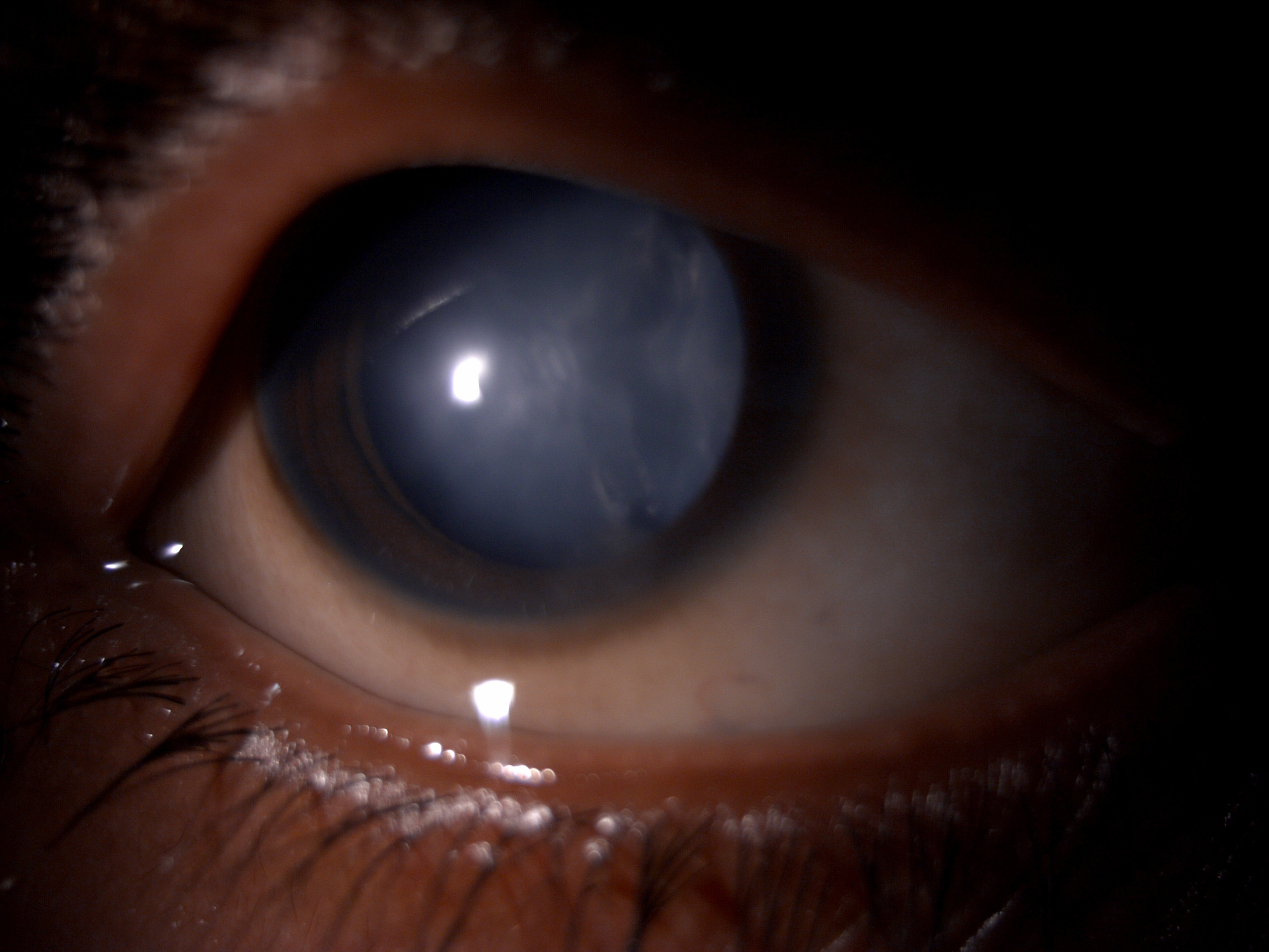
Charles Bonnet Syndrome. This image shows a cataractous lens in a patient with Charles Bonnet syndrome.
Contributed by B Gurnani, MD
References
Albqaeen W, Davies AC, Shim G. It Is All One-Sided: Monocular Blindness and Charles Bonnet Syndrome. Cureus. 2025 Sep:17(9):e92148. doi: 10.7759/cureus.92148. Epub 2025 Sep 12 [PubMed PMID: 41084641]
Christoph SEG, Boden KT, Siegel R, Seitz B, Szurman P, Schulz A. The prevalence of Charles-Bonnet syndrome in ophthalmic patients: A systematic review and meta-analysis. Brain research bulletin. 2025 Apr:223():111282. doi: 10.1016/j.brainresbull.2025.111282. Epub 2025 Mar 4 [PubMed PMID: 40049460]
Level 1 (high-level) evidenceBorsellino PJ, Schauer S, Rivas A, Aharonian K, Vida TA. Filling in the Blind Spot: Integrating Charles Bonnet Syndrome Screening in Ophthalmology. Clinical ophthalmology (Auckland, N.Z.). 2025:19():3619-3657. doi: 10.2147/OPTH.S547122. Epub 2025 Oct 3 [PubMed PMID: 41064591]
Patino CM, Ferreira JC. Inclusion and exclusion criteria in research studies: definitions and why they matter. Jornal brasileiro de pneumologia : publicacao oficial da Sociedade Brasileira de Pneumologia e Tisilogia. 2018 Apr:44(2):84. doi: 10.1590/s1806-37562018000000088. Epub [PubMed PMID: 29791550]
Pang L. Hallucinations Experienced by Visually Impaired: Charles Bonnet Syndrome. Optometry and vision science : official publication of the American Academy of Optometry. 2016 Dec:93(12):1466-1478 [PubMed PMID: 27529611]
El Haj M, Roche J, Jardri R, Kapogiannis D, Gallouj K, Antoine P. Clinical and neurocognitive aspects of hallucinations in Alzheimer's disease. Neuroscience and biobehavioral reviews. 2017 Dec:83():713-720. doi: 10.1016/j.neubiorev.2017.02.021. Epub 2017 Feb 21 [PubMed PMID: 28235545]
Jackson ML, Ferencz J. Cases: Charles Bonnet syndrome: visual loss and hallucinations. CMAJ : Canadian Medical Association journal = journal de l'Association medicale canadienne. 2009 Aug 4:181(3-4):175-6. doi: 10.1503/cmaj.090049. Epub [PubMed PMID: 19652169]
Level 3 (low-level) evidenceOzcan H, Yucel A, Ates O. Visual Hallucinations in an Old Patient after Cataract Surgery and Treatment. The Eurasian journal of medicine. 2016 Feb:48(1):62-4. doi: 10.5152/eurasianjmed.2015.14115. Epub [PubMed PMID: 27026767]
Teeple RC, Caplan JP, Stern TA. Visual hallucinations: differential diagnosis and treatment. Primary care companion to the Journal of clinical psychiatry. 2009:11(1):26-32 [PubMed PMID: 19333408]
Donovan NJ, Blazer D. Social Isolation and Loneliness in Older Adults: Review and Commentary of a National Academies Report. The American journal of geriatric psychiatry : official journal of the American Association for Geriatric Psychiatry. 2020 Dec:28(12):1233-1244. doi: 10.1016/j.jagp.2020.08.005. Epub 2020 Aug 19 [PubMed PMID: 32919873]
Level 3 (low-level) evidenceHusain MO, Chaudhry IB, Blakemore A, Shakoor S, Husain MA, Lane S, Kiran T, Jafri F, Memon R, Panagioti M, Husain N. Prevalence of depression and anxiety in patients with chronic obstructive pulmonary disease and their association with psychosocial outcomes: A cross-sectional study from Pakistan. SAGE open medicine. 2021:9():20503121211032813. doi: 10.1177/20503121211032813. Epub 2021 Oct 7 [PubMed PMID: 34659761]
Level 2 (mid-level) evidenceCinar N, Sahin S, Karsidag S. Eye-related visual hallucinations: consider 'Charles Bonnet syndrome'. Indian journal of ophthalmology. 2011 May-Jun:59(3):229-30. doi: 10.4103/0301-4738.81038. Epub [PubMed PMID: 21586846]
Level 3 (low-level) evidenceEkinci O. Successful treatment of Charles Bonnet syndrome after cerebellar stroke in a patient with binocular macular degeneration with low dose brexpiprazole: a case report. Acta neurologica Belgica. 2025 Dec:125(6):1681-1684. doi: 10.1007/s13760-025-02886-w. Epub 2025 Aug 31 [PubMed PMID: 40886207]
Level 3 (low-level) evidenceTsai SR, Lin WH. Terminal Glaucoma Mimicking Posterior Cerebral Artery Infarction: A Case Report. Cureus. 2025 Jul:17(7):e88358. doi: 10.7759/cureus.88358. Epub 2025 Jul 20 [PubMed PMID: 40837907]
Level 3 (low-level) evidenceBhat SP, Shipchandler A, Tokunaga C. Social Isolation as a Precipitating Factor for Charles Bonnet Syndrome in a Patient with Mild Visual Deterioration. Reports (MDPI). 2024 Aug 2:7(3):. doi: 10.3390/reports7030065. Epub 2024 Aug 2 [PubMed PMID: 40729288]
Jones L, Moosajee M. Charles Bonnet syndrome: taking another look at visual hallucinations in sight loss. Therapeutic advances in ophthalmology. 2025 Jan-Dec:17():25158414251359588. doi: 10.1177/25158414251359588. Epub 2025 Jul 29 [PubMed PMID: 40757182]
Level 3 (low-level) evidenceJahani S, Ardizi AE, Sahraian M. Charles Bonnet Syndrome (CBS) in a Patient With Secondary Progressive Multiple Sclerosis: A Case Report. Clinical case reports. 2025 Jul:13(7):e70588. doi: 10.1002/ccr3.70588. Epub 2025 Jul 6 [PubMed PMID: 40626174]
Level 3 (low-level) evidenceYun JS, Prieto G. Expanding the Spectrum of Charles Bonnet Syndrome: Severe Psychiatric Manifestations Associated With Total Vision Loss. Cureus. 2025 May:17(5):e84849. doi: 10.7759/cureus.84849. Epub 2025 May 26 [PubMed PMID: 40568259]
Valaparla VL, Bhattarai A, Karas PJ, Silveira DC. Coexistence of Charles Bonnet Syndrome and occipital epilepsy: A diagnostic challenge. Epilepsy & behavior reports. 2025 Jun:30():100764. doi: 10.1016/j.ebr.2025.100764. Epub 2025 Apr 3 [PubMed PMID: 40453184]
Mittal T, Haridas NJ. Charles Bonnet Syndrome in a Patient With Multiple Sclerosis: A Rare Presentation of Complex Visual Hallucinations Associated With Optic Neuritis. The primary care companion for CNS disorders. 2025 Jun 19:27(3):. pii: 24cr03865. doi: 10.4088/PCC.24cr03865. Epub 2025 Jun 19 [PubMed PMID: 40560825]
Teunisse RJ, Cruysberg JR, Hoefnagels WH, Verbeek AL, Zitman FG. Visual hallucinations in psychologically normal people: Charles Bonnet's syndrome. Lancet (London, England). 1996 Mar 23:347(9004):794-7 [PubMed PMID: 8622335]
Subedi B, Grossberg GT. Phantom limb pain: mechanisms and treatment approaches. Pain research and treatment. 2011:2011():864605. doi: 10.1155/2011/864605. Epub 2011 Aug 14 [PubMed PMID: 22110933]
Potts J. Response to Charles Bonnet Syndrome Special Collection. Therapeutic advances in ophthalmology. 2025 Jan-Dec:17():25158414251362980. doi: 10.1177/25158414251362980. Epub 2025 Aug 28 [PubMed PMID: 40893443]
Level 3 (low-level) evidenceNys J, Scheyltjens I, Arckens L. Visual system plasticity in mammals: the story of monocular enucleation-induced vision loss. Frontiers in systems neuroscience. 2015:9():60. doi: 10.3389/fnsys.2015.00060. Epub 2015 Apr 28 [PubMed PMID: 25972788]
Nan L, Yanbin H, Jingping Z. Acute Reversible Charles Bonnet Syndrome Following Eye Patch Placement. Neuro-ophthalmology (Aeolus Press). 2013:37(1):35-37. doi: 10.3109/01658107.2012.753913. Epub 2013 Jan 29 [PubMed PMID: 28179940]
Subhi Y, Schmidt DC, Bach-Holm D, Kolko M, Singh A. Prevalence of Charles Bonnet syndrome in patients with glaucoma: a systematic review with meta-analyses. Acta ophthalmologica. 2021 Mar:99(2):128-133. doi: 10.1111/aos.14567. Epub 2020 Aug 4 [PubMed PMID: 32749787]
Level 1 (high-level) evidenceYekta A, Hooshmand E, Saatchi M, Ostadimoghaddam H, Asharlous A, Taheri A, Khabazkhoob M. Global Prevalence and Causes of Visual Impairment and Blindness in Children: A Systematic Review and Meta-Analysis. Journal of current ophthalmology. 2022 Jan-Mar:34(1):1-15. doi: 10.4103/joco.joco_135_21. Epub 2022 Apr 16 [PubMed PMID: 35620376]
Level 1 (high-level) evidenceMenon GJ, Rahman I, Menon SJ, Dutton GN. Complex visual hallucinations in the visually impaired: the Charles Bonnet Syndrome. Survey of ophthalmology. 2003 Jan-Feb:48(1):58-72 [PubMed PMID: 12559327]
Level 3 (low-level) evidenceSchultz G, Melzack R. The Charles Bonnet syndrome: 'phantom visual images'. Perception. 1991:20(6):809-25 [PubMed PMID: 1816537]
Scott IU, Schein OD, Feuer WJ, Folstein MF. Visual hallucinations in patients with retinal disease. American journal of ophthalmology. 2001 May:131(5):590-8 [PubMed PMID: 11336933]
Level 2 (mid-level) evidenceNesher R, Nesher G, Epstein E, Assia E. Charles Bonnet syndrome in glaucoma patients with low vision. Journal of glaucoma. 2001 Oct:10(5):396-400 [PubMed PMID: 11711837]
Level 2 (mid-level) evidenceSchwartz TL, Vahgei L. Charles Bonnet syndrome in children. Journal of AAPOS : the official publication of the American Association for Pediatric Ophthalmology and Strabismus. 1998 Oct:2(5):310-3 [PubMed PMID: 10646756]
Klein R, Klein BE. The prevalence of age-related eye diseases and visual impairment in aging: current estimates. Investigative ophthalmology & visual science. 2013 Dec 13:54(14):ORSF5-ORSF13. doi: 10.1167/iovs.13-12789. Epub 2013 Dec 13 [PubMed PMID: 24335069]
Tian M, Zhang B. Identification of risk factors for the progression of age-related macular degeneration: a systematic review and meta-analysis of cohort studies. Frontiers in medicine. 2025:12():1544765. doi: 10.3389/fmed.2025.1544765. Epub 2025 Jul 23 [PubMed PMID: 40771483]
Level 1 (high-level) evidenceKhaled SM, Brederoo SG, Yehya A, Alabdulla M, Woodruff PW, Sommer IEC. Cross-cultural Differences in Hallucinations: A Comparison Between Middle Eastern and European Community-Based Samples. Schizophrenia bulletin. 2023 Feb 24:49(12 Suppl 2):S13-S24. doi: 10.1093/schbul/sbac086. Epub [PubMed PMID: 36840542]
Zhou C, Li S, Ye L, Chen C, Liu S, Yang H, Zhuang P, Liu Z, Jiang H, Han J, Jiang Y, Zhou L, Zhou X, Xiao J, Zhang C, Wen L, Lan C, Wang Y, Sun T, Jiang L, Xie P, Chen F, Liang G, Fu D, Zhang T, Shi X, Song Z, Liu X, Li S, Li P, Xu X, Wei Q, Wang W, Huang X, De Z, Deng A, Ding L, Pan X, Wen H, Zhang Z, Lv H, Zhang J, Tian X, Deng Z, Wang H, Wang F, Wang Y, Zhao H, Fang Y, Wu Y, Wu Y, Shen N, Li B, Li X, Dai H, Zhao N, Sun X, Zheng Z, Liu K, Xu X. Visual impairment and blindness caused by retinal diseases: A nationwide register-based study. Journal of global health. 2023 Nov 3:13():04126. doi: 10.7189/jogh.13.04126. Epub 2023 Nov 3 [PubMed PMID: 37921040]
O'Brien J, Taylor JP, Ballard C, Barker RA, Bradley C, Burns A, Collerton D, Dave S, Dudley R, Francis P, Gibbons A, Harris K, Lawrence V, Leroi I, McKeith I, Michaelides M, Naik C, O'Callaghan C, Olsen K, Onofrj M, Pinto R, Russell G, Swann P, Thomas A, Urwyler P, Weil RS, Ffytche D. Visual hallucinations in neurological and ophthalmological disease: pathophysiology and management. Journal of neurology, neurosurgery, and psychiatry. 2020 May:91(5):512-519. doi: 10.1136/jnnp-2019-322702. Epub 2020 Mar 25 [PubMed PMID: 32213570]
Cox TM, ffytche DH. Negative outcome Charles Bonnet syndrome. The British journal of ophthalmology. 2014 Sep:98(9):1236-9. doi: 10.1136/bjophthalmol-2014-304920. Epub 2014 May 13 [PubMed PMID: 24825847]
Vaphiades MS, Celesia GG, Brigell MG. Positive spontaneous visual phenomena limited to the hemianopic field in lesions of central visual pathways. Neurology. 1996 Aug:47(2):408-17 [PubMed PMID: 8757013]
Adachi N, Watanabe T, Matsuda H, Onuma T. Hyperperfusion in the lateral temporal cortex, the striatum and the thalamus during complex visual hallucinations: single photon emission computed tomography findings in patients with Charles Bonnet syndrome. Psychiatry and clinical neurosciences. 2000 Apr:54(2):157-62 [PubMed PMID: 10803809]
Baier B, de Haan B, Mueller N, Thoemke F, Birklein F, Dieterich M, Karnath HO. Anatomical correlate of positive spontaneous visual phenomena: a voxelwise lesion study. Neurology. 2010 Jan 19:74(3):218-22. doi: 10.1212/WNL.0b013e3181cb3e64. Epub [PubMed PMID: 20083797]
daSilva Morgan K, Collerton D, Firbank MJ, Schumacher J, Ffytche DH, Taylor JP. Visual cortical activity in Charles Bonnet syndrome: testing the deafferentation hypothesis. Journal of neurology. 2025 Feb 11:272(3):199. doi: 10.1007/s00415-024-12741-2. Epub 2025 Feb 11 [PubMed PMID: 39932561]
Mouchlianitis E, McCutcheon R, Howes OD. Brain-imaging studies of treatment-resistant schizophrenia: a systematic review. The lancet. Psychiatry. 2016 May:3(5):451-63. doi: 10.1016/S2215-0366(15)00540-4. Epub 2016 Mar 4 [PubMed PMID: 26948188]
Level 1 (high-level) evidenceAlex KD, Pehek EA. Pharmacologic mechanisms of serotonergic regulation of dopamine neurotransmission. Pharmacology & therapeutics. 2007 Feb:113(2):296-320 [PubMed PMID: 17049611]
Padungkiatsagul T, Leung LS, Moss HE. Retinal Diseases that Can Masquerade as Neurological Causes of Vision Loss. Current neurology and neuroscience reports. 2020 Sep 15:20(11):51. doi: 10.1007/s11910-020-01071-1. Epub 2020 Sep 15 [PubMed PMID: 32930896]
Mazade R, Alonso JM. Thalamocortical processing in vision. Visual neuroscience. 2017 Jan:34():E007. doi: 10.1017/S0952523817000049. Epub [PubMed PMID: 28965507]
Bai X, Edden RA, Gao F, Wang G, Wu L, Zhao B, Wang M, Chan Q, Chen W, Barker PB. Decreased γ-aminobutyric acid levels in the parietal region of patients with Alzheimer's disease. Journal of magnetic resonance imaging : JMRI. 2015 May:41(5):1326-31. doi: 10.1002/jmri.24665. Epub 2014 May 27 [PubMed PMID: 24863149]
Ko JH, Tang CC, Eidelberg D. Brain stimulation and functional imaging with fMRI and PET. Handbook of clinical neurology. 2013:116():77-95. doi: 10.1016/B978-0-444-53497-2.00008-5. Epub [PubMed PMID: 24112887]
Misch MR, Mitchell S, Francis PL, Sherborn K, Meradje K, McNeely AA, Honjo K, Zhao J, Scott CJ, Caldwell CB, Ehrlich L, Shammi P, MacIntosh BJ, Bilbao JM, Lang AE, Black SE, Masellis M. Differentiating between visual hallucination-free dementia with Lewy bodies and corticobasal syndrome on the basis of neuropsychology and perfusion single-photon emission computed tomography. Alzheimer's research & therapy. 2014:6(9):71. doi: 10.1186/s13195-014-0071-4. Epub 2014 Dec 5 [PubMed PMID: 25484929]
Zhang J, Waisbren E, Hashemi N, Lee AG. Visual hallucinations (Charles Bonnet syndrome) associated with neurosarcoidosis. Middle East African journal of ophthalmology. 2013 Oct-Dec:20(4):369-71. doi: 10.4103/0974-9233.119997. Epub [PubMed PMID: 24339694]
Level 3 (low-level) evidenceAhn SJ. Exploring neuropharmacokinetics: mechanisms, models, and clinical implications. Encephalitis (Seoul, Korea). 2025 Apr:5(2):36-52. doi: 10.47936/encephalitis.2024.00080. Epub 2025 Apr 8 [PubMed PMID: 40194559]
Alavijeh MS, Chishty M, Qaiser MZ, Palmer AM. Drug metabolism and pharmacokinetics, the blood-brain barrier, and central nervous system drug discovery. NeuroRx : the journal of the American Society for Experimental NeuroTherapeutics. 2005 Oct:2(4):554-71 [PubMed PMID: 16489365]
Pomares FB, Roy S, Funck T, Feier NA, Thiel A, Fitzcharles MA, Schweinhardt P. Upregulation of cortical GABAA receptor concentration in fibromyalgia. Pain. 2020 Jan:161(1):74-82. doi: 10.1097/j.pain.0000000000001707. Epub [PubMed PMID: 31569142]
Gurnani B, Kaur K. Post-market surveillance of ophthalmic implants: role of real-world evidence. Expert review of medical devices. 2025 Dec:22(12):1303-1308. doi: 10.1080/17434440.2025.2579578. Epub 2025 Oct 24 [PubMed PMID: 41123186]
Srinivas N, Maffuid K, Kashuba ADM. Clinical Pharmacokinetics and Pharmacodynamics of Drugs in the Central Nervous System. Clinical pharmacokinetics. 2018 Sep:57(9):1059-1074. doi: 10.1007/s40262-018-0632-y. Epub [PubMed PMID: 29464550]
Cogan DG. Visual hallucinations as release phenomena. Albrecht von Graefes Archiv fur klinische und experimentelle Ophthalmologie. Albrecht von Graefe's archive for clinical and experimental ophthalmology. 1973 Aug 23:188(2):139-50 [PubMed PMID: 4543235]
Burke W. The neural basis of Charles Bonnet hallucinations: a hypothesis. Journal of neurology, neurosurgery, and psychiatry. 2002 Nov:73(5):535-41 [PubMed PMID: 12397147]
Level 3 (low-level) evidenceBerrios GE, Brook P. The Charles Bonnet syndrome and the problem of visual perceptual disorders in the elderly. Age and ageing. 1982 Feb:11(1):17-23 [PubMed PMID: 7041567]
Level 3 (low-level) evidenceHolroyd S, Rabins PV, Finkelstein D, Nicholson MC, Chase GA, Wisniewski SC. Visual hallucinations in patients with macular degeneration. The American journal of psychiatry. 1992 Dec:149(12):1701-6 [PubMed PMID: 1443247]
Freiman TM, Surges R, Vougioukas VI, Hubbe U, Talazko J, Zentner J, Honegger J, Schulze-Bonhage A. Complex visual hallucinations (Charles Bonnet syndrome) in visual field defects following cerebral surgery. Report of four cases. Journal of neurosurgery. 2004 Nov:101(5):846-53 [PubMed PMID: 15540925]
Level 3 (low-level) evidenceFlint AC, Loh JP, Brust JC. Vivid visual hallucinations from occipital lobe infarction. Neurology. 2005 Sep 13:65(5):756 [PubMed PMID: 16157913]
Level 3 (low-level) evidenceLance JW. Simple formed hallucinations confined to the area of a specific visual field defect. Brain : a journal of neurology. 1976 Dec:99(4):719-34 [PubMed PMID: 828866]
Level 3 (low-level) evidenceLang UE, Stogowski D, Schulze D, Domula M, Schmidt E, Gallinat J, Tugtekin SM, Felber W. Charles Bonnet Syndrome: successful treatment of visual hallucinations due to vision loss with selective serotonin reuptake inhibitors. Journal of psychopharmacology (Oxford, England). 2007 Jul:21(5):553-5 [PubMed PMID: 17446204]
Level 3 (low-level) evidenceAlao AO, Hanrahan B. Charles Bonnet syndrome: visual hallucination and multiple sclerosis. International journal of psychiatry in medicine. 2003:33(2):195-9 [PubMed PMID: 12968834]
Level 3 (low-level) evidenceLepore FE. Spontaneous visual phenomena with visual loss: 104 patients with lesions of retinal and neural afferent pathways. Neurology. 1990 Mar:40(3 Pt 1):444-7 [PubMed PMID: 2314586]
Kölmel HW. Complex visual hallucinations in the hemianopic field. Journal of neurology, neurosurgery, and psychiatry. 1985 Jan:48(1):29-38 [PubMed PMID: 3973619]
Level 3 (low-level) evidenceRicard P. Vision loss and visual hallucinations: the Charles Bonnet syndrome. Community eye health. 2009 Mar:22(69):14 [PubMed PMID: 19506717]
Goldner JA. Metoprolol-induced visual hallucinations: a case series. Journal of medical case reports. 2012 Feb 15:6():65. doi: 10.1186/1752-1947-6-65. Epub 2012 Feb 15 [PubMed PMID: 22336000]
Level 2 (mid-level) evidenceDoane J, Stults B. Visual hallucinations related to angiotensin-converting enzyme inhibitor use: case reports and review. Journal of clinical hypertension (Greenwich, Conn.). 2013 Apr:15(4):230-3. doi: 10.1111/jch.12063. Epub 2013 Jan 25 [PubMed PMID: 23551721]
Level 3 (low-level) evidenceHigdon E, Twilla JD, Sands C. Moxifloxacin-Induced Visual Hallucinations: A Case Report and Review of the Literature. Journal of pharmacy practice. 2017 Jun:30(3):375-377. doi: 10.1177/0897190016637987. Epub 2016 Mar 21 [PubMed PMID: 27000139]
Level 3 (low-level) evidenceEssali N, Miller BJ. Psychosis as an adverse effect of antibiotics. Brain, behavior, & immunity - health. 2020 Dec:9():100148. doi: 10.1016/j.bbih.2020.100148. Epub 2020 Sep 19 [PubMed PMID: 34589893]
Padilla Peinado R, Esteban Fernández J, Rodríguez Álvarez S, Villa Albuguer T. Visual hallucinations related to use of ertapenem. Neurologia (Barcelona, Spain). 2015 Oct:30(8):520-1. doi: 10.1016/j.nrl.2014.02.008. Epub 2014 Mar 29 [PubMed PMID: 24684890]
Level 3 (low-level) evidenceCheng J, Paracha SS, Agrawal S, Wu Z, Sung CH. Recent Advances in Visual Dysfunction and Ocular Biomarkers in Neurological Disorders. Eye and brain. 2025:17():49-67. doi: 10.2147/EB.S516393. Epub 2025 Jun 14 [PubMed PMID: 40534708]
Level 3 (low-level) evidenceRaharja A, Whitefield L. Clinical approach to vision loss: a review for general physicians. Clinical medicine (London, England). 2022 Mar:22(2):95-99. doi: 10.7861/clinmed.2022-0057. Epub [PubMed PMID: 35304366]
Kaur K, Gurnani B. Contrast Sensitivity. StatPearls. 2026 Jan:(): [PubMed PMID: 35593849]
Shin HJ, Costello F. Imaging the optic nerve with optical coherence tomography. Eye (London, England). 2024 Aug:38(12):2365-2379. doi: 10.1038/s41433-024-03165-3. Epub 2024 Jul 3 [PubMed PMID: 38961147]
Prasad S. Diagnostic neuroimaging in neuro-ophthalmic disorders. Continuum (Minneapolis, Minn.). 2014 Aug:20(4 Neuro-ophthalmology):1023-62. doi: 10.1212/01.CON.0000453305.65851.1c. Epub [PubMed PMID: 25099107]
de Boer L, Poos JM, Van Den Berg E, De Houwer JFH, Swartenbroekx T, Dopper EGP, Boesjes P, Tahboun N, Bouzigues A, Foster PH, Ferry-Bolder E, Adams-Carr K, Russell LL, Convery RS, Rohrer JD, Seelaar H, Jiskoot LC. Montreal Cognitive Assessment vs the Mini-Mental State Examination as a Screening Tool for Patients With Genetic Frontotemporal Dementia. Neurology. 2025 Mar 11:104(5):e213401. doi: 10.1212/WNL.0000000000213401. Epub 2025 Feb 14 [PubMed PMID: 39951678]
Almutairi NM, Alahmadi S, Alharbi M, Gotah S, Alharbi M. The Association Between HbA1c and Other Biomarkers With the Prevalence and Severity of Diabetic Retinopathy. Cureus. 2021 Jan 6:13(1):e12520. doi: 10.7759/cureus.12520. Epub 2021 Jan 6 [PubMed PMID: 33564524]
Yang WH, Shao Y, Xu YW, Expert Workgroup of Guidelines on Clinical Research Evaluation of Artificial Intelligence in Ophthalmology (2023), Ophthalmic Imaging and Intelligent Medicine Branch of Chinese Medicine Education Association, Intelligent Medicine Committee of Chinese Medicine Education Association. Guidelines on clinical research evaluation of artificial intelligence in ophthalmology (2023). International journal of ophthalmology. 2023:16(9):1361-1372. doi: 10.18240/ijo.2023.09.02. Epub 2023 Sep 18 [PubMed PMID: 37724285]
Leopold IH. The phenylephrine saga--a drug dilemma. American journal of ophthalmology. 1978 Apr:85(4):572-4 [PubMed PMID: 655237]
Livingston G, Huntley J, Sommerlad A, Ames D, Ballard C, Banerjee S, Brayne C, Burns A, Cohen-Mansfield J, Cooper C, Costafreda SG, Dias A, Fox N, Gitlin LN, Howard R, Kales HC, Kivimäki M, Larson EB, Ogunniyi A, Orgeta V, Ritchie K, Rockwood K, Sampson EL, Samus Q, Schneider LS, Selbæk G, Teri L, Mukadam N. Dementia prevention, intervention, and care: 2020 report of the Lancet Commission. Lancet (London, England). 2020 Aug 8:396(10248):413-446. doi: 10.1016/S0140-6736(20)30367-6. Epub 2020 Jul 30 [PubMed PMID: 32738937]
Issa BA, Yussuf AD. Charles bonnet syndrome, management with simple behavioral technique. Journal of neurosciences in rural practice. 2013 Jan:4(1):63-5. doi: 10.4103/0976-3147.105618. Epub [PubMed PMID: 23546356]
Level 3 (low-level) evidenceSiatkowski RM, Zimmer B, Rosenberg PR. The Charles Bonnet syndrome. Visual perceptive dysfunction in sensory deprivation. Journal of clinical neuro-ophthalmology. 1990 Sep:10(3):215-8 [PubMed PMID: 2144541]
Level 3 (low-level) evidenceHolroyd S, Rabins PV. A three-year follow-up study of visual hallucinations in patients with macular degeneration. The Journal of nervous and mental disease. 1996 Mar:184(3):188-9 [PubMed PMID: 8600223]
Ukai S, Yamamoto M, Tanaka M, Takeda M. Treatment of typical Charles Bonnet syndrome with donepezil. International clinical psychopharmacology. 2004 Nov:19(6):355-7 [PubMed PMID: 15486523]
Level 3 (low-level) evidencedaSilva Morgan K, Schumacher J, Collerton D, Colloby S, Elder GJ, Olsen K, Ffytche DH, Taylor JP. Transcranial Direct Current Stimulation in the Treatment of Visual Hallucinations in Charles Bonnet Syndrome: A Randomized Placebo-Controlled Crossover Trial. Ophthalmology. 2022 Dec:129(12):1368-1379. doi: 10.1016/j.ophtha.2022.06.041. Epub 2022 Jul 9 [PubMed PMID: 35817197]
Level 1 (high-level) evidenceJones L, Ditzel-Finn L, Enoch J, Moosajee M. An overview of psychological and social factors in Charles Bonnet syndrome. Therapeutic advances in ophthalmology. 2021 Jan-Dec:13():25158414211034715. doi: 10.1177/25158414211034715. Epub 2021 Jul 30 [PubMed PMID: 34377938]
Level 3 (low-level) evidenceDas A, Babu GN, Gupta A, Kanaujia V, Paliwal VK. Vivid Visual Hallucinations in Visually Impaired: Charles Bonnet Syndrome - An Analog to "Phantom-Limb Phenomenon". Annals of Indian Academy of Neurology. 2020 Sep-Oct:23(5):734-735. doi: 10.4103/aian.AIAN_40_20. Epub 2020 Jun 5 [PubMed PMID: 33623292]
Kankanady Shivanand S. Review of Risk Factors, Pathophysiology, Management Principles, and Role of Medications. Case reports in psychiatry. 2024:2024():5456490. doi: 10.1155/2024/5456490. Epub 2024 Apr 9 [PubMed PMID: 38633731]
Level 3 (low-level) evidenceJones L, Jolly JK, Potts J, Callaghan T, Fisher K, Ip IB, Bridge H, Walker R, Moosajee M. From research to action: recommendations for Charles Bonnet syndrome care and policy. BMJ open ophthalmology. 2025 May 2:10(1):. doi: 10.1136/bmjophth-2024-002009. Epub 2025 May 2 [PubMed PMID: 40316414]
Sommer IE, Slotema CW, Daskalakis ZJ, Derks EM, Blom JD, van der Gaag M. The treatment of hallucinations in schizophrenia spectrum disorders. Schizophrenia bulletin. 2012 Jun:38(4):704-14. doi: 10.1093/schbul/sbs034. Epub 2012 Feb 24 [PubMed PMID: 22368234]
Raju NN, Kumar KSVRNP, Nihal G. Management of Medication-Induced Psychiatric Disorders. Indian journal of psychiatry. 2022 Mar:64(Suppl 2):S281-S291. doi: 10.4103/indianjpsychiatry.indianjpsychiatry_21_22. Epub 2022 Mar 23 [PubMed PMID: 35602361]
Boonstra FN, Bosch DGM, Geldof CJA, Stellingwerf C, Porro G. The Multidisciplinary Guidelines for Diagnosis and Referral in Cerebral Visual Impairment. Frontiers in human neuroscience. 2022:16():727565. doi: 10.3389/fnhum.2022.727565. Epub 2022 Jun 30 [PubMed PMID: 35845239]
Manera V, Rovini E, Wais P. Editorial: Early detection of neurodegenerative disorders using behavioral markers and new technologies: New methods and perspectives. Frontiers in aging neuroscience. 2023:15():1149886. doi: 10.3389/fnagi.2023.1149886. Epub 2023 Feb 28 [PubMed PMID: 36926633]
Level 3 (low-level) evidenceVarghese M, Kirpekar V, Loganathan S. Family Interventions: Basic Principles and Techniques. Indian journal of psychiatry. 2020 Jan:62(Suppl 2):S192-S200. doi: 10.4103/psychiatry.IndianJPsychiatry_770_19. Epub 2020 Jan 17 [PubMed PMID: 32055062]
van Nispen RM, Virgili G, Hoeben M, Langelaan M, Klevering J, Keunen JE, van Rens GH. Low vision rehabilitation for better quality of life in visually impaired adults. The Cochrane database of systematic reviews. 2020 Jan 27:1(1):CD006543. doi: 10.1002/14651858.CD006543.pub2. Epub 2020 Jan 27 [PubMed PMID: 31985055]
Level 1 (high-level) evidenceAbdollahi R. The Use of Non-Pharmacological Treatments Plus Drugs: A Promising Approach in the Management of Schizophrenic Patients. Iranian journal of psychiatry. 2025 Apr:20(2):142-144. doi: 10.18502/ijps.v20i2.18197. Epub [PubMed PMID: 40521281]
Nakao M, Shirotsuki K, Sugaya N. Cognitive-behavioral therapy for management of mental health and stress-related disorders: Recent advances in techniques and technologies. BioPsychoSocial medicine. 2021 Oct 3:15(1):16. doi: 10.1186/s13030-021-00219-w. Epub 2021 Oct 3 [PubMed PMID: 34602086]
Level 3 (low-level) evidenceHartney KE, Catalano G, Catalano MC. Charles Bonnet syndrome: are medications necessary? Journal of psychiatric practice. 2011 Mar:17(2):137-41. doi: 10.1097/01.pra.0000396067.87343.fb. Epub [PubMed PMID: 21430494]
Menon V, Ransing R, Praharaj SK. Management of Psychiatric Disorders in Patients with Hepatic and Gastrointestinal Diseases. Indian journal of psychiatry. 2022 Mar:64(Suppl 2):S379-S393. doi: 10.4103/indianjpsychiatry.indianjpsychiatry_18_22. Epub 2022 Mar 23 [PubMed PMID: 35602369]
Wieling W, Kaufmann H, Claydon VE, van Wijnen VK, Harms MPM, Juraschek SP, Thijs RD. Diagnosis and treatment of orthostatic hypotension. The Lancet. Neurology. 2022 Aug:21(8):735-746. doi: 10.1016/S1474-4422(22)00169-7. Epub [PubMed PMID: 35841911]
Lucas S, Ailani J, Smith TR, Abdrabboh A, Xue F, Navetta MS. Pharmacovigilance: reporting requirements throughout a product's lifecycle. Therapeutic advances in drug safety. 2022:13():20420986221125006. doi: 10.1177/20420986221125006. Epub 2022 Sep 27 [PubMed PMID: 36187302]
Level 3 (low-level) evidenceLevine AM. Visual hallucinations and cataracts. Ophthalmic surgery. 1980 Feb:11(2):95-8 [PubMed PMID: 7366949]
Level 3 (low-level) evidenceLapid MI, Burton MC, Chang MT, Rummans TA, Cha SS, Leavitt JA, Boeve BF. Clinical phenomenology and mortality in Charles Bonnet syndrome. Journal of geriatric psychiatry and neurology. 2013 Mar:26(1):3-9. doi: 10.1177/0891988712473800. Epub 2013 Feb 4 [PubMed PMID: 23385362]
Level 2 (mid-level) evidenceLee R, Wong TY, Sabanayagam C. Epidemiology of diabetic retinopathy, diabetic macular edema and related vision loss. Eye and vision (London, England). 2015:2():17. doi: 10.1186/s40662-015-0026-2. Epub 2015 Sep 30 [PubMed PMID: 26605370]
Cross N, van Steen C, Zegaoui Y, Satherley A, Angelillo L. Retinitis Pigmentosa: Burden of Disease and Current Unmet Needs. Clinical ophthalmology (Auckland, N.Z.). 2022:16():1993-2010. doi: 10.2147/OPTH.S365486. Epub 2022 Jun 20 [PubMed PMID: 35757022]
Kumar S, Soren S, Chaudhury S. Hallucinations: Etiology and clinical implications. Industrial psychiatry journal. 2009 Jul:18(2):119-26. doi: 10.4103/0972-6748.62273. Epub [PubMed PMID: 21180490]
Edinoff AN, Akuly HA, Hanna TA, Ochoa CO, Patti SJ, Ghaffar YA, Kaye AD, Viswanath O, Urits I, Boyer AG, Cornett EM, Kaye AM. Selective Serotonin Reuptake Inhibitors and Adverse Effects: A Narrative Review. Neurology international. 2021 Aug 5:13(3):387-401. doi: 10.3390/neurolint13030038. Epub 2021 Aug 5 [PubMed PMID: 34449705]
Level 3 (low-level) evidenceBeaulieu RA, Tamboli DA, Armstrong BK, Hogan RN, Mancini R. Reversible Charles Bonnet Syndrome After Oculoplastic Procedures. Journal of neuro-ophthalmology : the official journal of the North American Neuro-Ophthalmology Society. 2018 Sep:38(3):334-336. doi: 10.1097/WNO.0000000000000477. Epub [PubMed PMID: 27984353]
Willett SM, Maenner SK, Mayo JP. The perceptual consequences and neurophysiology of eye blinks. Frontiers in systems neuroscience. 2023:17():1242654. doi: 10.3389/fnsys.2023.1242654. Epub 2023 Aug 16 [PubMed PMID: 37654528]
Goldstein JE, Jackson ML, Fox SM, Deremeik JT, Massof RW, Low Vision Research Network Study Group. Clinically Meaningful Rehabilitation Outcomes of Low Vision Patients Served by Outpatient Clinical Centers. JAMA ophthalmology. 2015 Jul:133(7):762-9. doi: 10.1001/jamaophthalmol.2015.0693. Epub [PubMed PMID: 25856370]
Jan T, Del Castillo J. Visual hallucinations: charles bonnet syndrome. The western journal of emergency medicine. 2012 Dec:13(6):544-7. doi: 10.5811/westjem.2012.7.12891. Epub [PubMed PMID: 23357937]
Minakaran N, de Carvalho ER, Petzold A, Wong SH. Optical coherence tomography (OCT) in neuro-ophthalmology. Eye (London, England). 2021 Jan:35(1):17-32. doi: 10.1038/s41433-020-01288-x. Epub 2020 Nov 25 [PubMed PMID: 33239763]
Gurnani B, Kaur K. Comment on: Surgeon adoption of immediate sequential bilateral cataract surgery in the United States from 2018 to 2022. Journal of cataract and refractive surgery. 2025 Aug 1:51(8):747. doi: 10.1097/j.jcrs.0000000000001704. Epub [PubMed PMID: 41060140]
Level 3 (low-level) evidenceKaur K, Gurnani B. Low Vision Aids. StatPearls. 2026 Jan:(): [PubMed PMID: 36256771]
Gurnani B, Kaur K, Sivakumar P, Bhandari S. Retrospective analysis of low vision assistive products - A 6-year review. Saudi journal of ophthalmology : official journal of the Saudi Ophthalmological Society. 2023 Jan-Mar:37(1):32-37. doi: 10.4103/sjopt.sjopt_253_21. Epub 2023 Mar 9 [PubMed PMID: 36968774]
Level 2 (mid-level) evidencePerlmutter MS, Bhorade A, Gordon M, Hollingsworth H, Engsberg JE, Carolyn Baum M. Home lighting assessment for clients with low vision. The American journal of occupational therapy : official publication of the American Occupational Therapy Association. 2013 Nov-Dec:67(6):674-82. doi: 10.5014/ajot.2013.006692. Epub [PubMed PMID: 24195901]
Heinze N, Jones L. Social functioning in adults with visual impairment from minority ethnic communities in the United Kingdom. Frontiers in public health. 2024:12():1277472. doi: 10.3389/fpubh.2024.1277472. Epub 2024 Feb 1 [PubMed PMID: 38362219]
Wiggins MN, Landes RD, Bhaleeya SD, Uwaydat SH. Primary care physicians' knowledge of the ophthalmic effects of diabetes. Canadian journal of ophthalmology. Journal canadien d'ophtalmologie. 2013 Aug:48(4):265-8. doi: 10.1016/j.jcjo.2013.03.011. Epub [PubMed PMID: 23931464]
Ucan Gunduz G, Yalcinbayir O, Gelisken O. Awareness of Charles Bonnet syndrome among ophthalmologists: a survey study. International ophthalmology. 2025 May 26:45(1):207. doi: 10.1007/s10792-025-03575-6. Epub 2025 May 26 [PubMed PMID: 40418493]
Level 3 (low-level) evidenceShah P, Schwartz SG, Gartner S, Scott IU, Flynn HW Jr. Low vision services: a practical guide for the clinician. Therapeutic advances in ophthalmology. 2018 Jan-Dec:10():2515841418776264. doi: 10.1177/2515841418776264. Epub 2018 Jun 11 [PubMed PMID: 29998224]
Level 3 (low-level) evidence