 Tools
Tools
 Hearing Loss Assessment in Children
Introduction
Congenital and childhood hearing loss is among the most common sensory disorders, affecting approximately 1 to 3 per 1000 newborns, with prevalence increasing throughout childhood.[1][2][3] By adolescence, up to 20% of children demonstrate some degree of hearing loss. Early identification is essential because untreated hearing loss can adversely affect speech and language development, academic performance, vestibular function, psychosocial well-being, and overall quality of life, including increased risks of anxiety, depression, and low self-esteem.[4][5][6][7] Early auditory stimulation during critical periods of neuroplasticity is essential for optimal development of the cortical auditory pathways.[8]
Universal newborn hearing screening (UNHS) was endorsed by the National Institutes of Health in 1993 and subsequently formalized through successive Joint Committee on Infant Hearing guidelines position statements, most recently updated in 2019 [JEHDI. Year 2019 Position Statement: Principles and Guidelines for Early Hearing Detection and Intervention Programs]. Results from one study found that UNHS protocols resulted in 1 hearing loss diagnosis for every 811 infants screened without high-risk factors. One study found that using Universal Newborn Hearing Screening protocols resulted in one hearing loss diagnosis for every 811 babies screened without high-risk factors.[9][10] The established 1-3-6 benchmark advocates for screening by 1 month, diagnostic confirmation by 3 months, and intervention by 6 months.[11] The 2019 Joint Committee on Infant Hearing position statement recommends that programs meeting these targets strive for an accelerated 1-2-3 timeline.
Despite a national screening rate exceeding 98%, loss to follow-up remains a significant challenge.[12] Furthermore, the deployment of UNHS programs varies by setting and specific circumstances. Program implementation should account for local resource availability, screening accuracy, follow-up systems, and cost-effectiveness considerations. Screening protocols commonly employ automated auditory brainstem response, with implementation varying by institutional resources, patient population, and follow-up infrastructure. Results from one study showed that additional testing on day 7 of life was beneficial for determining the ideal timing of neonatal hearing screening using transient evoked otoacoustic emissions.[13]
Hearing loss may be unilateral or bilateral. Although unilateral hearing loss is frequently considered less problematic than bilateral hearing loss, more recent data have demonstrated its substantial clinical and educational significance. Symmetrical auditory input to both ears is essential for the development of binaural hearing pathways early in life.[14] Reduced binaural input impairs sound localization and speech perception in noisy environments through decrements in binaural squelch.[15] Children with unilateral hearing loss have a 10-fold higher risk of grade repetition than children with normal hearing (35% vs 3.5%, respectively). Up to 40% require additional educational assistance.[15][16] Unilateral hearing loss may also progress to bilateral loss, underscoring the need for ongoing surveillance.[17] Inner ear malformations, including enlarged vestibular aqueduct and Mondini spectrum anomalies, are commonly identified among children with congenital unilateral hearing loss.[18] Additional etiologies include genetic mutations, congenital cytomegalovirus infection, neonatal intensive care unit–related risk factors, ototoxic exposure, and other structural abnormalities.[19][20]
Anatomy and Physiology
Register For Free And Read The Full Article
 Search engine and full access to all medical articles
- 10 free questions in your specialty
- Free CME/CE Activities
- Free daily question in your email
- Save favorite articles to your dashboard
- Emails offering discounts
Learn more about a Subscription to StatPearls Point-of-Care
Anatomy and Physiology
Hearing requires coordinated structural and neural processes to convert a mechanical sound wave into an electrical stimulus that the brain can interpret. Because of differences in the surface area of the tympanic membrane and the stapes and the processes of ossicular coupling and acoustic coupling, sound pressure is amplified through the middle ear space such that the mechanical transmission of sound at the stapes is 82.5 times greater than at the tympanic membrane.[21] Middle ear amplification produces a total pressure gain of approximately 20 to 30 dB at the oval window, primarily due to the area ratio between the tympanic membrane and the stapes footplate and the ossicular lever mechanics.
The cochlea is the spiral-shaped inner ear structure responsible for hearing. The structure is divided into 3 fluid-filled compartments: the scala vestibuli (superior), scala media (cochlear duct), and scala tympani (inferior). See "Neuroanatomy, Ear," for further information. The scala vestibuli and scala media are separated by the vestibular (Reissner) membrane, while the basilar membrane separates the scala media and scala tympani. The scala vestibuli and scala tympani contain perilymph, a sodium-rich fluid similar to extracellular fluid. In contrast, the scala media contains potassium-rich endolymph, which is similar in composition to intracellular fluid and is essential for hair cell transduction. Within the scala media, the organ of Corti rests on the basilar membrane and contains inner and outer hair cells and supporting cells and is overlaid by the tectorial membrane, which interacts with hair cell stereocilia during sound transduction.
Transmission of mechanical energy through the middle ear results in movement of the stapes footplate at the oval window, generating pressure waves within the cochlear fluids.[22] The resulting pressure fluctuations produce a traveling wave along the basilar membrane, leading to frequency-dependent maximal displacement along the cochlear spiral. Shearing forces between the basilar membrane and the overlying tectorial membrane deflect the stereocilia of the organ of Corti, opening mechanically gated ion channels and depolarizing hair cells, which are tonotopically organized along the cochlea. Low-frequency sounds preferentially stimulate the apical cochlea, while high-frequency sounds stimulate the basal region. Place-based frequency organization forms the basis of cochlear tonotopy.[23]
Depolarization of inner hair cells triggers neurotransmitter release onto spiral ganglion neurons, generating action potentials that travel via the auditory (cochlear) nerve to the cochlear nucleus in the brainstem. Auditory information is then relayed bilaterally through the superior olivary complex, lateral lemniscus, and inferior colliculus, followed by transmission to the medial geniculate body of the thalamus and ultimately to the primary auditory cortex (the Heschl gyrus) in the superior temporal lobe.[24]
The 2 primary modalities used in newborn hearing screening are otoacoustic emissions (OAE) and automated auditory brainstem response.[25] OAEs are low-level acoustic signals generated by the electromotile activity of cochlear outer hair cells in response to sound stimulation and are recorded using a probe placed in the external auditory canal. The presence of OAEs reflects normal outer hair cell function and, indirectly, cochlear integrity.[26][27][28] Current newborn hearing screening programs follow the 1-3-6 benchmark: screening by 1 month of age, diagnostic confirmation by 3 months, and initiation of appropriate intervention by 6 months to optimize language and neurodevelopmental outcomes.[29]
The most commonly used OAE subtypes in newborn screening are transient evoked OAE and distortion product otoacoustic emissions. Transient evoked OAE are elicited using brief broadband stimuli and provide a global assessment of cochlear function across a wide frequency range; they are typically present by approximately 30 weeks of gestation. In contrast, distortion product OAE are generated by 2 simultaneous pure-tone stimuli and provide more frequency-specific information. However, they are generally less robust at lower frequencies (less than approximately 4 kHz) in clinical screening settings.[30][31][32]
The presence of OAEs is used in newborn hearing screening as an indicator of normal outer hair cell function and is designated as a pass result. In contrast, the absence of OAEs is classified as a refer result requiring further evaluation. An absent OAE may be associated with either conductive pathology, such as middle ear effusion, or sensorineural hearing loss, typically greater than mild to moderate levels (approximately 30 to 40 dB hearing level or greater). However, the presence of OAEs does not exclude hearing loss because OAEs assess only cochlear outer hair cell function and do not evaluate auditory nerve or central auditory pathway integrity.[33][34]
The auditory brainstem response (ABR) test evaluates neural conduction along the auditory pathway.[35] Scalp electrodes record time-locked electrical activity generated in response to acoustic stimuli, producing a series of waves reflecting activity along successive brainstem structures. Wave 1 originates from the distal auditory nerve, wave 2 from the proximal auditory nerve and cochlear nucleus region, wave 3 primarily reflects activity within the superior olivary complex, wave 4 primarily reflects activity from the lateral lemniscus, and wave 5 primarily reflects activity from the inferior colliculus. Prolonged latencies or reduced amplitudes in specific waves may indicate delayed neural conduction and help localize pathology along the auditory pathway. ABR testing is also used to estimate auditory thresholds and assess neural synchrony. See Image. Auditory Brainstem Response Test.
Auditory neuropathy spectrum disorder (ANSD) accounts for a subset of patients with abnormal or dyssynchronous ABR findings, including some with patterns of severe to profound hearing loss.[36] In ANSD, cochlear outer hair cell function is typically preserved, resulting in present OAEs in many patients, whereas neural transmission along the auditory pathway is impaired, leading to absent or markedly abnormal ABR responses at appropriate stimulus intensities. Diagnostic findings typically include preserved or near-normal OAEs with absent or severely abnormal ABR responses. ANSD may be unilateral or bilateral and is bilateral in most cases. Acoustic reflexes are often absent or elevated; however, this finding is variable and is not required for diagnosis.
Indications
All newborns should undergo universal hearing screening before hospital discharge as part of early hearing detection and intervention programs.[29] Infants who pass initial screening should continue routine surveillance of auditory, speech, and language development within the primary care medical home. Newborns who do not pass the initial screening should undergo repeat testing, ideally several hours after the initial assessment and, when feasible, before discharge.[11][37] Screening in well-baby nurseries is commonly performed using either OAE or automated auditory brainstem response, depending on institutional protocol.
Infants admitted to the neonatal intensive care unit, particularly those requiring care for 5 days or longer, should undergo automated auditory brainstem response screening because of their increased risk of neural hearing disorders, including ANSD.[38] Additional risk factors for hearing loss include hyperbilirubinemia requiring exchange transfusion, culture-proven sepsis, exposure to ototoxic medications, congenital infections, neonatal infections, craniofacial abnormalities, and syndromic conditions associated with hearing impairment.[3][18] Newborns requiring neonatal intensive care unit care have a substantially increased risk of hearing loss compared with infants cared for exclusively in the well-baby nursery.[39] Although the risk of ANSD in otherwise healthy newborns is low, infants with significant risk factors warrant more comprehensive physiologic screening.
Beyond the newborn period, referral for formal audiologic evaluation is indicated whenever concerns regarding hearing, speech and language development, or auditory responsiveness are raised by parents or caregivers, teachers, speech-language pathologists, or healthcare professionals.[40] Although parental concern may underestimate subtle hearing deficits, caregiver suspicion remains an important clinical indicator, with reported detection rates of approximately 61% among parents or guardians who suspect hearing loss.[40][41] Referral is also warranted for children demonstrating delayed speech or language acquisition, academic difficulties, recurrent otitis media, inattentiveness, or behavioral concerns suggestive of impaired auditory function.[42] Many children with speech delays appear physically healthy and show no other apparent anomalies or disabilities.[43][44] Differentiating typical developmental variations from speech delays that could benefit from early intervention can therefore be challenging for clinicians.[45]
Contraindications
Newborn and pediatric hearing screening has no absolute contraindications because these assessments are noninvasive and well-tolerated. However, certain clinical circumstances may limit the feasibility or interpretability of routine screening, including significant external auditory canal obstruction, medical instability, or congenital external ear malformations. Infants with congenital aural atresia, severe microtia, or other visible structural ear abnormalities may not be appropriate candidates for routine physiologic screening in the newborn nursery, as results may be unobtainable or predictably abnormal due to conductive pathway abnormalities.[46] Prompt referral for a comprehensive diagnostic audiologic evaluation is recommended for these infants and often includes auditory brainstem response testing and an otolaryngologic assessment to determine hearing status and guide early intervention.
Equipment
Audiologic evaluation requires access to equipment capable of performing physiologic and behavioral hearing assessments, including OAE, auditory brainstem response, tympanometry, acoustic reflex testing, and comprehensive age-appropriate audiometric testing. Selection of the appropriate testing modality depends on the child’s developmental age, cognitive ability, and capacity to participate reliably in behavioral assessment. Visual reinforcement audiometry is typically used for infants and young children, approximately 6 to 24 months of developmental age, who can sit upright independently and demonstrate sufficient head and neck control to localize an auditory stimulus. Independent sitting and sufficient head and neck control are generally achieved by approximately 6 months of corrected age.[47]
Conditioned play audiometry is most appropriate for children aged approximately 2 to 5 years who are conditioned to perform a play-based task in response to auditory stimuli. By approximately 5 years of age, conventional pure-tone audiometry is generally used, although younger children may undergo this testing if they can reliably complete structured behavioral testing. Additionally, acoustic reflex testing is commonly incorporated into the diagnostic evaluation of older children and adults to assess middle ear function and the integrity of the auditory brainstem pathway. However, clinicians use this testing less frequently in newborns and very young children due to developmental and testing limitations.
Personnel
The evaluation and treatment of children with hearing loss are best accomplished through an interdisciplinary team approach. The care team should include an audiologist experienced in pediatric audiologic assessment, an otolaryngologist, and a pediatrician to ensure comprehensive diagnostic evaluation, medical treatment, and longitudinal developmental surveillance. Genetic consultation and counseling should be considered when ANSD or a hereditary etiology is suspected because a substantial proportion of patients with ANSD have identifiable genetic abnormalities.[26] Comprehensive audiologic monitoring is also recommended for children with known genetic or systemic disorders associated with an increased risk of hearing loss, including sickle cell disease.[48] Children with nonsyndromic sensorineural hearing loss have a 2- to 3-fold increased risk of associated ocular abnormalities.[49] Accordingly, ophthalmologic evaluation should be considered as part of the comprehensive diagnostic evaluation to identify coexisting visual deficits that may further affect language acquisition and overall developmental outcomes.[50]
Preparation
Successful audiologic assessment depends on selecting developmentally appropriate testing methods and ensuring optimal patient preparation because unreliable participation may compromise test validity. For older infants and children undergoing behavioral audiometric assessment, cooperation can be optimized by ensuring that the child is well-rested, adequately fed, and comfortable during testing. Routine comfort measures, including diaper changes or other age-appropriate care immediately before testing, may further improve participation and the reliability of results.
For newborns undergoing nonsedated auditory brainstem response testing, preparation is particularly important to facilitate natural sleep during the examination. Infants should ideally arrive awake and hungry rather than be fed immediately beforehand. After the recording electrodes are placed and test preparation is completed, the infant should be fed and allowed to fall asleep naturally for the duration of the assessment. Natural sleep minimizes movement artifact, improves recording quality, and maximizes the likelihood of obtaining accurate and interpretable results.
Technique or Treatment
Several modalities are available for the screening and diagnostic evaluation of pediatric hearing loss. Universal newborn hearing screening is most commonly performed using otoacoustic emissions and automated auditory brainstem response testing. Infants and children who do not pass initial screening or who present with risk factors or clinical concern for hearing impairment typically undergo further diagnostic evaluation using tympanometry, ABR, age-appropriate behavioral audiometric testing, and conventional pure-tone audiometry when developmentally appropriate. Additional diagnostic studies, including imaging and genetic evaluation, may be indicated in selected patients to identify underlying structural or hereditary etiologies. The roles of these diagnostic modalities in the comprehensive assessment of pediatric hearing loss are discussed below.
Otoacoustic Emissions
A probe is placed within the child’s external auditory canal and functions as both a sound stimulus generator and a recording microphone. Standard screening protocols assess responses across multiple frequencies, typically ranging from 2000 to 5000 Hz. The two most commonly used screening modalities are transient-evoked otoacoustic emissions and distortion-product otoacoustic emissions, both of which are acceptable for newborn hearing screening. However, transient-evoked otoacoustic emissions are more commonly used in many screening programs. Test results are qualitative and reported as either a pass or refer result based on the presence or absence of reproducible OAE, which reflects the functional integrity of cochlear outer hair cells. A pass result suggests normal peripheral cochlear function, whereas a refer result indicates the need for repeat screening or further diagnostic evaluation.
Auditory Brainstem Response
Insert earphones are placed within the child’s external auditory canals, and surface electrodes are positioned on the scalp and mastoid or forehead to record electrophysiologic activity. For newborns and young infants undergoing nonsedated testing, feeding and routine comfort measures, such as diaper changes, are often used to facilitate natural sleep and minimize movement artifact during the examination. Acoustic stimuli, typically clicks or tone bursts delivered at varying intensities, are presented through the earphones. These stimuli evoke time-locked electrical responses along the auditory pathway, which are recorded by the electrodes and displayed as waveforms on the monitoring system. The resulting auditory brainstem response is characterized by waves 1 through 5, each corresponding to neural activity generated at successive anatomic levels of the auditory pathway, as described in the Anatomy and Physiology section.
Tympanometry
An immittance probe with a soft-sealing tip is placed in the external auditory canal of the test ear to create an airtight seal. During testing, air pressure within the ear canal is systematically varied while a probe tone is presented, allowing measurement of acoustic admittance of the tympanic membrane and middle ear system. These measurements assess middle ear mobility and pressure dynamics. The standard probe tone for tympanometry is 226 Hz and is routinely used in children older than 6 months. In infants younger than 6 months, a 1000 Hz probe tone is often preferred because it provides greater diagnostic accuracy due to the unique acoustic properties and mass-dominant middle-ear mechanics of early infancy.
A tympanogram typically reports ear canal volume (cm³), peak middle ear pressure (decapascals [daPa]), and static compliance (mL or mmho). A type A tympanogram reflects normal middle ear function, generally demonstrating peak pressure between −100 and +100 daPa with normal compliance values. Type Ad tympanograms indicate excessive compliance and may suggest ossicular discontinuity or a hypermobile tympanic membrane. In contrast, type A tympanograms demonstrate reduced compliance consistent with a stiffened middle ear system, such as may occur with otosclerosis or tympanosclerosis.
Type B tympanograms are flat, with no identifiable compliance peak. When accompanied by normal ear canal volume, this pattern typically suggests middle ear effusion; when associated with increased ear canal volume, it may indicate tympanic membrane perforation or a patent tympanostomy tube. Type C tympanograms show peak compliance at significantly negative pressures (typically < −100 daPa), suggesting negative middle ear pressure and Eustachian tube dysfunction.
Acoustic Reflex Testing
Acoustic reflex testing is not routinely performed in newborn hearing screening but may be included as part of a comprehensive immittance evaluation in selected pediatric or diagnostic evaluations. During testing, an immittance probe is placed in the test ear to measure changes in middle ear compliance. In contrast, acoustic stimuli are delivered either ipsilaterally or contralaterally via an earphone in the opposite ear. In neonates, a 1000 Hz probe tone is typically preferred for immittance measurements because of the acoustic characteristics of the infant's middle ear. In contrast, a 226 Hz probe tone is more commonly used in older infants and children.
Acoustic reflexes are elicited using pure-tone stimuli, commonly at 500, 1000, 2000, and 4000 Hz, beginning at moderate intensities and increasing in small increments until a stapedial muscle contraction is detected as a measurable change in middle ear compliance. Reflex thresholds are typically recorded in dB hearing level. Intensities greater than approximately 100 to 105 dB hearing level are generally avoided unless clinically indicated, such as when conductive hearing loss is suspected. To ensure reliability, responses are typically repeated or confirmed by rechecking at 5 dB increments. Acoustic reflex testing may be limited or uninterpretable in the presence of abnormal middle ear function, such as a type B tympanogram, due to impaired transmission of sound through the middle ear system.
Behavioral Audiometry
Between approximately 6 and 24 months of developmental age, children are typically assessed using visual reinforcement audiometry.[47] Successful testing requires that the child be able to sit independently with adequate head and neck control to localize an auditory stimulus. Testing may be performed using soundfield speakers or insert earphones, depending on the level of ear-specific information required.
During VRA, visual reinforcers are positioned approximately 90° to the left and right of the child. These reinforcers may include illuminated toys or video-based displays. Auditory stimuli of varying frequencies and intensities are presented, and when the child correctly orients toward the side from which the stimulus is presented, a visual reward is delivered. The conditioned response allows estimation of behavioral hearing thresholds.
For children aged approximately 2 to 5 years, conditioned play audiometry is the primary behavioral testing method. During conditioned play audiometry, auditory stimuli are delivered via headphones or insert earphones across a range of frequencies and intensities. The child is conditioned to perform a specific play-based task in response to hearing a sound, such as placing a block in a bucket, stacking a ring, or completing a simple game activity. Responses are reinforced by positive feedback, enabling the systematic determination of frequency-specific hearing thresholds in each ear.
Pure-tone Audiometry
Pure-tone audiometry is considered the gold standard behavioral method for assessing hearing thresholds in cooperative patients. Pure-tone audiometry is typically feasible in children aged years of age or older, although successful testing may be possible earlier, depending on developmental readiness and attention span. During testing, headphones or insert earphones are placed over or within the patient’s ears to deliver pure-tone stimuli at standardized frequencies and varying intensities to each ear independently. The patient is instructed to provide a consistent behavioral response when a tone is detected, commonly by pressing a response button. In pediatric populations, alternative response methods such as raising a hand, repeating a word, or clapping may be used depending on developmental level and task comprehension. Air conduction thresholds are obtained across standard test frequencies, and bone conduction testing may be performed when indicated to differentiate conductive from sensorineural hearing loss. Thresholds are determined using a descending-intensity technique until the lowest level of reliable response is established for each frequency in each ear.
Imaging
Both CT and MRI may be used to evaluate pediatric hearing loss, depending on the clinical scenario. However, current evidence and expert consensus generally do not support routine imaging in children with bilateral sensorineural hearing loss, particularly when no additional concerning clinical features are present.[26] Imaging is more commonly indicated for unilateral hearing loss or ANSD; these studies may help identify structural abnormalities in the cochlea, vestibular system, or auditory nerve. In unilateral hearing loss, the diagnostic yield of clinically significant findings has been reported to be approximately 37% for CT and 35% for MRI.[51] MRI is particularly important in the preoperative evaluation of children being considered for cochlear implants, as it allows assessment of cochlear nerve integrity and inner ear anatomy. Findings such as cochlear nerve aplasia or severe cochlear nerve hypoplasia may preclude candidacy for cochlear implants or significantly alter it, and may influence surgical planning and expected outcomes.
Genetic and Laboratory Testing
Patients with bilateral sensorineural hearing loss or ANSD should be considered for genetic evaluation, particularly in patients with congenital onset, early identification, or clinical suspicion of an inherited etiology. Genetic causes account for approximately 50% to 60% of congenital hearing loss, with the majority being nonsyndromic.[52] The most common nonsyndromic genetic cause is a mutation in the gap junction protein β 2 gene (GJB2) (connexin 26), which plays a central role in cochlear gap junction function.[53]
Syndromic forms of genetic hearing loss may follow autosomal dominant or autosomal recessive inheritance patterns. Common autosomal dominant syndromic causes include Waardenburg syndrome, branchio-oto-renal syndrome, and neurofibromatosis type 2. Common autosomal recessive syndromic causes include Usher syndrome, Pendred syndrome, and Jervell and Lange-Nielsen syndrome. Genetic testing provides a high diagnostic yield in appropriately selected patients and is second only to comprehensive audiometric evaluation in identifying an etiology for bilateral sensorineural hearing loss. In contrast, isolated asymmetric hearing loss is less commonly genetic in origin, although genetic etiologies should still be considered in selected patients based on clinical context and associated findings.
Approximately half of congenital hearing loss is attributed to nongenetic causes, with congenital cytomegalovirus (CMV) infection as the most common identifiable infectious etiology. CMV accounts for a significant proportion of congenital or early-onset hearing loss, with estimates varying by population. Diagnostic testing is most accurate within the first 3 weeks of life when using saliva or urine polymerase chain reaction testing. After this period, confirmation typically relies on retrospective testing of dried blood spot samples. Treatment of congenital CMV depends on clinical presentation. Antiviral therapy with valganciclovir is currently used in selected symptomatic patients and has been associated with improved audiologic and neurodevelopmental outcomes in results from some studies; however, its use in isolated asymptomatic hearing loss remains an area of ongoing research and is not universally approved for this indication.[54]
In Summary
Pediatric hearing evaluation involves a combination of physiologic, behavioral, and diagnostic techniques selected according to the child’s developmental age and clinical context. Universal newborn hearing screening is most commonly performed using OAE and AABR, which assess cochlear outer hair cell function and neural synchrony of the auditory pathway, respectively. Infants who do not pass screening or who present with risk factors undergo further diagnostic evaluation, including tympanometry, acoustic reflex testing, and ABR, to localize potential conductive or sensorineural pathology and estimate hearing thresholds. With increasing developmental maturity, behavioral testing becomes central to assessment, including visual reinforcement audiometry in infants approximately 6 to 24 months of age, conditioned play audiometry in young children and preschool-aged children, and conventional pure-tone audiometry in children typically older than 5 years who can provide reliable responses. These behavioral methods are complemented by immittance testing and physiologic measures to characterize middle ear function and cochlear integrity. In selected patients, imaging and genetic testing are incorporated to identify structural or hereditary etiologies of hearing loss and to guide prognosis and intervention planning. Together, these modalities provide a comprehensive framework for early detection, diagnosis, and treatment of pediatric hearing impairment.
Complications
Audiologic assessment in children is generally safe and noninvasive, with no significant inherent procedural risks associated with most testing modalities. The primary limitations of pediatric hearing evaluation concern test accuracy, particularly the risk of false-positive and, more importantly, false-negative results. False-positive findings may lead to additional diagnostic testing and contribute to caregiver anxiety; however, they do not typically result in long-term harm. In contrast, false-negative results may delay the diagnosis of hearing loss, with potential downstream effects on speech, language, cognitive, and psychosocial development.
True procedural complications are rare and generally limited to minor, transient issues associated with instrumentation of the external auditory canal, such as mild irritation or, in very uncommon cases, tympanic membrane trauma. Such risks are minimal and primarily associated with probe or earphone placement rather than the physiologic testing itself. Patient-related factors, including behavioral variability, limited cooperation, or developmental delay, may reduce test reliability and increase the likelihood of inaccurate or incomplete results. When these factors affect testing, repeat testing or alternative assessment strategies may be required to ensure diagnostic accuracy.
Clinical Significance
Significant adverse developmental, linguistic, and psychosocial effects of untreated hearing loss have been well documented, particularly when diagnosis and intervention are delayed. When identified early, timely intervention can substantially improve auditory access and support speech and language development during critical periods of neuroplasticity. Primary rehabilitative options include conventional air-conduction hearing aids, bone-anchored hearing systems, typically fitted with a softband in children younger than 5 years of age, contralateral routing of signal devices for unilateral hearing loss, and remote microphone and frequency modulation systems to improve the signal-to-noise ratio in educational settings.[55]
Children with severe to profound sensorineural hearing loss should be evaluated for cochlear implants.[18] Hearing aids can provide meaningful amplification and may improve auditory thresholds by approximately 20 to 30 dB in appropriately selected patients; however, their effectiveness is limited in severe to profound hearing loss due to insufficient amplification of speech frequencies.[56] In contrast, results from studies have shown that cochlear implants significantly improve speech perception outcomes, with many pediatric recipients achieving open-set word recognition and substantial gains in functional hearing. Outcomes are highly variable and influenced by factors such as age at implantation, duration of deafness, and the presence of additional disabilities. Cochlear implants can result in hearing thresholds similar to those of control patients, with word reception scores of 85%.[57] Reduced developmental delays have also been reported in patients with cochlear implantation, with 95% of implanted patients not requiring learning adaptations or full-time educational support.[54]
Delayed diagnosis or failure to complete follow-up after abnormal newborn screening can result in missed opportunities during periods of heightened neural plasticity.[58] Early auditory stimulation through hearing aids or cochlear implantation has been associated with measurable changes in cortical auditory pathway development and improved language outcomes. Despite advances in universal newborn hearing screening programs, loss to follow-up remains a significant challenge, with national data indicating that more than 1 in 4 infants with referred screening results may not complete diagnostic evaluation in a timely manner.[59]
Although imaging and genetic testing may provide important etiologic information in selected patients, they are not indicated in all cases of pediatric hearing loss and should be guided by clinical context. With adherence to Early Hearing Detection and Intervention, [CDC. EHDI 1-3-6 Benchmarks] screening by 1 month, diagnostic confirmation by 3 months, and intervention by 6 months. Many children with hearing loss can achieve favorable communication and developmental outcomes. In regions with effective screening programs, the average age at diagnosis has decreased from approximately 2.5 years to between 3 and 6 months, reflecting significant improvements in early detection.[60]
Enhancing Healthcare Team Outcomes
The evaluation and treatment of pediatric hearing loss require a structured, interprofessional, and patient-centered approach involving clinicians, nurses, audiologists, pharmacists, speech-language pathologists, genetic counselors, and otolaryngologists. Effective care depends on timely communication and coordinated decision-making across disciplines to ensure early identification, diagnostic confirmation, and rapid initiation of appropriate interventions. Clinicians are responsible for clinical recognition, referral, and medical treatment, while audiologists perform and interpret diagnostic and behavioral hearing assessments. Nurses play a key role in care coordination, patient and caregiver education, and ensuring adherence to follow-up testing and intervention schedules. Pharmacists may contribute to medication safety monitoring, particularly in patients receiving ototoxic agents or antiviral therapies such as valganciclovir.
Clinicians have an ethical responsibility to minimize delays in diagnosis and treatment because untreated hearing loss during critical periods of neurodevelopment can result in irreversible deficits in speech, language, and cognitive development. Shared decision-making with caregivers is essential to align intervention strategies with family goals, cultural considerations, and long-term developmental planning. Interprofessional communication through structured referral pathways, shared documentation systems, and standardized screening protocols is critical for reducing loss to follow-up and improving continuity of care. A coordinated, team-based model enhances patient safety, improves developmental outcomes, and supports equitable access to early hearing detection and intervention services.
Media
(Click Image to Enlarge)
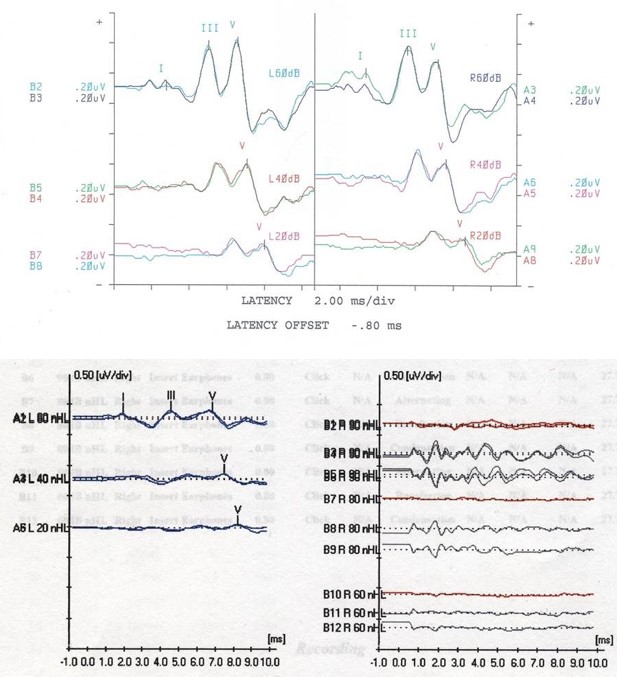
Auditory Brainstem Response Test. A normal ABR in the top figure with prominent peaks was seen from testing in both ears. The bottom figure demonstrates classic findings in a child with auditory neuropathy, with diminished or absent ABR peaks
Contributed by J Sommerfeldt, MD
References
Butcher E, Dezateux C, Cortina-Borja M, Knowles RL. Prevalence of permanent childhood hearing loss detected at the universal newborn hearing screen: Systematic review and meta-analysis. PloS one. 2019:14(7):e0219600. doi: 10.1371/journal.pone.0219600. Epub 2019 Jul 11 [PubMed PMID: 31295316]
Level 1 (high-level) evidenceGustafson SJ, Corbin NE. Pediatric Hearing Loss Guidelines and Consensus Statements-Where Do We Stand? Otolaryngologic clinics of North America. 2021 Dec:54(6):1129-1142. doi: 10.1016/j.otc.2021.07.003. Epub 2021 Sep 15 [PubMed PMID: 34535279]
Level 3 (low-level) evidenceFarinetti A, Raji A, Wu H, Wanna B, Vincent C. International consensus (ICON) on audiological assessment of hearing loss in children. European annals of otorhinolaryngology, head and neck diseases. 2018 Feb:135(1S):S41-S48. doi: 10.1016/j.anorl.2017.12.008. Epub 2018 Feb 1 [PubMed PMID: 29366866]
Level 3 (low-level) evidenceLieu JEC, Kenna M, Anne S, Davidson L. Hearing Loss in Children: A Review. JAMA. 2020 Dec 1:324(21):2195-2205. doi: 10.1001/jama.2020.17647. Epub [PubMed PMID: 33258894]
Idstad M, Tambs K, Aarhus L, Engdahl BL. Childhood sensorineural hearing loss and adult mental health up to 43 years later: results from the HUNT study. BMC public health. 2019 Feb 8:19(1):168. doi: 10.1186/s12889-019-6449-2. Epub 2019 Feb 8 [PubMed PMID: 30736854]
Ronner EA, Benchetrit L, Levesque P, Basonbul RA, Cohen MS. Quality of Life in Children with Sensorineural Hearing Loss. Otolaryngology--head and neck surgery : official journal of American Academy of Otolaryngology-Head and Neck Surgery. 2020 Jan:162(1):129-136. doi: 10.1177/0194599819886122. Epub 2019 Nov 5 [PubMed PMID: 31684823]
Level 2 (mid-level) evidenceEiserman WD, Shisler L. Identifying Children With Hearing Loss: The Key Role of School Nurses in Evidence-Based Screenings. NASN school nurse (Print). 2025 Jan:40(1):24-27. doi: 10.1177/1942602X241268715. Epub 2024 Aug 31 [PubMed PMID: 39215607]
Persic D, Thomas ME, Pelekanos V, Ryugo DK, Takesian AE, Krumbholz K, Pyott SJ. Regulation of auditory plasticity during critical periods and following hearing loss. Hearing research. 2020 Nov:397():107976. doi: 10.1016/j.heares.2020.107976. Epub 2020 Apr 20 [PubMed PMID: 32591097]
Connolly JL, Carron JD, Roark SD. Universal newborn hearing screening: are we achieving the Joint Committee on Infant Hearing (JCIH) objectives? The Laryngoscope. 2005 Feb:115(2):232-6 [PubMed PMID: 15689741]
Patel H, Feldman M. Universal newborn hearing screening. Paediatrics & child health. 2011 May:16(5):301-10 [PubMed PMID: 22547950]
American Academy of Pediatrics, Joint Committee on Infant Hearing. Year 2007 position statement: Principles and guidelines for early hearing detection and intervention programs. Pediatrics. 2007 Oct:120(4):898-921 [PubMed PMID: 17908777]
Johnson E, Ronis S. Measuring Hearing, Vision, and Developmental Screening and Follow-Up: Obstacles and Opportunities. Pediatrics. 2026 Jun 1:157(6):. pii: e2025075798. doi: 10.1542/peds.2025-075798. Epub [PubMed PMID: 42161373]
Feroz S, Irshad M, Shamsi SA, Sohail E, Hamid A, Rohan M. Evaluation of optimal postnatal timing for the screening of neonatal hearing via transient evoked oto-acoustic emission (TEOAE): a retrospective cross-sectional study. Irish journal of medical science. 2026 Apr:195(2):961-968. doi: 10.1007/s11845-025-04238-2. Epub 2025 Dec 23 [PubMed PMID: 41432879]
Level 2 (mid-level) evidenceMoore DR. Anatomy and physiology of binaural hearing. Audiology : official organ of the International Society of Audiology. 1991:30(3):125-34 [PubMed PMID: 1953442]
Lieu JE, Tye-Murray N, Fu Q. Longitudinal study of children with unilateral hearing loss. The Laryngoscope. 2012 Sep:122(9):2088-95. doi: 10.1002/lary.23454. Epub 2012 Aug 1 [PubMed PMID: 22865630]
Level 2 (mid-level) evidenceLieu JE. Speech-language and educational consequences of unilateral hearing loss in children. Archives of otolaryngology--head & neck surgery. 2004 May:130(5):524-30 [PubMed PMID: 15148171]
Malesci R, Laria C, Freda G, Vecchio VD, Mallardo A, Serra N, Auletta G, Fetoni AR. Hearing Outcomes in Children with Unilateral Hearing Loss. The Benefits of Rehabilitative Strategies: Preliminary Results. Audiology research. 2025 Apr 2:15(2):. doi: 10.3390/audiolres15020037. Epub 2025 Apr 2 [PubMed PMID: 40277582]
Bagatto M, DesGeorges J, King A, Kitterick P, Laurnagaray D, Lewis D, Roush P, Sladen DP, Tharpe AM. Consensus practice parameter: audiological assessment and management of unilateral hearing loss in children. International journal of audiology. 2019 Dec:58(12):805-815. doi: 10.1080/14992027.2019.1654620. Epub 2019 Sep 5 [PubMed PMID: 31486692]
Level 3 (low-level) evidenceSingh S, Maheshwari A, Boppana S. CMV-induced Hearing Loss. Newborn (Clarksville, Md.). 2023 Oct-Dec:2(4):249-262. doi: 10.5005/jp-journals-11002-0081. Epub 2024 Jan 5 [PubMed PMID: 38348106]
Singh NK, Kumar N, Hage N, Ramamourthy B, Nayani D, Srinivasan D, Haokip M. Neonatal Hearing Loss: Mechanisms, Clinical Implications, and Global Perspectives. NeoReviews. 2026 May 1:27(5):e263-e278. doi: 10.1542/neo.27-5-009. Epub [PubMed PMID: 42061897]
Level 3 (low-level) evidencePickles JO. Auditory pathways: anatomy and physiology. Handbook of clinical neurology. 2015:129():3-25. doi: 10.1016/B978-0-444-62630-1.00001-9. Epub [PubMed PMID: 25726260]
Level 3 (low-level) evidenceUgarteburu M, Withnell RH, Cardoso L, Carriero A, Richter CP. Mammalian middle ear mechanics: A review. Frontiers in bioengineering and biotechnology. 2022:10():983510. doi: 10.3389/fbioe.2022.983510. Epub 2022 Oct 10 [PubMed PMID: 36299283]
Ruben RJ. The Developing Concept of Tonotopic Organization of the Inner Ear. Journal of the Association for Research in Otolaryngology : JARO. 2020 Feb:21(1):1-20. doi: 10.1007/s10162-019-00741-3. Epub 2020 Feb 4 [PubMed PMID: 32020418]
Vincent PFY, Young ED, Edge ASB, Glowatzki E. Auditory Hair Cells and Spiral Ganglion Neurons Regenerate Synapses with Refined Release Properties In Vitro. bioRxiv : the preprint server for biology. 2023 Dec 2:():. pii: 2023.10.05.561095. doi: 10.1101/2023.10.05.561095. Epub 2023 Dec 2 [PubMed PMID: 38076928]
Ngui LX, Tang IP, Prepageran N, Lai ZW. Comparison of distortion product otoacoustic emission (DPOAE) and automated auditory brainstem response (AABR) for neonatal hearing screening in a hospital with high delivery rate. International journal of pediatric otorhinolaryngology. 2019 May:120():184-188. doi: 10.1016/j.ijporl.2019.02.045. Epub 2019 Feb 27 [PubMed PMID: 30844634]
Liming BJ, Carter J, Cheng A, Choo D, Curotta J, Carvalho D, Germiller JA, Hone S, Kenna MA, Loundon N, Preciado D, Schilder A, Reilly BJ, Roman S, Strychowsky J, Triglia JM, Young N, Smith RJ. International Pediatric Otolaryngology Group (IPOG) consensus recommendations: Hearing loss in the pediatric patient. International journal of pediatric otorhinolaryngology. 2016 Nov:90():251-258. doi: 10.1016/j.ijporl.2016.09.016. Epub 2016 Sep 15 [PubMed PMID: 27729144]
Level 3 (low-level) evidenceVerstappen G, Foulon I, Van den Houte K, Heuninck E, Van Overmeire B, Gordts F, Topsakal V. Analysis of congenital hearing loss after neonatal hearing screening. Frontiers in pediatrics. 2023:11():1153123. doi: 10.3389/fped.2023.1153123. Epub 2023 May 15 [PubMed PMID: 37255573]
Mehl AL, Thomson V. The Colorado newborn hearing screening project, 1992-1999: on the threshold of effective population-based universal newborn hearing screening. Pediatrics. 2002 Jan:109(1):E7 [PubMed PMID: 11773575]
Deng X, Ema S, Mason C, Nash A, Carbone E, Gaffney M. Receipt and Timeliness of Newborn Hearing Screening and Diagnostic Services Among Babies Born in 2017 in 9 States. Journal of public health management and practice : JPHMP. 2022 Jan-Feb 01:28(1):E100-E108. doi: 10.1097/PHH.0000000000001232. Epub [PubMed PMID: 32956290]
Kanji A, Khoza-Shangase K, Moroe N. Newborn hearing screening protocols and their outcomes: A systematic review. International journal of pediatric otorhinolaryngology. 2018 Dec:115():104-109. doi: 10.1016/j.ijporl.2018.09.026. Epub 2018 Sep 25 [PubMed PMID: 30368368]
Level 1 (high-level) evidenceManz K, Nennstiel U, Marzi C, Mansmann U, Brockow I. Quality measures of two-stage newborn hearing screening: systematic review and meta-analysis. Frontiers in public health. 2025:13():1566478. doi: 10.3389/fpubh.2025.1566478. Epub 2025 Apr 16 [PubMed PMID: 40308920]
Level 1 (high-level) evidenceMackey AR, Bussé AML, Del Vecchio V, Mäki-Torkko E, Uhlén IM. Protocol and programme factors associated with referral and loss to follow-up from newborn hearing screening: a systematic review. BMC pediatrics. 2022 Aug 5:22(1):473. doi: 10.1186/s12887-022-03218-0. Epub 2022 Aug 5 [PubMed PMID: 35932008]
Level 1 (high-level) evidenceY S, R G, Y D, Bz J, S K, V N, M K. Predicting hearing loss in children according to the referrer and referral cause. International journal of pediatric otorhinolaryngology. 2020 Jan:128():109685. doi: 10.1016/j.ijporl.2019.109685. Epub 2019 Sep 15 [PubMed PMID: 31610440]
Fabus R, Berg AL, Serpanos YC, Cooley Hidecker MJ. The Effectiveness of Parental Questionnaires in the Assessment of Speech-Language and Auditory Function in Children. Folia phoniatrica et logopaedica : official organ of the International Association of Logopedics and Phoniatrics (IALP). 2017:69(5-6):261-270. doi: 10.1159/000488054. Epub 2018 May 7 [PubMed PMID: 29734179]
Young A, Cornejo J, Spinner A. Auditory Brainstem Response. StatPearls. 2026 Jan:(): [PubMed PMID: 33231991]
De Siati RD, Rosenzweig F, Gersdorff G, Gregoire A, Rombaux P, Deggouj N. Auditory Neuropathy Spectrum Disorders: From Diagnosis to Treatment: Literature Review and Case Reports. Journal of clinical medicine. 2020 Apr 10:9(4):. doi: 10.3390/jcm9041074. Epub 2020 Apr 10 [PubMed PMID: 32290039]
Level 3 (low-level) evidenceChiong CM. Newborn Hearing Screening and Beyond: A Continuing Journey in the Philippines. Acta medica Philippina. 2023:57(9):7-14. doi: 10.47895/amp.v57i9.8836. Epub 2023 Sep 27 [PubMed PMID: 39483801]
Colella-Santos MF, Hein TA, de Souza GL, do Amaral MI, Casali RL. Newborn hearing screening and early diagnostic in the NICU. BioMed research international. 2014:2014():845308. doi: 10.1155/2014/845308. Epub 2014 Jun 9 [PubMed PMID: 24999481]
Pourarian S, Khademi B, Pishva N, Jamali A. Prevalence of hearing loss in newborns admitted to neonatal intensive care unit. Iranian journal of otorhinolaryngology. 2012 Summer:24(68):129-34 [PubMed PMID: 24303398]
Reynolds G, Werfel KL, Vachio M, Lund EA. Early Experiences of Parents of Children who are Deaf or Hard of Hearing: Navigating through Identification, Intervention, and Beyond. Journal of early hearing detection and intervention. 2023:8(1):56-68. doi: 10.26077/6d9d-06f3. Epub [PubMed PMID: 39193584]
Swierniak W, Gos E, Skarzynski PH, Czajka N, Skarzynski H. The accuracy of parental suspicion of hearing loss in children. International journal of pediatric otorhinolaryngology. 2021 Feb:141():110552. doi: 10.1016/j.ijporl.2020.110552. Epub 2020 Dec 11 [PubMed PMID: 33341715]
Liang WHK, Gn LWE, Tan YCD, Tan GH. Speech and language delay in children: a practical framework for primary care physicians. Singapore medical journal. 2023 Dec:64(12):745-750. doi: 10.4103/singaporemedj.SMJ-2022-051. Epub [PubMed PMID: 38047330]
Miniscalco C, Nygren G, Hagberg B, Kadesjö B, Gillberg C. Neuropsychiatric and neurodevelopmental outcome of children at age 6 and 7 years who screened positive for language problems at 30 months. Developmental medicine and child neurology. 2006 May:48(5):361-6 [PubMed PMID: 16608544]
Tokgöz-Yılmaz S, Özcebe E, Türkyılmaz MD, Köse A, Sennaroğlu G, Orhon F, Ulukol B. Evaluation of hearing and speech-language in preschool children: how important, why we should perform? The Turkish journal of pediatrics. 2013 Nov-Dec:55(6):606-11 [PubMed PMID: 24577978]
Aldoseri R, Salem I, Isa F, Almansoori H, Almansoori S, Binsanad N, Faisal A, Nasser M, Alshehabi M. Incidence of Hearing Loss in Preschoolers Presenting With Delayed Speech. Cureus. 2024 Dec:16(12):e75958. doi: 10.7759/cureus.75958. Epub 2024 Dec 18 [PubMed PMID: 39830545]
Truong MT, Liu YC, Kohn J, Chinnadurai S, Zopf DA, Tribble M, Tanner PB, Sie K, Chang KW. Integrated microtia and aural atresia management. Frontiers in surgery. 2022:9():944223. doi: 10.3389/fsurg.2022.944223. Epub 2022 Dec 26 [PubMed PMID: 36636584]
Bonino AY, Hemann A, Mood D, Kay E, Pancoast ES, Sommerfeldt KK. Visual Reinforcers Designed for Children with Developmental Disabilities. Journal of early hearing detection and intervention. 2021:6(1):69-76. doi: 10.26077/b0fc-4c2d. Epub [PubMed PMID: 33898753]
Bois E, Francois M, Benkerrou M, Van Den Abbeele T, Teissier N. Hearing loss in children with sickle cell disease: A prospective French cohort study. Pediatric blood & cancer. 2019 Jan:66(1):e27468. doi: 10.1002/pbc.27468. Epub 2018 Sep 24 [PubMed PMID: 30251366]
Batson S, Kelly K, Morrison D, Virgin F. Ophthalmologic Abnormalities in Children with Congenital Sensorineural Hearing Loss. Journal of binocular vision and ocular motility. 2019 Jul-Sep:69(3):126-130. doi: 10.1080/2576117X.2019.1625629. Epub 2019 Jun 17 [PubMed PMID: 31206347]
Johnston DR, Curry JM, Newborough B, Morlet T, Bartoshesky L, Lehman S, Ennis S, O'Reilly RC. Ophthalmologic disorders in children with syndromic and nonsyndromic hearing loss. Archives of otolaryngology--head & neck surgery. 2010 Mar:136(3):277-80. doi: 10.1001/archoto.2010.13. Epub [PubMed PMID: 20231647]
Level 2 (mid-level) evidenceRopers FG, Pham ENB, Kant SG, Rotteveel LJC, Rings EHHM, Verbist BM, Dekkers OM. Assessment of the Clinical Benefit of Imaging in Children With Unilateral Sensorineural Hearing Loss: A Systematic Review and Meta-analysis. JAMA otolaryngology-- head & neck surgery. 2019 May 1:145(5):431-443. doi: 10.1001/jamaoto.2019.0121. Epub [PubMed PMID: 30946449]
Level 1 (high-level) evidenceAdams B, Lichten L, Heyward A, Govil N. The effect of sample type on genetic testing completion in pediatric congenital hearing loss patients. Journal of community genetics. 2026 Jan 24:17(1):21. doi: 10.1007/s12687-025-00834-y. Epub 2026 Jan 24 [PubMed PMID: 41578117]
Wingard JC, Zhao HB. Cellular and Deafness Mechanisms Underlying Connexin Mutation-Induced Hearing Loss - A Common Hereditary Deafness. Frontiers in cellular neuroscience. 2015:9():202. doi: 10.3389/fncel.2015.00202. Epub 2015 May 29 [PubMed PMID: 26074771]
Geers AE, Brenner CA, Tobey EA. Long-term outcomes of cochlear implantation in early childhood: sample characteristics and data collection methods. Ear and hearing. 2011 Feb:32(1 Suppl):2S-12S. doi: 10.1097/AUD.0b013e3182014c53. Epub [PubMed PMID: 21832885]
Hagr A. BAHA: Bone-Anchored Hearing Aid. International journal of health sciences. 2007 Jul:1(2):265-76 [PubMed PMID: 21475438]
Geers AE, Nicholas JG, Sedey AL. Language skills of children with early cochlear implantation. Ear and hearing. 2003 Feb:24(1 Suppl):46S-58S [PubMed PMID: 12612480]
Sharma SD, Cushing SL, Papsin BC, Gordon KA. Hearing and speech benefits of cochlear implantation in children: A review of the literature. International journal of pediatric otorhinolaryngology. 2020 Jun:133():109984. doi: 10.1016/j.ijporl.2020.109984. Epub 2020 Mar 9 [PubMed PMID: 32203759]
Yuan D, Tournis E, Ryan ME, Lai CM, Geng X, Young NM, Wong PCM. Early-stage use of hearing aids preserves auditory cortical structure in children with sensorineural hearing loss. Cerebral cortex (New York, N.Y. : 1991). 2024 Apr 1:34(4):. doi: 10.1093/cercor/bhae145. Epub [PubMed PMID: 38610087]
Cheung A, Chen T, Rivero R, Hartman-Joshi K, Cohen MB, Levi JR. Assessing Loss to Follow-up After Newborn Hearing Screening in the Neonatal Intensive Care Unit: Sociodemographic Factors That Affect Completion of Initial Audiological Evaluation. Ear and hearing. 2022 Mar/Apr:43(2):577-581. doi: 10.1097/AUD.0000000000001120. Epub [PubMed PMID: 34524152]
Elden LM, Potsic WP. Screening and prevention of hearing loss in children. Current opinion in pediatrics. 2002 Dec:14(6):723-30 [PubMed PMID: 12436045]
Level 3 (low-level) evidence